Оплодотворение.ppt
- Количество слайдов: 24


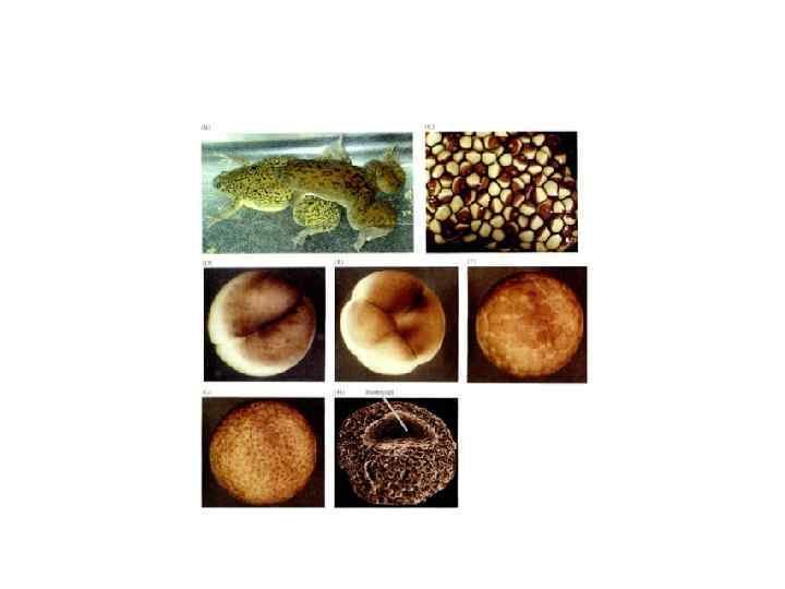
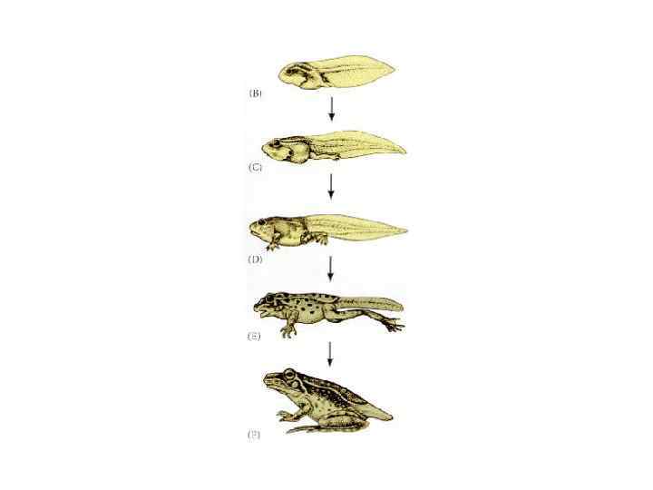
Жизненный цикл амфибий
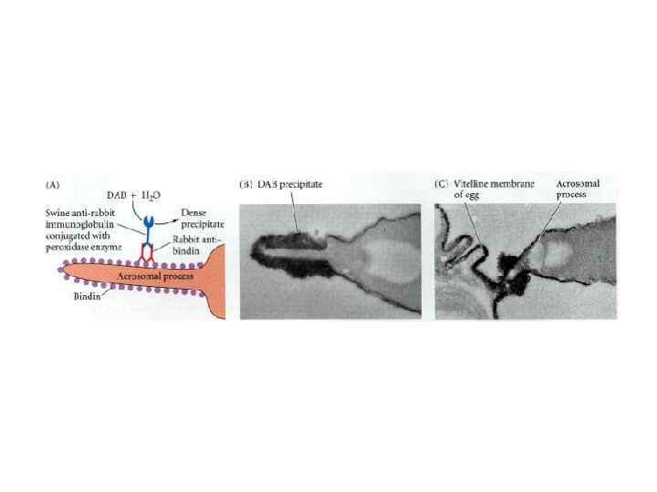

аллурин

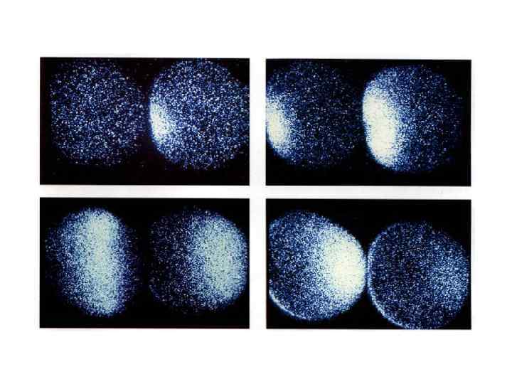
Быстрый блок полиспермии
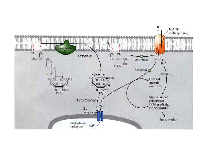

Cl outflux

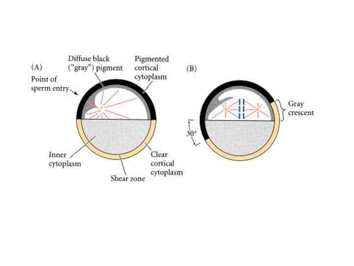
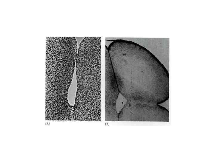
Кортикальная реакция

Event Approximate time postinseminationa EARLY RESPONSES Sperm-egg binding 0 sec Fertilization potential rise (fast block to polyspermy) within 1 sec Sperm-egg membrane fusion within 6 sec Calcium increase first detected 6 sec Cortical vesicle exocytosis (slow block to polyspermy) 15 - 60 sec LATE RESPONSES Activation of NAD kinase starts at 1 min Increase in NADH and NADPH starts at 1 min Increase in O 2 consumption starts at 1 min Sperm entry 1 - 2 min Acid efflux 1 - 5 min Increase in p. H (remains high) 1 - 5 min Sperm chromatin decondensation 2 -12 min Sperm nucleus migration to egg center 2 - 12 min Egg nucleus migration to sperm nucleus 5 - 10 min Activation of protein synthesis starts at 5 -10 min Activation of amino acid transport starts at 5 10 min Initiation of DNA synthesis 20 - 40 min Mitosis 60 - 80 min First cleavage 85 -95 min
 during the early development of the frog Xenopus laevis.")
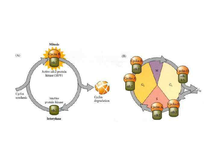
Levels of maturation-promoting factor (MPF) during the early development of the frog Xenopus laevis. The normal maturational signal is the hormone progesterone, which stimulates the ovulation of the oocytes and the initiation of meiosis. (After Murray and Kirschner, 1989. )
 Cyclin and")
Close Window The development of cell cycle regulation in Drosophila embryogenesis. (A) Cyclin and cdc 25 (string) protein are both abundant prior to fertilization. Therefore, during the first seven cell cycles, MPF kinase activity is constant and the nuclear divisions proceed as rapidly as the enzymes and substrates function. As cyclin becomes degraded, its synthesis (from m. RNAs stored in the cytoplasm) becomes limiting at cycle 8. By cycle 14, the maternal m. RNA for cyclin is gone, and it must be synthesized from nuclear genes. Moreover, the degradation of string protein mandates new synthesis from the nucleus. Pre-MPF accumulates but isn't activated until the string phosphatase cleaves the T-14 and Y-15 phosphates from the cdc 2 kinase. The mechanisms that relate MPF activity to the completion of DNA synthesis and the initiation of cytokinesis are currently being investigated. (After Edgar et al. , 1994. )
 - гетеродимер из cyclin.")
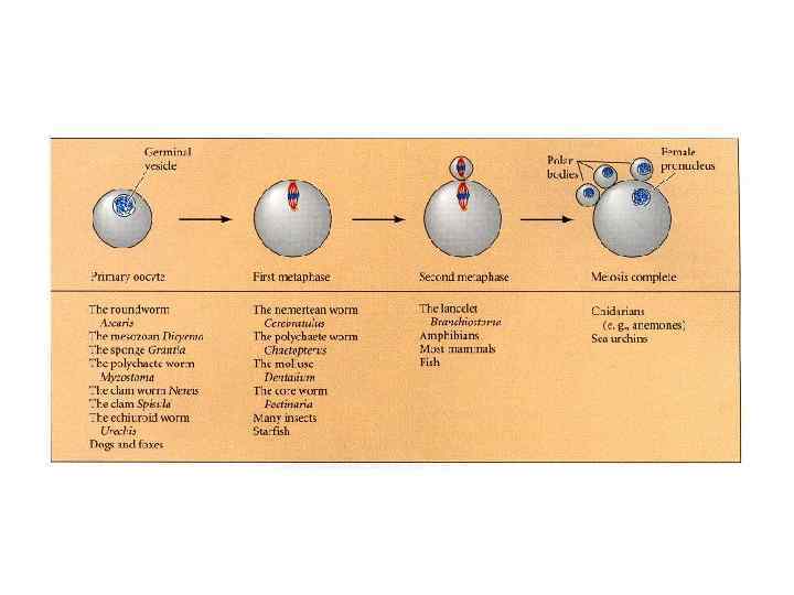
Метафаза r коррелирует с активность MPF ( mitosis promoting factor) - гетеродимер из cyclin. B и cyclin-dependent kinase(Cdc 2 или Сdk 1). Комплекс стабилизируется CFS (cytostatic factor), присутствие которого доказано на этой стадии ( экстракт арестует деления). В митозе есть негативная обратная связь, когда активация MPF ведет к его инактивации. Здесь MPF ингибируется в профазе. I ( на ст II роста) путем фосфорилирования Cdc 2 ( есть киназа и фосфатаза) CSF защищает циклин от убиквитинового комплекса ( состоящего из 12 белков) , комплекс называется anaphase promoting complex (APC). Endogenous meiotic inhibitor 2 (Emi 2) блокирует работу АРС. Emi подвергается убиквитиновому разрушению после фосфорилирования калмодулин-зависимой киназой при активации кальмодулина всплеском Ca после оплодотворения.

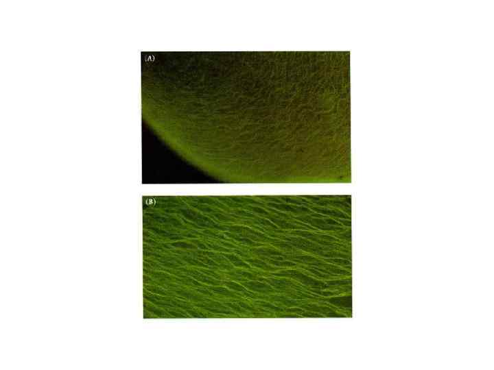
EMTB-3 GFP Transgenic Zebrafish Embryos Allow Live Imaging of Microtubule Organization in Large Cells Orange arrows indicate positions of centrosomes. (A) Shortly after fertilization, sperm aster expands throughout the cell. The scale bar represents 200 mm. (B) Before metaphase, sperm aster breaks down and first mitotic spindle forms. (C) During anaphase-telophase, astral microtubules grow out, and centrosomes move apart. An interaction zone forms in the plane where sister asters contact each other (between blue arrows). (D) Centrosomes separate and align in the direction of the future spindle during late telophase (see enlargement). The centrosomes in the left aster are out of focus. Nuclei (green arrow) follow centrosomes, lagging behind. (E) Second mitotic spindles assemble after cytokinesis
 pull with dynein")
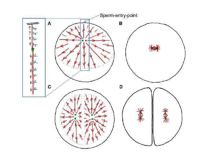
Model for Cleavage Plane Determination in Large Cells. Astral microtubules (red) pull with dynein (blue)on cytoplasm to determine center and longest axis for cell division. (A) Sperm enters at periphery. Cellular boundary causes asymmetry in sperm aster. Numbers of dynein bound is proportional to microtubule length, resulting in net force on centrosome toward cell’s center, but strongest stress on duplicated centrosomes (green) is perpendicular to movement. (B) Sperm aster breaks down; small first mitotic spindle forms. (C) At onset of anaphase, asters expand but do not grow into each other. The free interaction zone between the microtubules generates the asymmetry in the aster, leading to a net force on the pair of centrosomes toward the future centers of the daughter cells. The forces on the individual centrosomes cause the largest stress perpendicular to this movement, resulting in the alignment of the linked centrosomes with the aster’s longest axis. (D) The cytokinetic furrow divided the cell into two, where the telophase asters overlapped, cutting through the sperm entry point. Asters break down; small mitotic spindles form at center and alongest axis of daughter cells
Оплодотворение.ppt