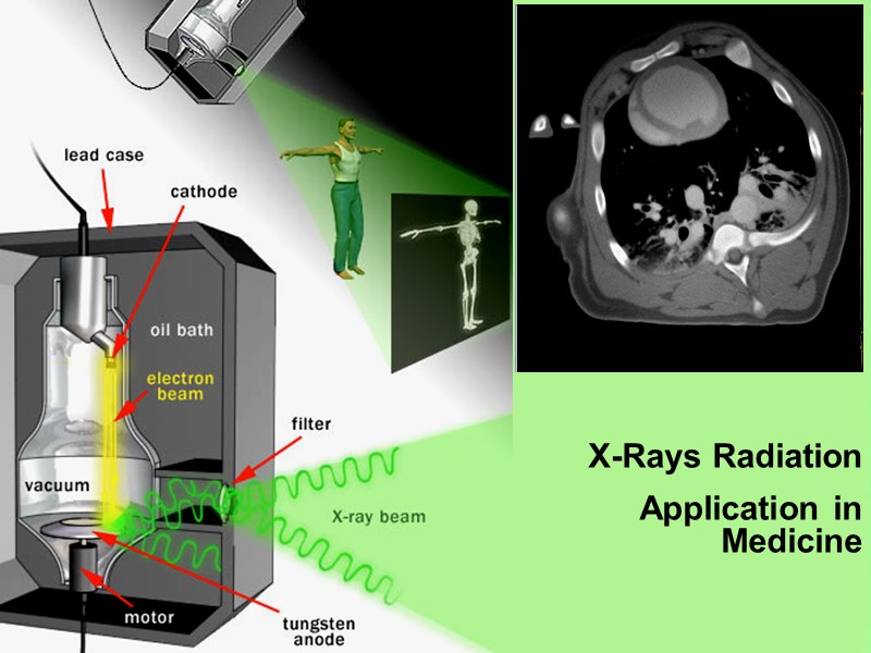
 X-Rays Radiation Application in Medicine
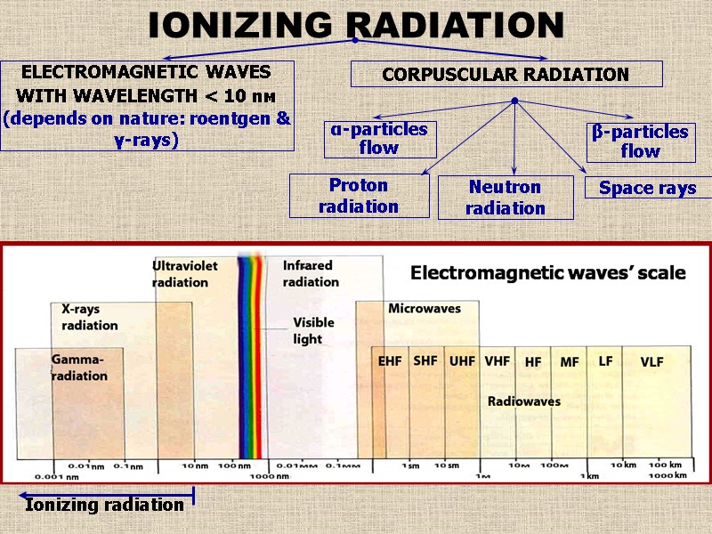 IONIZING RADIATION ELECTROMAGNETIC WAVES WITH WAVELENGTH < 10 nм (depends on nature: roentgen & γ-rays) CORPUSCULAR RADIATION α-particles flow Proton radiation Space rays β-particles flow Neutron radiation
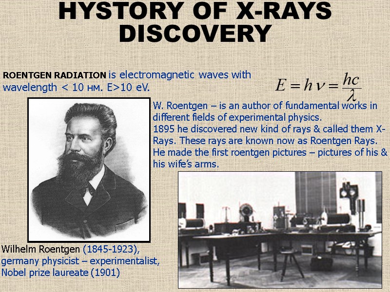 W. Roentgen – is an author of fundamental works in different fields of experimental physics. 1895 he discovered new kind of rays & called them X-Rays. These rays are known now as Roentgen Rays. He made the first roentgen pictures – pictures of his & his wife’s arms. ROENTGEN RADIATION is electromagnetic waves with wavelength < 10 нм. Е>10 еV. Wilhelm Roentgen (1845-1923), germany physicist – experimentalist, Nobel prize laureate (1901) HYSTORY OF X-RAYS DISCOVERY
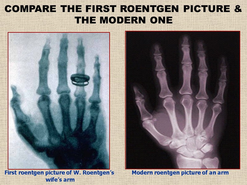 First roentgen picture of W. Roentgen’s wife’s arm COMPARE THE FIRST ROENTGEN PICTURE & THE MODERN ONE Modern roentgen picture of an arm
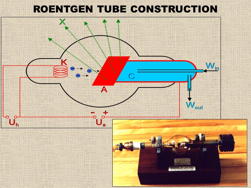 ROENTGEN TUBE CONSTRUCTION
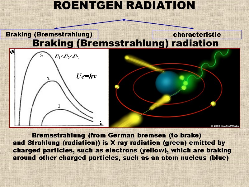 ROENTGEN RADIATION Braking (Bremsstrahlung) characteristic Braking (Bremsstrahlung) radiation Bremsstrahlung (from German bremsen (to brake) and Strahlung (radiation)) is X ray radiation (green) emitted by charged particles, such as electrons (yellow), which are braking around other charged particles, such as an atom nucleus (blue)
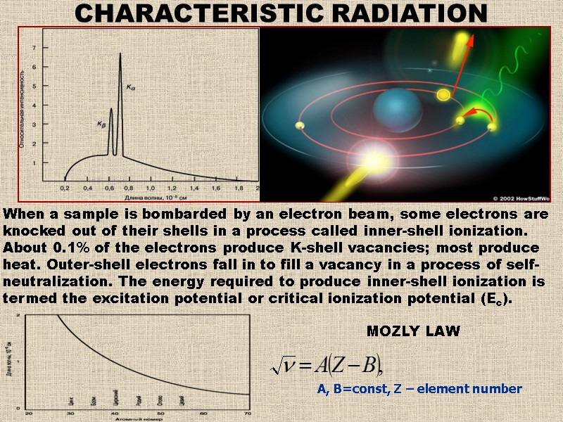 А, В=const, Z – element number CHARACTERISTIC RADIATION When a sample is bombarded by an electron beam, some electrons are knocked out of their shells in a process called inner-shell ionization. About 0.1% of the electrons produce K-shell vacancies; most produce heat. Outer-shell electrons fall in to fill a vacancy in a process of self-neutralization. The energy required to produce inner-shell ionization is termed the excitation potential or critical ionization potential (Ec). MOZLY LAW
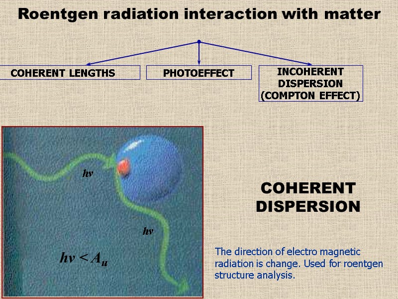 Roentgen radiation interaction with matter COHERENT LENGTHS INCOHERENT DISPERSION (COMPTON EFFECT) PHOTOEFFECT COHERENT DISPERSION The direction of electro magnetic radiation is change. Used for roentgen structure analysis.
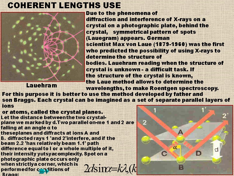 Due to the phenomena of diffraction and interference of X-rays on a crystal on a photographic plate, behind the crystal, symmetrical pattern of spots (Lauegram) appears. German scientist Max von Laue (1879-1960) was the first who predicted the possibility of using X-rays to determine the structure of bodies. Lauehram reading when the structure of crystal is unknown - a difficult task. If the structure of the crystal is known, the Laue method allows to determine the wavelengths, to make Roentgen spectroscopy. Let the distance between the two crystal-plane we marked by d.Two parallel on-me 1 and 2 are falling at an angle α to theseplanes and diffracts at ions A and B. diffracted rays 1 'and 2'interfere, and if the beam 2.2 'has relatively beam 1.1' path difference equal to l or a whole multiple of it, their intensity yutsyacomplexity. Spot on a photographic plate occurs only when strictlya corner, which is performed for conditions of Bragg: For this purpose it is better to use the method developed by father and son Braggs. Each crystal can be imagined as a set of separate parallel layers of ions or atoms, called the crystal planes. COHERENT LENGTHS USE
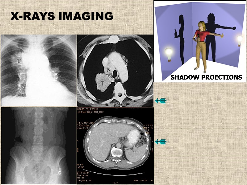 X-RAYS IMAGING SHADOW PROECTIONS
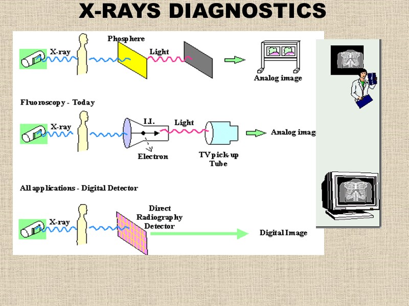 X-RAYS DIAGNOSTICS
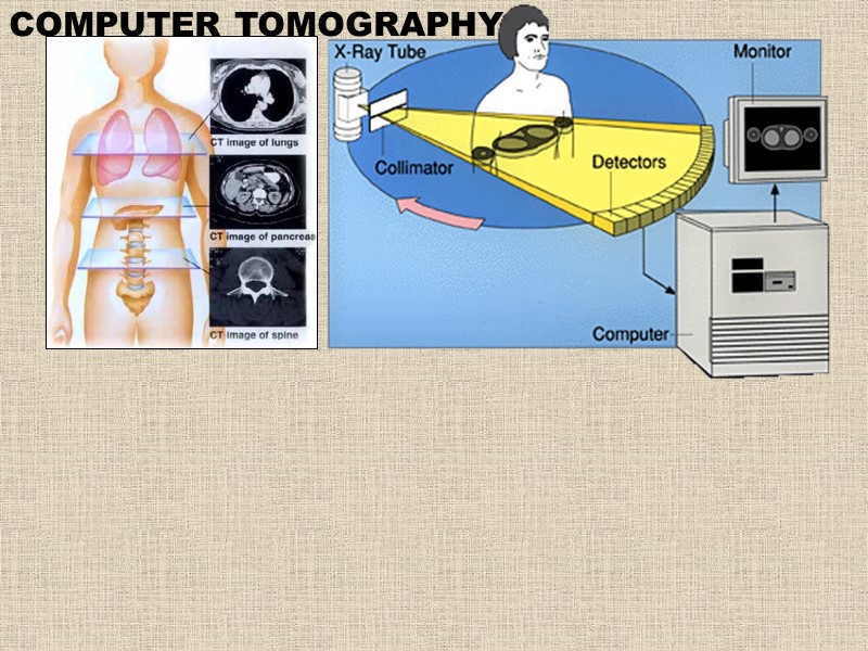 COMPUTER TOMOGRAPHY
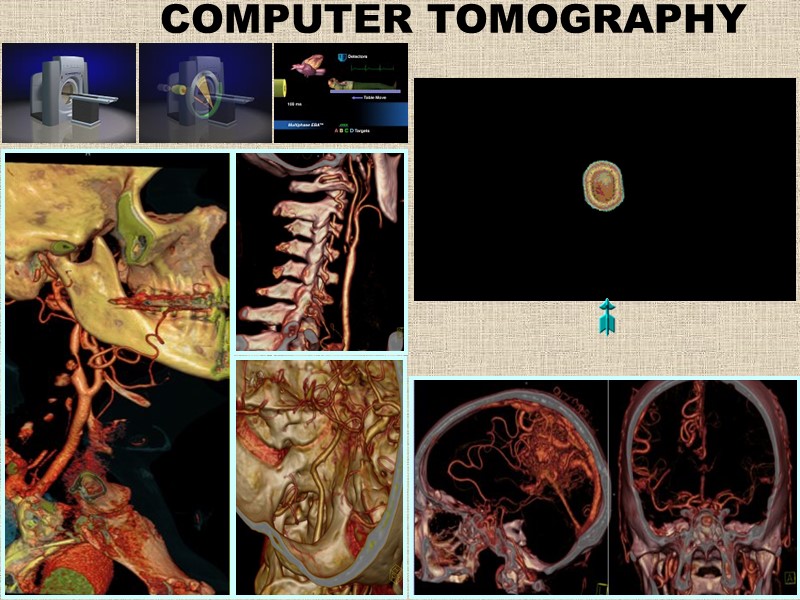 COMPUTER TOMOGRAPHY
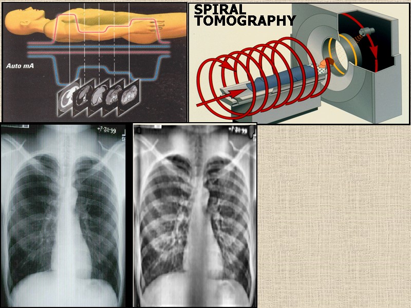 SPIRAL TOMOGRAPHY
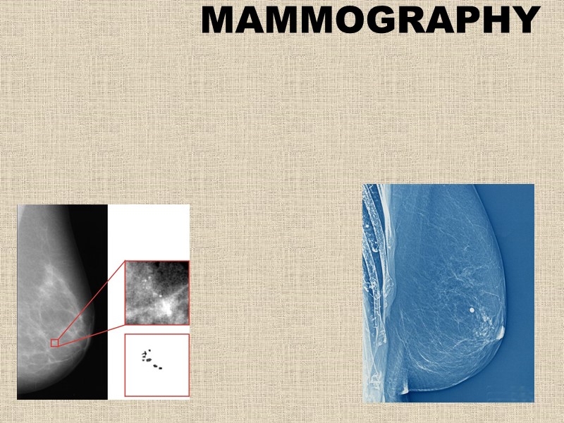 MAMMOGRAPHY
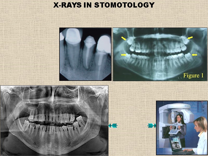 X-RAYS IN STOMOTOLOGY
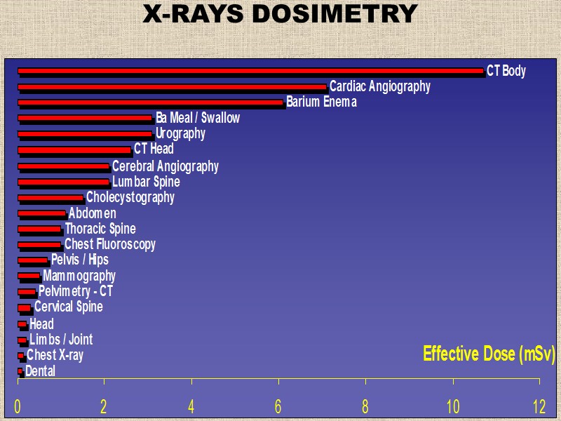 X-RAYS DOSIMETRY


