

ЛЮМИН2.ppt
- Количество слайдов: 41
 What is fluorescence? FLUORESCENCE is the light emitted by an atom or molecule after a finite duration subsequent to the absorption of electromagnetic energy. Specifically, the emitted light arises from the transition of the excited species from its first excited electronic singlet level to its ground electronic level.
 ions electric fields Fluorescent Probe temperature p. H viscosity polarity Fluorescence Probes are essentially molecular stopwatches which monitor dynamic events which occur during the excited state lifetime – such as movements of proteins or protein domains
 Experimental Systems Accessible to Fluorescence Molecular structure and dynamics Actin filament Animals Cell organization and function Actin filaments In endothelia cell Engineered surfaces GFP in a mouse High throughput Drug discovery GM
 light Light Propagation Direction") Polarization Unpolarized (natural) light Light Propagation Direction
Polarization Unpolarized (natural) light Light Propagation Direction
 light Polarized light") Polarizer Unpolarized (natural) light Polarized light
Polarizer Unpolarized (natural) light Polarized light
 Поляризация люминесценции Z Electric vector of exciting light Exciting light X O Y
 Z Electric vector of exciting light Exciting light X O Y
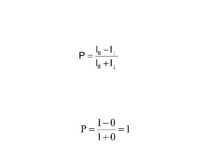
 If the emitted light is totally polarized in the perpendicular direction then: The limits of polarization are thus +1 to -1
 or : P r 0. 50 0. 40 0. 30 0. 22 0. 10 0. 069
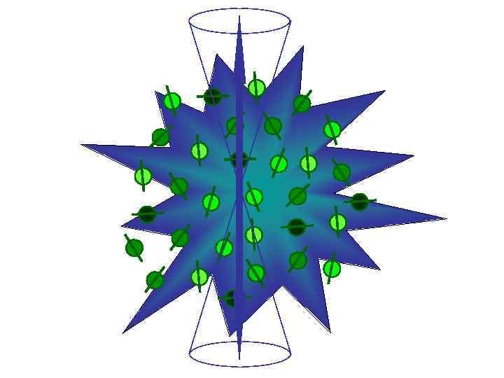
 Z + Electric vector of exciting light Exciting light X O Y -
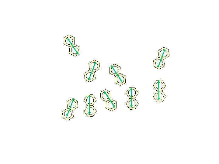
 Specifically, the probability of the absorption is proportional to the cosine squared (cos 2 ) of the angle between the exciting light and the transition dipole. cos 2 function Absorption Dipole Exciting light o 0 o 45 o 90
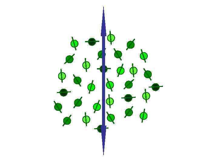
 h Potential dipoles Excited state dipoles
 + -
+ -
 : S 0 S 2 S 0 S 1 S 0 S 1 S 0 200 250 300 S 0 S 2 S 0 S 1
 S 0 -S 1
 Average direction
 S 0 -S 2
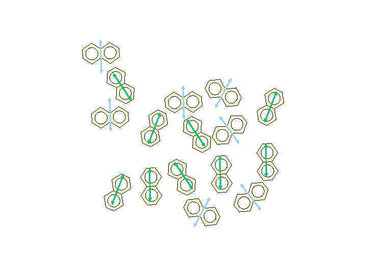
 Average direction
.") Consider the excitation polarization spectrum for phenol (in glycerol at - 70 C).
Consider the excitation polarization spectrum for phenol (in glycerol at - 70 C).
 In cases where there are multiple overlapping absorption bands at various angles, the excitation polarization spectrum can be somewhat complex as shown below for indole.
 Excitation polarization spectra of rhodamine B embedded in a Lucite matrix at room temperature. Emission was viewed through a cut-on filter passing wavelengths longer than 560 nm; slits were ~4 nm.
 Another example is protoporphyrin IX in glycerol at – 20 C
 Note: in the case of multi-photon excitation the limits differ
 Absorption dipole Emission dipole t=0 Emission dipole t>0
 Absorption dipole Emission dipole t=0 Emission dipole t>0 :
 t=0 t ABSORPTION w ORIENTATION AUTOCORRELATION FUNCTION probability that a molecule having a certain orientation at time zero is oriented at angle with respect to its initial orientation EMISSION
 Длительные процессы свечения Фосфоресценция. Внутренняя и интекомбинационная конверсия
 The Perrin-Jablonski Diagram The life history of an excited state electron in a luminescent probe Internal conversion 10 -12 s S 2 S 1 Absorption 10 -15 s S 0 Inter-system Crossing 10 -10 s T 1 Fluorescence 10 -9 s Radiationless Decay <10 -9 s Phosphorescence 10 -3 s Key points: üExcitation spectra are mirror images of the emission spectra üEmission has lower energy compared to absorption üTriplet emission is lower in energy compared to singlet emission üMost emission/quenching/FRET/chemical reactions occur from the lowest vibrational level of [S]1 GM
 Фосфоресценция
, Iвозб S 1 ◠◡◠◡ S") Процесс hv 0 + S 0 S 1 (возбуждение), Iвозб S 1 ◠◡◠◡ S 0 + тепло (внутренняя конверсия) kвк [S 1 ] S 1 ◠◡◠◡ T 1 + тепло (интеркомбинационная KST [S 1 ] конверсия) T 1 ◠◡◠◡ S 0 + тепло (интеркомбинационная KT [T 1 ] конверсия) T 1 S 0 + hvфос (фосфоресценция), Kфос [T 1] S 1 S 0 + hvфл (флуоресценция), Kфл [S 1 ] (замедленная флуоресценция) Kе [T 1 ]
Процесс hv 0 + S 0 S 1 (возбуждение), Iвозб S 1 ◠◡◠◡ S 0 + тепло (внутренняя конверсия) kвк [S 1 ] S 1 ◠◡◠◡ T 1 + тепло (интеркомбинационная KST [S 1 ] конверсия) T 1 ◠◡◠◡ S 0 + тепло (интеркомбинационная KT [T 1 ] конверсия) T 1 S 0 + hvфос (фосфоресценция), Kфос [T 1] S 1 S 0 + hvфл (флуоресценция), Kфл [S 1 ] (замедленная флуоресценция) Kе [T 1 ]
 Замедленная люминесценция • Типа Е
 Замедленная люминесценция • Типа П
 ЗФ-1, ФОС-2
 КОНЕЦ
КОНЕЦ