Адгезионные молекулы.ppt
- Количество слайдов: 31
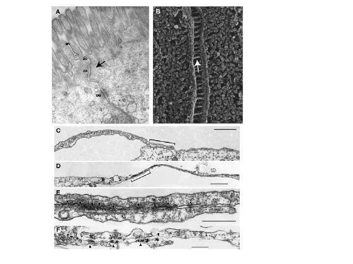
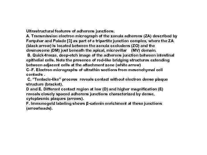 Ultrastructural features of adherens junctions. A. Transmission electron micrograph of the zonula adherens (ZA) described by Farquhar and Palade [3] as part of a tripartite junction complex, where the ZA (black arrow) is located between the zonula occludens (ZO) and the desmosome (DM) just beneath the apical, microvillar (MV) domain. B. Quick-freeze, deep-etch image of the adherens junction between intestinal epithelial cells. Note the presence of rod-like bridging structures extending between adjacent cells at the attachment zone (white arrow) C–F. Electron micrographs of ultrathin sections from mesenchymal cell contacts. C. “Tentacle-like” process reveals contact without electron dense plaque structure (bracket). D and E. Different contact region at low (D) and higher magnification (E) reveals closely spaced adherens junctions characterized by dense, cytoplasmic plaques (arrows). F. Immunogold labeling shows β-catenin enrichment at these junctions (arrowheads).
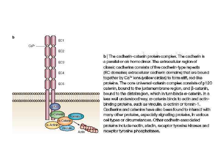
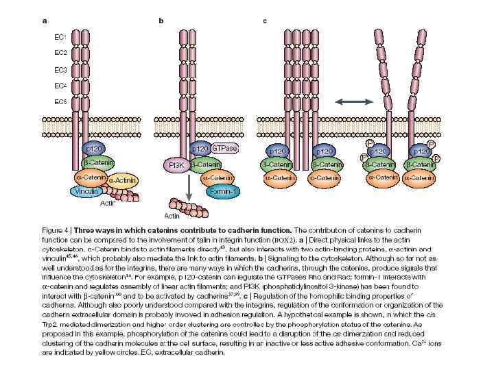
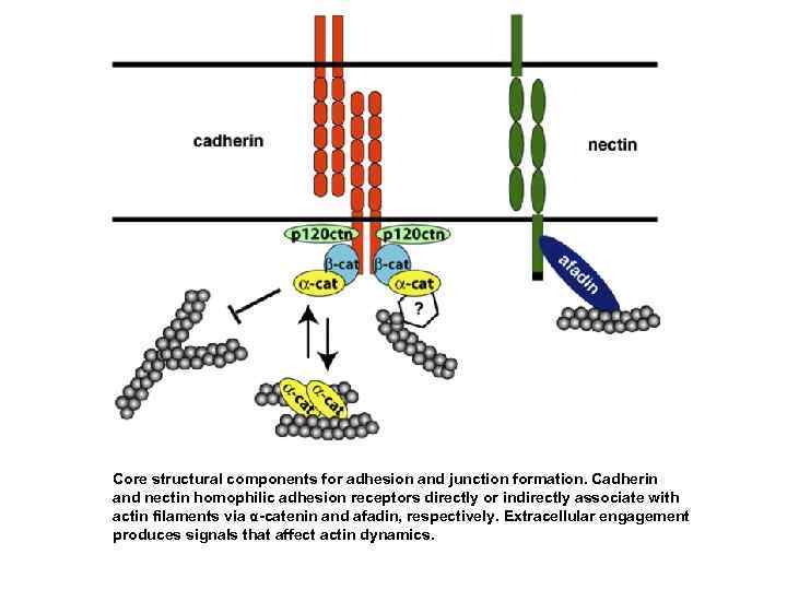 Core structural components for adhesion and junction formation. Cadherin and nectin homophilic adhesion receptors directly or indirectly associate with actin filaments via α-catenin and afadin, respectively. Extracellular engagement produces signals that affect actin dynamics.
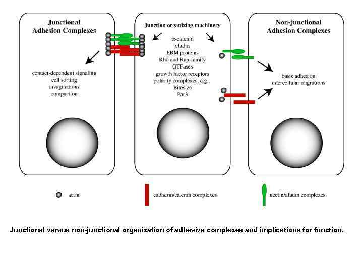 Junctional versus non-junctional organization of adhesive complexes and implications for function.
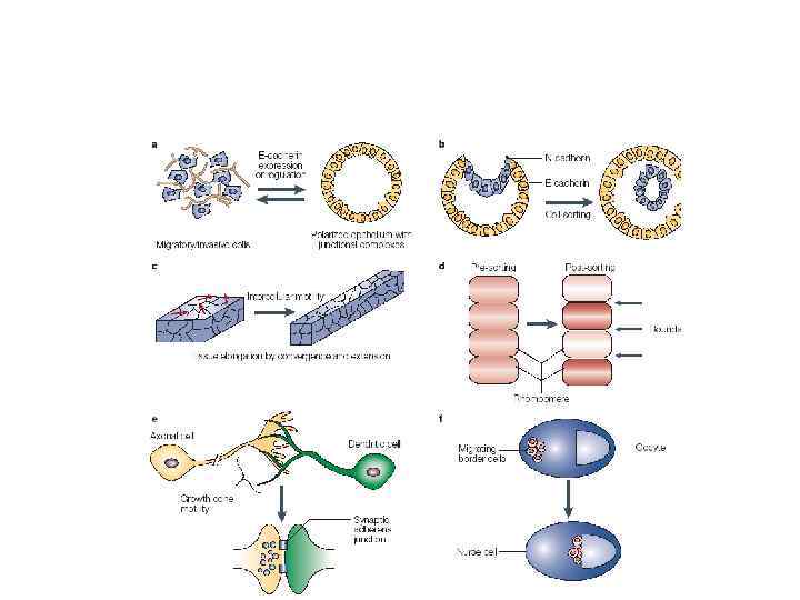
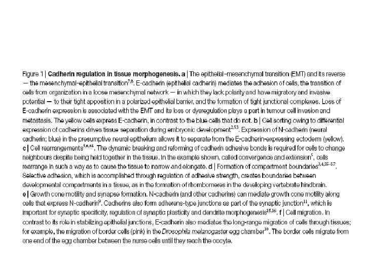
 In adult tissues, cadherins control • orderly turnover of rapidly growing tissues such as the lining of the gut and the epidermis, • the plasticity and regulation of neuronal synapses, • the physiological regulation of epithelial and endothelial cell • junctions to allow controlled passage of solutes, water and lymphoid cells across the cell layer, • the maintenance of stable tissue organization to prevent the dissociation and spread of tumour cells
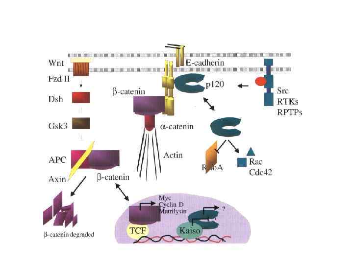
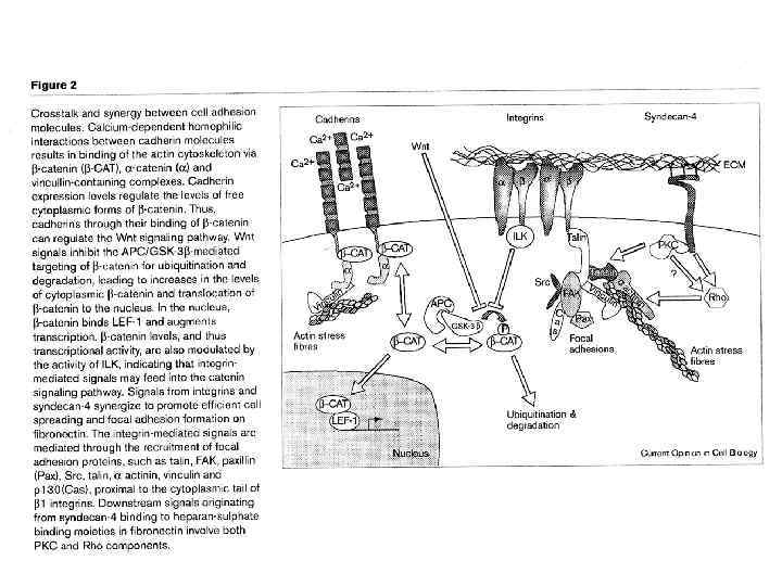
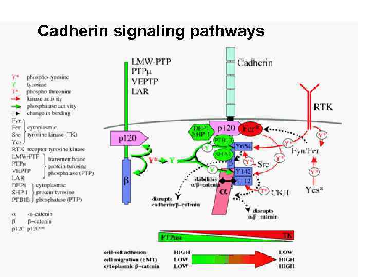 Cadherin signaling pathways
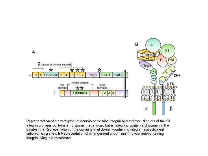 Representation of a prototypical αI-domain-containing integrin heterodimer. Nine out of the 18 integrin α chains contains an αI domain, as shown, but all integrins contain a βI domain in the β subunit. a Representation of the domains in αI domain-containing integrin (stars divalent cation-binding sites. b Representation of arrangement of domains in αI-domain-containing integrin kying in a membrane
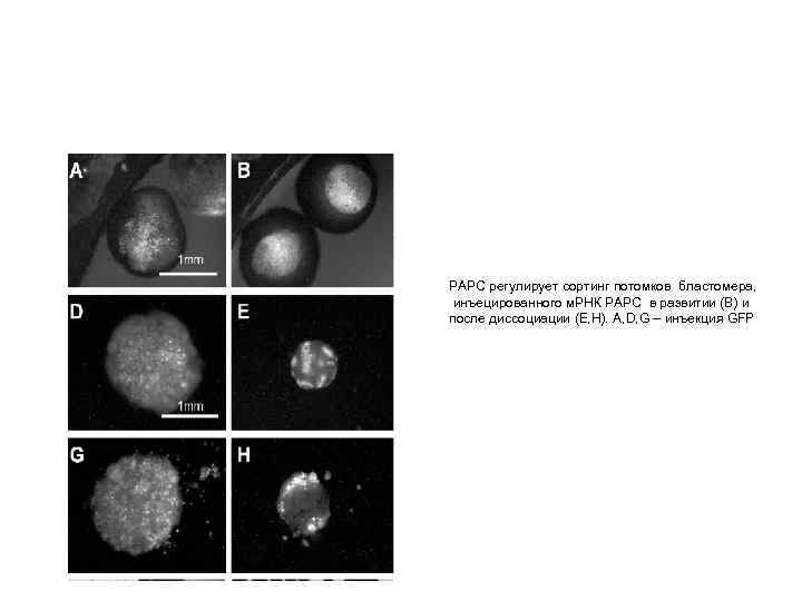 РАРС регулирует сортинг потомков бластомера, инъецированного м. РНК РАРС в развитии (В) и после диссоциации (Е, Н). A, D, G – инъекция GFP
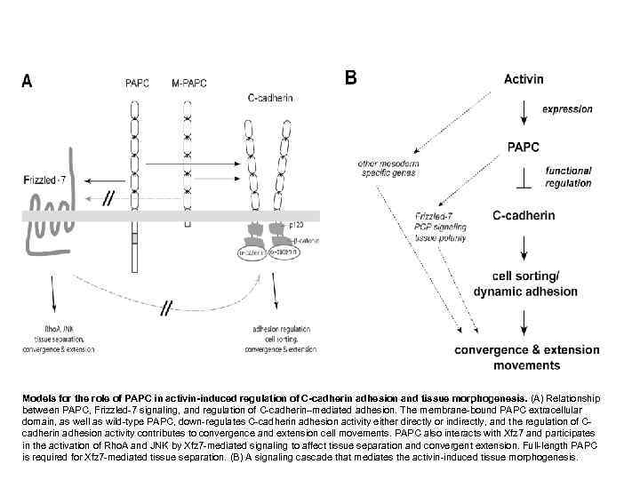 Models for the role of PAPC in activin-induced regulation of C-cadherin adhesion and tissue morphogenesis. (A) Relationship between PAPC, Frizzled-7 signaling, and regulation of C-cadherin–mediated adhesion. The membrane-bound PAPC extracellular domain, as well as wild-type PAPC, down-regulates C-cadherin adhesion activity either directly or indirectly, and the regulation of C- cadherin adhesion activity contributes to convergence and extension cell movements. PAPC also interacts with Xfz 7 and participates in the activation of Rho. A and JNK by Xfz 7 -mediated signaling to affect tissue separation and convergent extension. Full-length PAPC is required for Xfz 7 -mediated tissue separation. (B) A signaling cascade that mediates the activin-induced tissue morphogenesis.
