
Transthoracic Echocardiography. Standard Imaging of A.


- Размер: 16.4 Mегабайта
- Количество слайдов: 49
Описание презентации Transthoracic Echocardiography. Standard Imaging of A. по слайдам
Transthoracic Echocardiography. Standard Imaging of
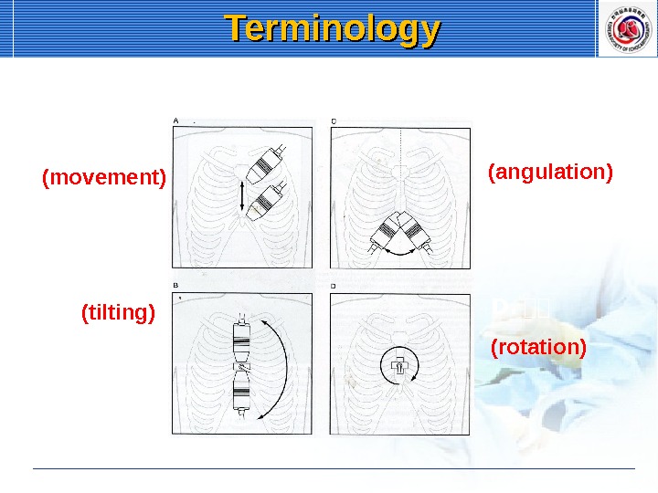
A. 이이 (movement) B. 이이이 (tilting) C. 이이 (angulation) D. 이이 (rotation) Terminology
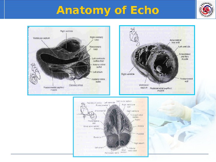
Anatomy of Echo
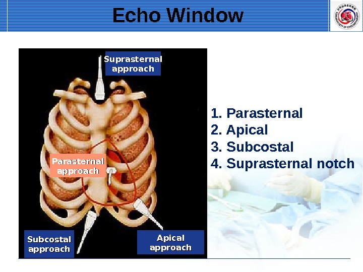
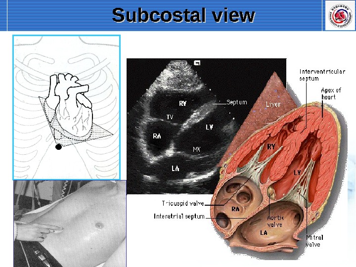
Suprasternal approach Parasternal approach Subcostal approach Apical approach Echo Window 1. Parasternal 2. Apical 3. Subcostal 4. Suprasternal notch
Basic views of Echocardiography Apical view Subcostal view Suprasternal view
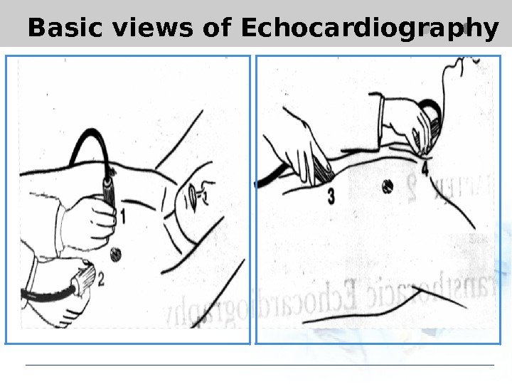
Basic views of Echocardiography
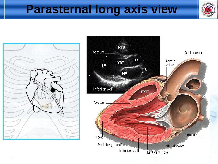
Parasternal long axis view
Parasternal long axis view
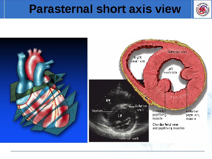
Parasternal short axis view
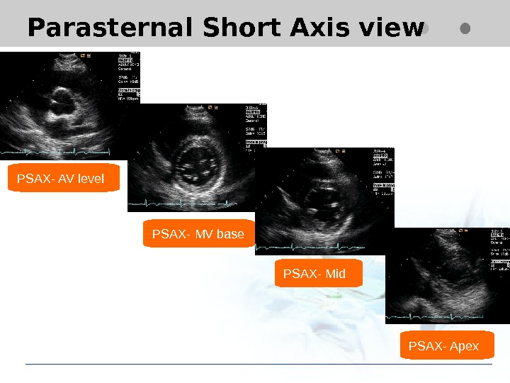
Parasternal Short Axis view PSAX- AV level PSAX- Mid PSAX- MV base PSAX- Apex
Parasternal short axis view
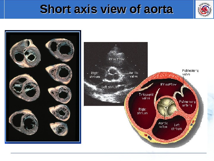
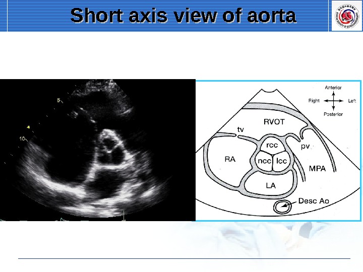
Short axis view of aorta
Short axis view of aorta
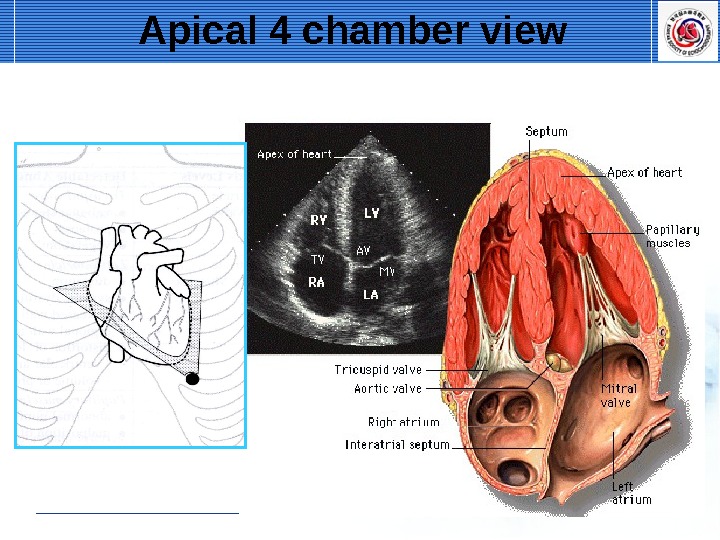
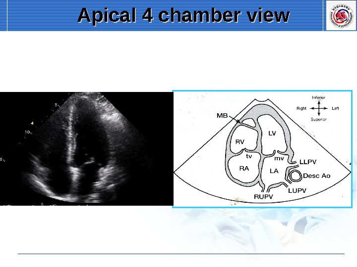
Apical 4 chamber view
Apical 4 chamber view
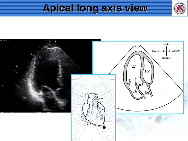
Apical long axis view
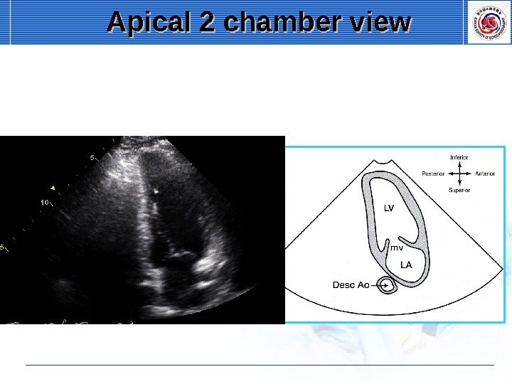
Apical 2 chamber view
Apical 2 chamber view
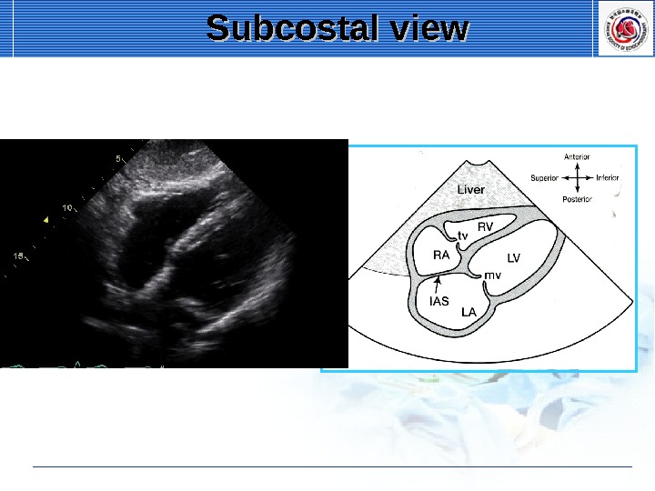
Subcostal view
Subcostal view
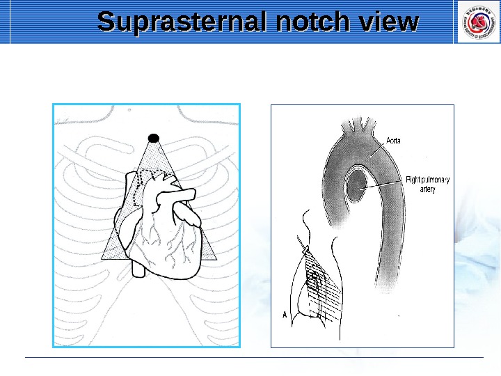
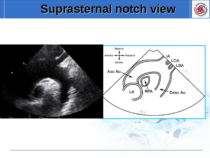
Suprasternal notch view
Suprasternal notch view
Measurement of Cardiac Chambers
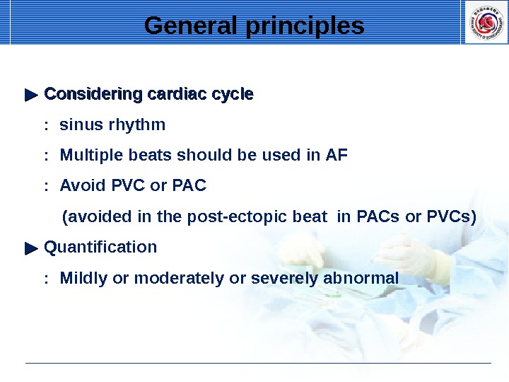
▶▶ Considering cardiac cycle : sinus rhythm : Multiple beats should be used in AF : Avoid PVC or PAC (avoided in the post-ectopic beat in PACs or PVCs) ▶▶ Quantification : Mildly or moderately or severely abnormal General principles
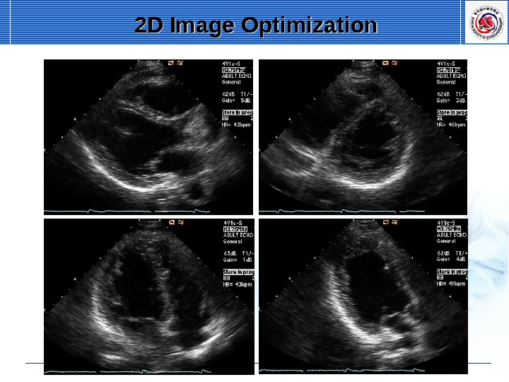
• Respiration (at end-expiration) • Image at minimum depth necessary • Highest possible transducer frequency • Adjust gains, dynamic range, transmit • Frame rate ≥ 30/s • Harmonic imaging • B-color imaging General principles
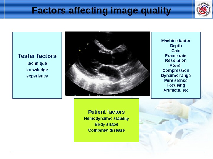
Factors affecting image quality Tester factors technique knowledge experience Machine factor Depth Gain Frame rate Resolution Power Compression Dynamic range Persistence Focusing Artifacts, etc Patient factors Hemodynamic stability Body shape Combined disease
2 D Image Optimization
2 D Image Optimization
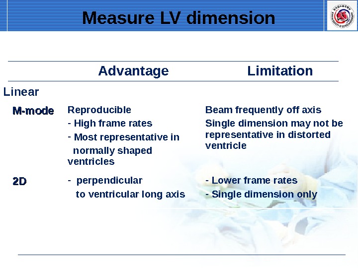
Measure LV dimension Advantage Limitation Linear M-mode Reproducible — High frame rates — Most representative in normally shaped ventricles Beam frequently off axis Single dimension may not be representative in distorted ventricle 2 D 2 D — perpendicular to ventricular long axis — Lower frame rates — Single dimension only
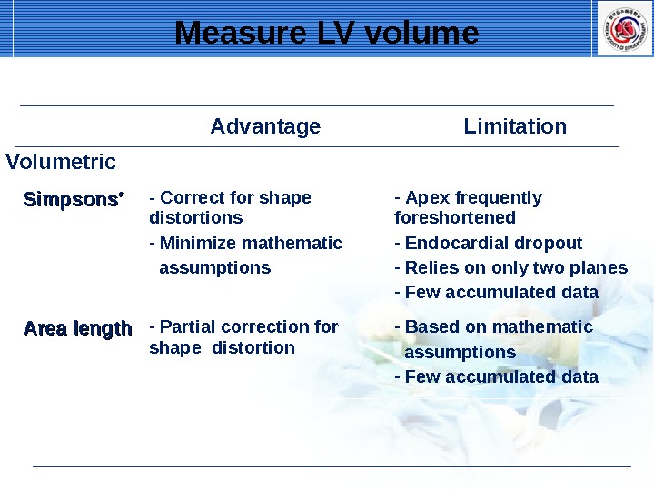
Advantage Limitation Volumetric Simpsons′ — Correct for shape distortions — Minimize mathematic assumptions — Apex frequently foreshortened — Endocardial dropout — Relies on only two planes — Few accumulated data Area length — Partial correction for shape distortion — Based on mathematic assumptions — Few accumulated data. Measure LV volume
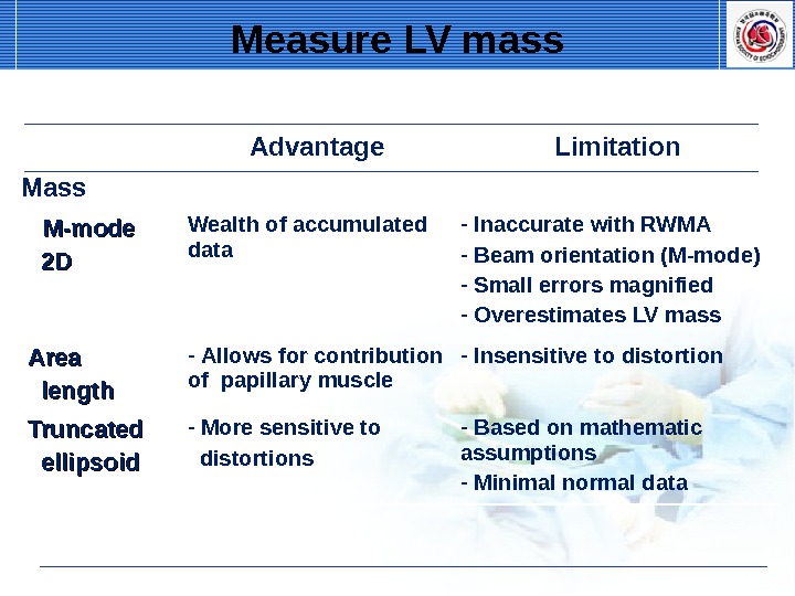
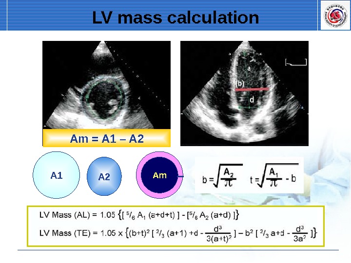
Advantage Limitation Mass M-mode 2 D 2 D Wealth of accumulated data — Inaccurate with RWMA — Beam orientation (M-mode) — Small errors magnified — Overestimates LV mass Area length — Allows for contribution of papillary muscle — Insensitive to distortion Truncated ellipsoid — More sensitive to distortions — Based on mathematic assumptions — Minimal normal data. Measure LV mass
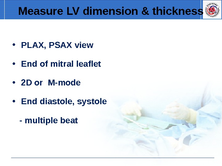
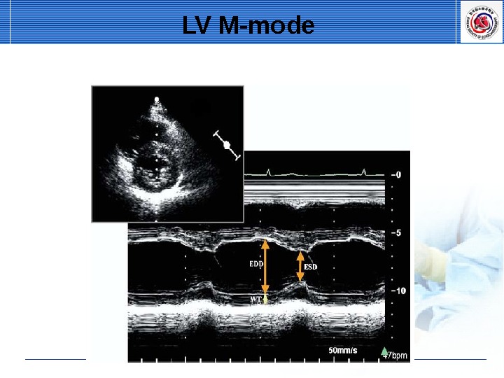
• PLAX, PSAX view • End of mitral leaflet • 2 D or M-mode • End diastole, systole — multiple beat. Measure LV dimension & thickness
LV M-mode EDD ES
LV M-mode
Oblique parasternal images 이 이이이. LV
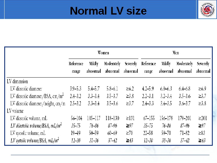
Normal LV size
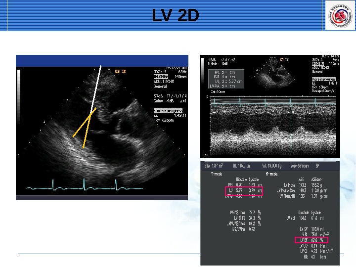
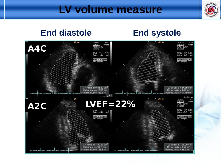
LV volume ▶▶ Manual measurements : Mid-papillary short axis view , A 4 C, and A 2 C view : Trace endocardial border ▶▶ End diastole : QRS starting point, pre-MV closure, or biggest dimension during cardiac cycle ▶▶ End systole : Pre-MV opening, or smallest dimension during cardiac cycle
LV volume measure End diastole End systole A 2 CA 4 C LVEF=22%
LV mass calculation A 2 A 1 Am. Am = A 1 –
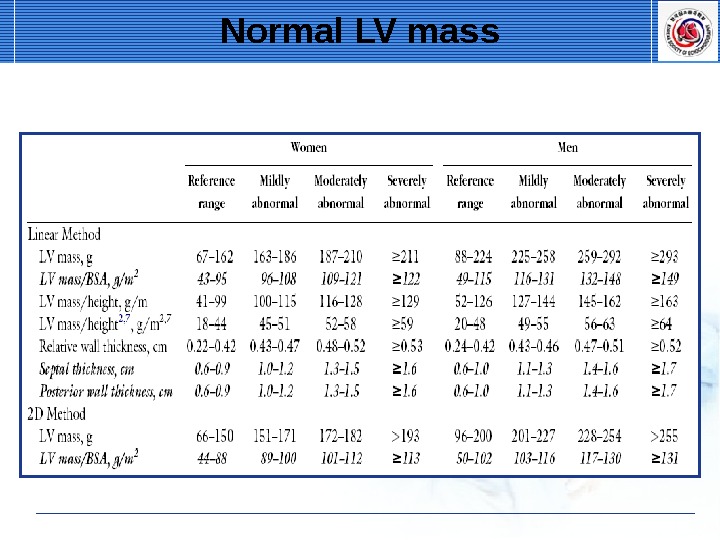
Normal LV mass
Measure LA size ▶▶ LV end systole, maximal LA size ▶▶ Avoid foreshortening of LA ▶▶ LA length in true long axis of the LA ▶▶ Excluded pulmonary veins and L
▶▶ Measured from the leading edge of the posterior aortic wall to the leading edge of the posterior LA wall — measure end systole Measure LA size
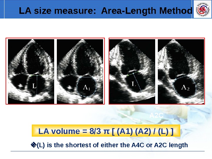
A 4 C A 2 C LA volume = 8/3 π [ (A 1) (A 2) / (L) ] ※※ (L) is the shortest of either the A 4 C or A 2 C length. LA size measure: Area-Length Method
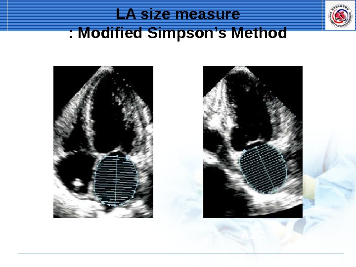
A 4 C A 2 CLA size measure : Modified Simpson’s Method
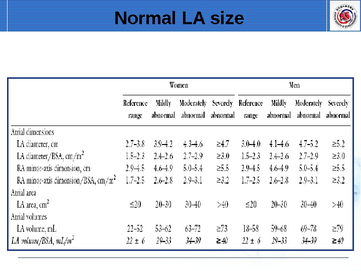
Normal LA size
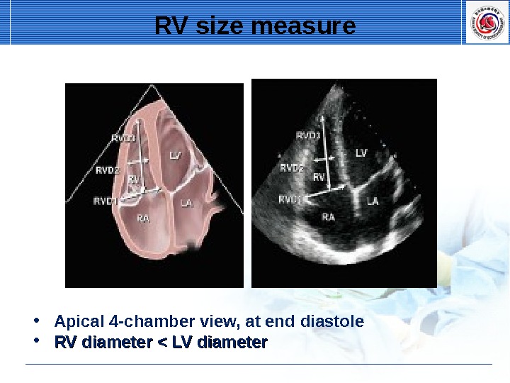
RV size measure • Apical 4 -chamber view, at end diastole • RV diameter < LV diameter
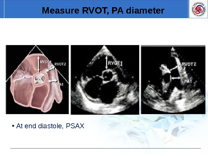
• At end diastole, PSAX Measure RVOT, PA diameter
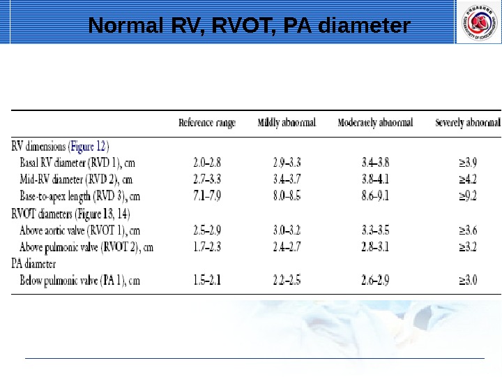
Normal RV, RVOT, PA diameter