c2a37bbd46ae3ad2c208f929bbfcde3c.ppt
- Количество слайдов: 40


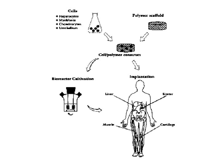
Tissue Engineering Expand number in culture Seed onto an appropriate scaffold with suitable growth factors and cytokines Remove cells from the body. Re-implant engineered tissue repair damaged site Place into culture

Tissue Engineering Hydrogels Nanofibrous scaffolds Self-assembling scaffolds Solid freeform fabricated scaffolds Differentiated cells Adult stem cells Embryonic stem cells Cells Scaffolds Tissue Engineering Biorectors Dynamic cell seeding Improved mass transfer Mechanical stimuli Signals Small molecules Growth factors/polypeptides Nucleic acids (DNA, si. RNA, and antisense oligonucleotides) Nanotechnology and Tissue Engineering: The Scaffold, CRC Press; 1 edition (June 16, 2008)

Nanotecnologia La nanotecnologia è una branca della scienza e dell’ingegneria che tratta di strutture e dispositivi in scala nanometrica. Caratterizzazione v Microscopia ottica v Microscopia elettronica v Microscopia a forza atomica Preparazione v Litografia (Optical, E-beam, NIP, DPL) v Etching (Wet Etching, Plasma Etching) v Deposizione (Evaporation, PECVD, Electrochemical Deposition, Sputtering, etc. ) v Epitaxial growth (MOCVD, MBE, etc. )

Perchè applichiamo la Nanotecnologia a in TE? Tissue Engineering is a methodology + Nanotechnology is a tool Cells on microfibrous scaffolds have a polarized relationship, with one side of the cell attached to the scaffold, the other exposed to physiological media. In comparison, it is likely that cells are more naturally constrained by nanofibrous scaffolds.

Scaffold Nanofibrosi • Electrospinning • Self-Assembly

Electrospinning • Questo processo coinvolge l’espulsione di un fluido contenente un polimero carico su di una superficie di carica opposta. • Polimeri differenti possono essere impiegati • E’ possibile controllare la grandezza delle fibre e la loro struttura

Research on Parameters of Electrospinning Process • Solution properties Ø Ø Ø Viscosity Conductivity Surface tension Polymer molecular weight Dipole moment Dielectric constant • Controlled variables Ø Ø Ø Flow rate Electric field strength Distance between tip and collector Needle tip design Collector composition and geometry • Ambient parameters Ø Temperature Ø Humidity Ø Air velocity Tissue Engineering. May 2006, 12(5): 1197 -1211.
 – Highly crystalline, hydrophilic, byproduct is glycolic")
Research on Materials • Polyglycolic acid (PGA) – Highly crystalline, hydrophilic, byproduct is glycolic acid • Polylactic acid (PLA) – Hydrophobic, lower melting temperature, byproduct is lactic acid • Polydioxanone (PDO) – Highly crystalline • Polycaprolactone (PCL) – Semi-crystalline properties, easily co-polymerized, byproduct caproic acid • Blends – – PGA-PLA PGA-PCL PLA-PCL PDO-PCL • • • Elastin Gelatin collagen Fibrillar collagen Collagen blends Fibrinogen • Synthetic polymers v PGA, PLA and PLGA most commonly used v PDO most similar to Elastin collagen blend (limited by shape memory) v PCL most elastic and mixed frequenlty with other material s v Provide nanoscale physical features • Natural polymers v Collagen Type I & III + PDO: best possible match for blood vessels Advanced Drug Delivery Reviews Volume 59, Issue 14, 10 December 2007, Pages 1413 -1433

Self Assembly Figure 1: Fabrication of various peptide materials. Figure 2: Self-assembling peptides form a three -dimensional scaffold woven from nanofibers ~ 10 nm in diameter. (a) Representation of self-assembling peptide. (b) Electron micrograph of three-dimensional scaffold formed in vitro. (c) Rat hippocampal neurons form active nerve connections; each green dot represents a single synapsis. (d) Neural cells from a rat hippocampal tissue slide migrate on the three-dimensional peptide scaffold. Cells on the polymer membrane (left) and on the peptide scaffold (right) are shown. Both glial cells (green) and neural progenitors (red) migrate into the three-dimensional peptide scaffold. (e) Brain damage repair in hamster. The peptide scaffold was injected into the optic nerve, which was first severed with a knife. The cut was sealed by the migrating cells after 2 days. A great number of neurons form synapses. (f) Chondrocytes from young and adult bovine encapsulated in the peptide scaffold. These cells not only produce a large amount of glycosaminoglycans (purple) and type II collagen (yellow), characteristic materials found in cartilage, but also a cartilage-like tissue in vitro 53. (g) Adult rat liver progenitor cells encapsulated in the peptide scaffold. The cells on the two-dimensional dish did not produce cytochrome P 450–type enzymes (left). However, cells in three-dimensional scaffolds showed cytochrome P 450 activity (right). Nature Biotechnology 21, 1171 - 1178 (2003)

Self-assembly • Associazione spontanea di pochi o numerosi componenti molecolari che porta alla formazione di strutture supramolecolari (strati, film, membrane…). • Il prodotto finale è ottenuto spontaneamente mescolando insieme I componenti in rapporti ottimizzati ed in condizioni particolari (solvente, temperatura, p. H, …) • The formation of supermolecules results from the recognition-directed spontaneous association of a welldefined and limited number of molecular components under the intermolecular control of the non-covalent interactions that hold them together. J. -M. Lehn, Science 2002, 295, 2400

Self-organization can be considered as ordered self-assembly. It concerns systems presenting a spontaneous emergence of order in either space or time or both. Structure of the LH 2 light-harvesting antenna system of Rhodopseudomonas acidophila which contains rings of 18 (a) and 9 (b) bacteriochlorophyll molecules.

Self-assembly and self-organization • Self-assembly and self-organization of a supramolecular architecture are both multistep processes implying information and instructed components. • They may follow a sequence and a hierarchy of assembly steps, and require reversibility of the connecting events, i. e. , kinetic lability and rather weak bonding (compared with covalent bonds), in order to allow the full exploration of the energy hypersurface of the system. • In other words, the product formation must be completely reversible and represent thermodynamic minimum for the system. In essence, all the information necessary for the assembly to occur is coded into the constituent parts.

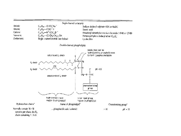
Self-assembly of amphiphiles v In amphiphilic molecules, such as surfactants, lipids and certain copolymers, one end contains a hydrophilic group while the rest of the molecule is hydrophobic, usually a long hydrocarbon chain. v The hydrophilic and hydrophobic interactions rely on the structure of the water H-bonds adopted around the dissolved end groups. Amphiphiles such as surfactants and lipids can associate into a variety of structures in aqueous solutions. These can transform from one to another by changing the solution conditions such as the electrolyte or lipid concentration, p. H or temperature. Most single chained surfactants form micelles, while most double chained surfactants form bilayers.

Self Assembly Figure 3: Lipid, peptide and protein scaffold nanowires. Figure 4: Microlenses and fiber-optics fabricated from protein scaffolds. Nature Biotechnology 21, 1171 - 1178 (2003)

Self-Assembling Peptide Scaffolds for Regenerative Medicine SAPNS heals the brain in young animals. SAPNS allows axons to regenerate through the lesion site in brain. PNAS March 28, 2006 vol. 103 no. 13 5054 -5059

Phase separation • • • This process involves dissolving of a polymer in a solvent at a high temperature followed by a liquid–liquid or solid–liquid phase separation induced by lowering the solution temperature Capable of wide range of geometry and dimensions include pits, islands, fibers, and irregular pore structures Simpler than self-assembly a) powder, b) scaffolds with continuous network, c) foam with closed pores SEM of nanofibrous scaffold with interconnected spherical macropores Advanced Drug Delivery Reviews Volume 59, Issue 14, 10 December 2007, Pages 1413 -1433

Carbon Nanotube Cell tracking and labeling Sensing cellular behavior Augmenting cellular behavior Cytotoxicity Murine myoblast stem cells incubated with DNA-encapsulated nanotubes neuron bridging an array of carbon nanotubes thereby creating neural networks. Biomaterials Volume 28, Issue 2, January 2007, Pages 344 -353

1985: Richard Smalley scopre che, in particolari situazioni, gli atomi di C compongono delle strutture ordinate di forma sferica, i fullereni. La struttura, dopo un successivo rilassamento, tende ad arrotolarsi su sé stessa, ottenendo la tipica struttura cilindrica: questi sono i nanotubi di carbonio. I nanotubi possono essere visti, analogamente al fullerene, come una delle forme allotropiche del carbonio. Le molecole di fullerene, costituite interamente di carbonio, assumono una forma simile a una sfera cava, a un ellissoide o ad un tubolare. I fullereni di forma simile a una sfera o a un ellissoide sono chiamati buckyball mentre quelli di forma tubolare sono chiamati buckytube o nanotubi di carbonio.

I fullereni sono strutturalmente simili alla grafite costituita di anelli esagonali collegati tra loro su un piano, ma si differenziano per alcuni anelli di forma pentagonale o a volte ettagonale che impediscono una struttura planare.

Il più piccolo e il più diffuso fullerene in cui nessuna coppia di pentagoni condivide un bordo, poiché questa condivisione risulterebbe destabilizzante, è il buckminsterfullerene. La struttura del buckminster-fullerene è quella di un icosaedro troncato costituito da esagoni e pentagoni, come un pallone da calcio, ai cui vertici si trova un atomo di carbonio e i cui bordi rappresentano i legami. Il nome di questo fullerene fa riferimento alla somiglianza con le cupole geodetiche predilette dall'architetto Richard Buckminster Fuller. Alcune molecole di fullerene sono piuttosto stabili a temperatura e pressione ambiente, nonostante siano energeticamente sfavorite rispetto ad altri allotropi del carbonio quali la grafite ed il diamante. La definizione di stabilità non può essere attribuita a tutta la categoria: alle molecole più stabili, come il C 60, si accompagnano una miriade di altre, a struttura più labile o del tutto instabile.

Il corpo del nanotubo è formato da soli esagoni, mentre le strutture di chiusura sono formate da esagoni e pentagoni, come i fullereni. Per questa ragione i nanotubi possono essere considerati come una specie di fullereni giganti. Il diametro di un nanotubo è compreso tra un minimo di 0, 7 nm e un massimo di 10 nm. L'elevatissimo rapporto tra lunghezza e diametro (nell'ordine di 10 4) consente di considerarli come delle nanostrutture virtualmente monodimensionali e conferisce a queste molecole delle proprietà veramente peculiari.

Recentemente i nanotubi di carbonio sono stati utilizzati anche per applicazioni biomediche grazie a diversi tipi di funzionalizzazioni. Le varie funzionalizzazioni hanno permesso di renderli solubili in acqua favorendo la biocompatibilità e riducendone drasticamente la citotossicità. I nanotubi hanno perciò avuto modo di essere impiegati come carriers di farmaci, potenziali agenti per il trattamento del tumore al rene e per aumentare l'attività neuronale. Nel campo diagnostico sono stati recentemente studiati in vitro ed in vivo, in modelli suini, come possibili agenti di contrasto ecografici.

Block Coploymer Synthetic scheme of block copolymers. In vitro release profile of FITClabelled dextran (Mr 20, 000) from PEO–PLLA–PEO (Mr 5, 000– 2, 040– 5, 000) triblock copolymer. Injectable drug-delivery system Gel–sol transition curves. Science 30 May 1997: Vol. 276. no. 5317, pp. 1401 - 1404 Nature 388, 860 -862 (28 August 1997)

Printing Technology • Nanoimprinting Lithography • Organ Printing • Contact Printing

Nanoimprinting Lithography Prof. Stephen Y. Chou Thermal-sensitive Polymer Optical-sensitive Polymer

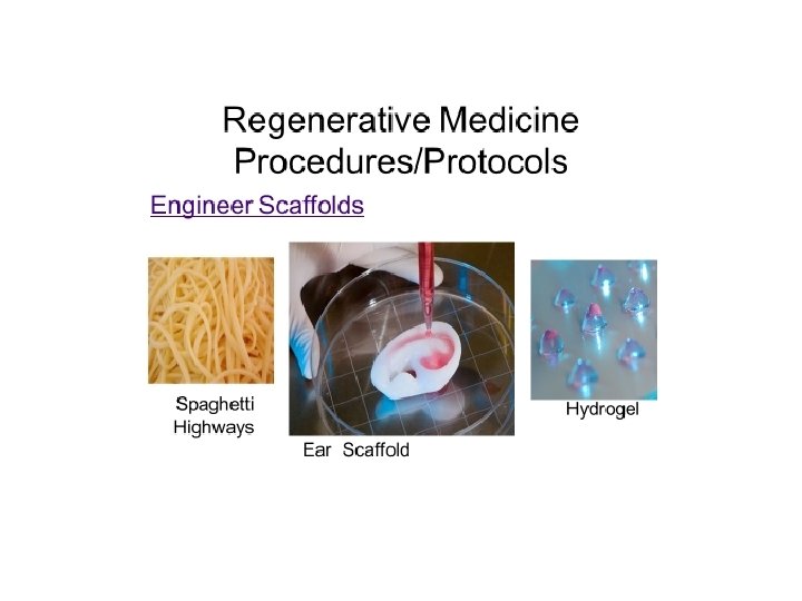
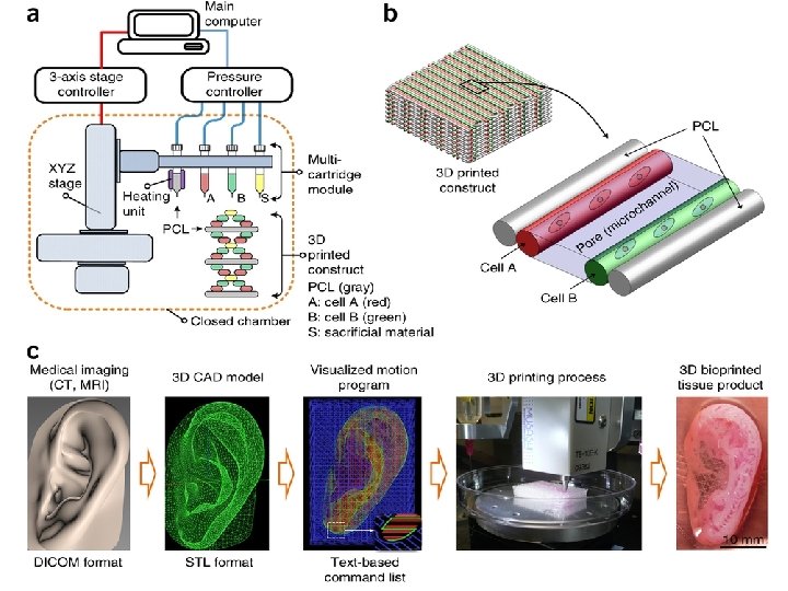
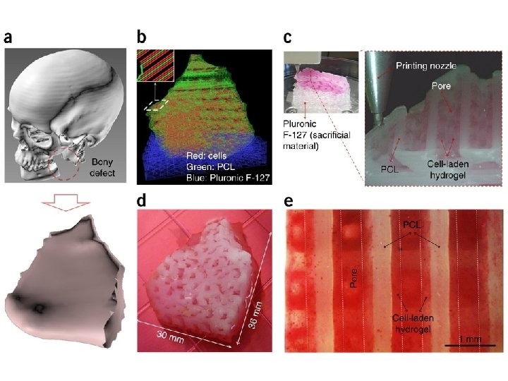
Alla Cornell University, nello stato di New York, il dottor Lawrence Bonassar del Dipartimento di Ingegneria Biomedica ha utilizzato una stampante 3 D per creare il lobo di un orecchio. Il primo passo è stato la scansione della testa del paziente al fine di creare un modello computerizzato. La Wake Forest Institute for Regenerative Medicine in California è stata la prima a trapiantare vesciche prodotte in laboratorio. Ora sono impegnati a generare tessuti con stampanti in 3 D da trapiantare su persone ustionate. Il direttore dell'istituto, il chirurgo Anthony Atala, ha mostrato in un video un esperimento in fase iniziale: una stampante 3 D che stampa un rene.
 • Uses 3 -D Print Technology •")
Regenerative Medicine Innovator Jordan Miller (Rice University) • Uses 3 -D Print Technology • Engineers Blood Vessels Using Sugar
 • • Leader in")
Regenerative Medicine Innovator Ramille N. Shah, Ph. D (Northwestern University) • • Leader in Field of 3 D-Printable Materials Engineers new 3 D-Inks Creates Porous Scaffolds Technique: Additive Manufacturing (Nanofiber Scaffold For Cartilage Regeneration)

Regenerative Medicine Challenges 1. Design of Biomaterials that Function in the Body 2. Getting Enough Cells and Cell Types for Engineering Tissues and Organs 3. Vascularity: Engineering Blood Vessels that Supply Nutrients, Oxygen and Signals to Bioengineered Tissues and Organs 4. Cost of Tissue and Organ Development Procedures

Inchiostri a base di cellule, tessuti progettati al computer e poi "stampati" A realizzare le prime bio-stampanti 3 D sono state due società, la californiana Organovo e l'australiana Invetech. Il professor Gabor Forgacs, dell'Università del dipartimento di Fisica Biologica del Missouri-Columbia, dal 2009 attraverso la nascita della Organovo sta studiando le applicazioni della stampa in 3 D nella medicina rigenerativa. Nel bio-ink (bionchiostro) ci sono decine di migliaia di cellule umane: si tratta di cellule del paziente che vengono usate per creare i nuovi tessuti, cosa che dovrebbe evitare il problema del rigetto immunitario. Ottenuto tessuto epatico del paziente.

I file generati da una risonanza magnetica possono essere stampati con titanio, ottenendo una sezione di cranio per un paziente che aveva avuto un incidente automobilistico

Organ printing: computer-aided jet-based 3 D tissue engineering Fig. 1. Fusion of embryonic myocardial ring. Myocardium rings were cut from Stage 15– 16 HH chick ventricle, containing only myocardium, endocardium and some intervening matrix. Isolated rings beat steadily for several days; (a) adjacent apposed rings fused overnight and (b) beat as one. (c). Schematic representation of principle of organ printing technology: placing of cell aggregates layer by layer in solidifying thermo-reversible gel with sequential cell aggregate fusion and morphing into 3 D tube. Fig. 2. Cell printer and images of printed cells and tissue constructs. Fig. 3. (a) Printed bagel-like ring that consists of several layers of sequentially (layer-by-layer) deposited collagen type 1 gel. (b) Manually printed living tube with radial branches from the chick 27 stage HH embryonic heart cushion tissue placed in 3 D collagen type 1 gel. Trends Biotechnol. 2003 Apr; 21(4): 157 -61.
, pp 16774– 16775 Advanced")
Contact Printing J. Am. Chem. Soc. , 2005, 127 (48), pp 16774– 16775 Advanced Materials Volume 19 Issue 24, Pages 4338 - 4342

References § Nanotechnology and Tissue Engineering: The Scaffold, CRC Press; 1 edition (June 16, 2008) § Quynh P. Pham, Upma Sharma, Ph. D. , Dr. Antonios G. Mikos, Electrospinning of Polymeric Nanofibers for Tissue Engineering Applications: A Review, Tissue Engineering. May 2006, 12(5): 1197 -1211. § Catherine P. Barnes, Scott A. Sell, Eugene D. Boland, David G. Simpson, Gary L. Bowlin, Nanofiber technology: Designing the next generation of tissue engineering scaffolds, Advanced Drug Delivery Reviews, Volume 59, Issue 14, Intersection of Nanoscience and Modern Surface Analytical Methodology, 10 December 2007, Pages 1413 -1433, ISSN 0169 -409 X, DOI: 10. 1016/j. addr. 2007. 04. 022. § Shuguang Zhang, Fabrication of novel biomaterials through molecular self-assembly, Nature Biotechnology 21, 1171 - 1178 (2003) § Rutledge G. Ellis-Behnke, Yu-Xiang Liang, Si-Wei You, David K. C. Tay, Shuguang Zhang, Kwok-Fai So, and Gerald E. Schneider, Nano neuro knitting: Peptide nanofiber scaffold for brain repair and axon regeneration with functional return of vision PNAS 2006 103 (13) 5054 -5059 § Benjamin S. Harrison, Anthony Atala, Carbon nanotube applications for tissue engineering, Biomaterials, Volume 28, Issue 2, Cellular and Molecular Biology Techniques for Biomaterials Evaluation, January 2007, Pages 344 -353, ISSN 0142 -9612, DOI: 10. 1016/j. biomaterials. 2006. 07. 044. § Miri Park, Christopher Harrison, Paul M. Chaikin, Richard A. Register, Douglas H. Adamson, Block Copolymer Lithography: Periodic Arrays of ~1011 Holes in 1 Square Centimeter, Science 30 May 1997: Vol. 276. no. 5317, pp. 1401 - 1404 § Byeongmoon Jeong, You Han Bae, Doo Sung Lee and Sung Wan Kim, Biodegradable block copolymers as injectable drug -delivery systems, Nature 388, 860 -862 (28 August 1997) § Evelyn K. F. Yim, Ron M. Reano, Stella W. Pang, Albert F. Yee, Christopher S. Chen, Kam W. Leong, Nanopattern-induced changes in morphology and motility of smooth muscle cells, Biomaterials, Volume 26, Issue 26, September 2005, Pages 5405 -5413, ISSN 0142 -9612, DOI: 10. 1016/j. biomaterials. 2005. 01. 058. § Mironov V, Boland T, Trusk T, Forgacs G, Markwald RR. Organ printing: computer-aided jet-based 3 D tissue engineering. Trends Biotechnol. 2003 Apr; 21(4): 157 -61. § Yu, A. A. ; Stellacci, F. , Contact Printing beyond Surface Roughness: Liquid Supramolecular Nano-Stamping, Advanced Materials, 19, 4338 -4342, 2007 § Yu A. A. , Savas T. , Cabrini S. , di. Fabrizio E. , Smith H. I. , Stellacci F. , High resolution printing of DNA features on poly(methyl methacrylate) substrates using supramolecular nano-stamping, J. Am. Chem. Soc. , 127, 16774 -16775, 2005
c2a37bbd46ae3ad2c208f929bbfcde3c.ppt