


Theme: Anatomy of skull, teeth bones and the slime layer of oral cavity. COMPILED BY: COLLEGE TEACHER A. K. KABIRAYEVA.

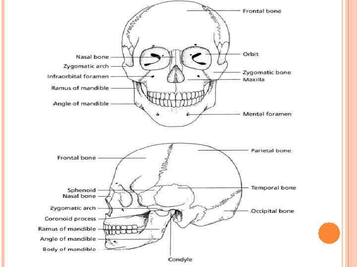
The skull is the topmost part of the bony skeleton of the body, the head, and is made up of two main areas: Cranium – the hollow cavity that surrounds the brain Face – the front vertical part of the skull, containing the orbital, nasal and oral cavities and the jaws All of the bones of the skull except the lower jaw, the mandible, are fixed to each other by immovable joints called sutures.

ANATOMY OF THE CRANIUM IS COMPOSED OF EIGHT BONES, WHICH ARE SEPARATED BY THE CRANIAL SUTURES – THESE ARE VISIBLE ON A DRY SKULL AS THE ZIGZAGGED JOINTS BETWEEN THE BONY PLATES.

THE BONES THEMSELVES ARE AS FOLLOWS: Frontal bone – single plate at the front of the cranium above the eyes, forming the forehead Parietal bones – pair of plates forming the top and the greater area of the sides of the cranium Temporal bones – pair of fan-shaped plates in the temple region of the lower sides of the cranium, in front of the ears Occipital bone – single plate at the back and partial underside of the cranium Sphenoid bone – single plate forming the majority of the base of the cranium Ethmoid bone – single plate at the lower front of the cranium, immediately behind the nose

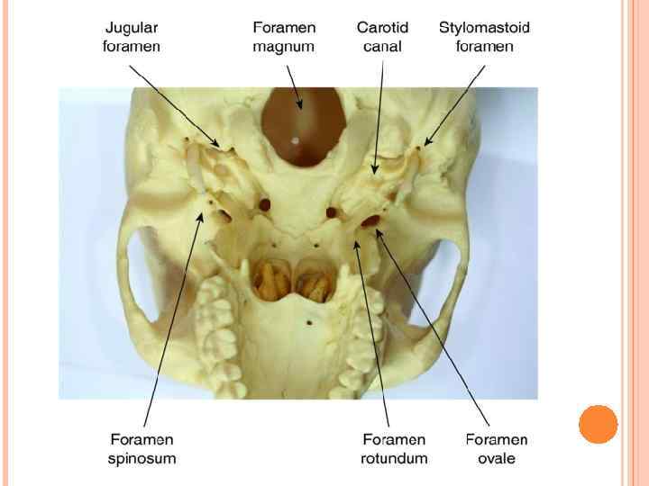
The main function of the cranium is to encase and protect the brain. All of the sensory nerve cells running from the body to the brain, and all of the motor nerves running from the brain to the body have to pass in and out of this bony cavity, and they do so through many natural openings in the underside of the cranium, called foramina (singular – foramen). Similarly, all of the blood vessels supplying the head and neck structures pass through these same foramina or between natural spaces between adjacent bones, called fissures.

ANATOMY OF THE FACE The face is composed of 14 bones that are all separated from each other by sutures, as in the cranium. The only exception is the mandible, forming the lower jaw, which articulates with the temporal bone at the hinged temporomandibular joint (TMJ).

The bones themselves are as follows: Vomer – single bone behind the nasal cavity, that articulates with cranial and other facial bones to connect the two regions of the skull together Lacrimal bones – pair of fragile bony plates forming the inner wall of the orbital cavities Nasal bones – pair of bones forming the bridge of the nose Nasal turbinates – pair of fragile curled bones projecting into the nasal cavity, which increase the contact of inspired air with the nasal mucosa – this aids debris removal before inhalation to the lungs, and warms the air Palatine bones – pair of bony plates forming the posterior section of the hard palate, and the side wall of the nasal cavity Zygomatic bones – pair of facial bones that articulate with the cranium posteriorly (frontal, temporal and sphenoid bones), and extend anteriorly into the zygomatic arch (cheek bone) to articulate with the maxilla Maxilla – pair of bones forming the upper jaw, the lower border of the orbital cavities, the base of the nose and the anterior portion of the hard palate Mandible – single horseshoe-shaped bone forming the lower jaw, with its posterior vertical bony struts articulating with the cranium at

FACIAL BONES.

ORAL ANATOMY The facial bones making up the oral cavity and its associated structures are of the utmost importance to all dental care professionals (DCPs). The bones will each be discussed in detail, with their associated structures covered later in this chapter.

MAXILLA – ANATOMICAL LANDMARKS.

PALATE – ANATOMICAL LANDMARKS.

PALATE – ANATOMICAL LANDMARKS.

THE MANDIBLE The mandible is the single, horseshoe-shaped bone that forms the lower jaw, and is the only moveable bone of the skull.

Its front horizontal portion extends into the alveolar process, which holds the lower teeth in situ, while its two posterior vertical struts allow articulation with the temporal bone at the temporomandibular joint (TMJ) and allows the insertion at various points for the muscles of mastication.

Looking at the mandible from an anterior view, the following anatomical landmarks are visible: Mental symphysis – the fused midline point of the two halves of the mandibular processes, as they formed in the embryo Mental protuberance – the most anterior point of the bone, forming the chin Mental foramen – bony opening located between the roots of the lower premolar teeth, allowing entry of the sensory nerve supplying the anterior teeth to the second premolar, and their buccal and labial soft tissues Body of mandible – the base of bone that supports the full length of the lower alveolar process

MANDIBLE – ANTERIOR ANATOMICAL LANDMARKS.

MANDIBLE – LATERAL ANATOMICAL LANDMARKS

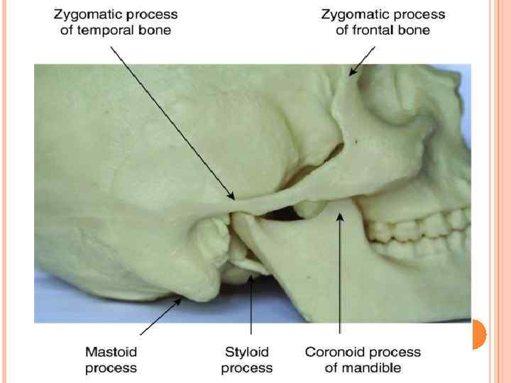
LOOKING AT THE MANDIBLE FROM A LATERAL VIEW, THE FOLLOWING ADDITIONAL ANATOMICAL LANDMARKS ARE VISIBLE: Angle of mandible – the corner of bone where the horizontal section turns upwards to form the vertical bony strut of the mandible Ramus of mandible – the vertical bony strut of the mandible, and the area of insertion of a muscle of mastication Head of condyle – the articulation point of the mandible with the temporal bone, at the TMJ, and the point of insertion of some muscles of mastication Coronoid process – the front bony projection of the ramus, and a point of insertion of a muscle of mastication Sigmoid notch – the dipped area between the condyle and the coronoid process, at the top of the ramus Coronoid notch – the concave anterior surface of the ramus, as it slopes to join the body of the mandible External oblique line – the crest of bone at the point where the ramus and the body join together, at the base of the coronoid notch, and the point where the long buccal nerve crosses from the lateral surface of the body to the medial surface, carrying sensory nerves from the buccal soft tissues of the lower molar teeth

Teeth are composed of calcium, phosphorus, and other minerals. Bones contain calcium, phosphorus, sodium and other minerals, but mostly consist of the protein collagen. Collagen is a living, growing tissue that gives bones their a flexible framework that allows them to withstand pressure. Calcium fills in the space around that framework and makes the bone strong enough to support the body's weight.
 The oral mucosa is the")
SLIME LAYER OF ORAL CAVITY ORAL. ( MUCOSA ) The oral mucosa is the mucous membrane lining the inside of the mouth and consists of stratified squamous epithelium termed oral epithelium and an underlying connective tissue termed lamina propria. [1] The oral cavity has sometimes been described as a mirror that reflects the health of the individual.

CLASSIFICATION It can be divided into three main categories based on function and histology: Masticatory mucosa, keratinized stratified squamous epithelium, found on the dorsum of the tongue, hard palate and attached gingiva. Lining mucosa, nonkeratinized stratified squamous epithelium, found almost everywhere else in the oral cavity, including the: Buccal mucosa refers to the inside lining of the cheeks and is part of the lining mucosa. Labial mucosa refers to the inside lining of the lips and is part of the lining mucosa. Alveolar mucosa refers to the mucosa between the gums and the buccal/labial mucosa. Specialized mucosa, specifically in the regions of the taste buds on lingual papillae on the dorsal surface of the tongue that contains nerve endings for general sensory reception and taste perception. [4]

Oral mucosa consists of two layers, the surface stratified squamous epithelium and the deeper lamina propria. In keratinized oral mucosa, the epithelium consists of four layers: Stratum basale (basal layer) Stratum spinosum (prickle layer) Stratum granulosum (granular layer) Stratum corneum (keratinized layer)