Функции толостой кишки.ppt
- Количество слайдов: 33
 The physiology of different colon regions
 The physiology of different colon regions • The ascending colon is specialized for processing chyme delivered from the terminal ileum. • The transverse colon is specialized for the storage and removal of water & electrolytes from feces. • The descending colon is a conduit between the transverse and sigmoid colon. • This region has the neural program for power propulsion that is involved in defecation reflex.
 The physiology of different colon regions • The rectosigmoid region, anal canal, and pelvic floor musculature maintains fecal continence. • The sigmoid and rectum are reservoirs with a capacity of up to 500 ml. • The puborectalis muscle and external anal sphincter comprise a functional unit that maintain continence. • Fibers of puborectalis join behind the anorectum and pass around it to form a U-shaped sling (physiological valve).
 Secretion in the colon
 Secretion in the colon • • • It is mainly mucus, no digestive enzymes. The mucus has the following functions 1 - It helps to lubricate feces. 2 - It neutralizes against any acids present. 3 - It protects against irritation. 4 - It provides a binding medium for fecal matter. •
 Secretion of water & electrolytes • Whenever a segment of large intestine becomes irritated as occurs in bacterial infection, the mucosa secretes large amount of water & electrolytes in addition to the alkaline mucus. • This dilute the irritating factors and causes rapid movement of the feces toward the anus.
 Absorption in the colon • Most of absorption in the colon occurs in the proximal half of the colon (absorptive colon). Whereas the distal colon function for storage (storage colon). • 1 - Water absorption, about 0. 5 - 1. 5 L/day is absorbed. The net water loss is 150 -200 ml/day. The large intestine can absorb a maximum of 5 to 8 liters of fluid and electrolytes each day • 2 - Na+ absorption, about 60 mmol/day is actively absorbed) in the presence of Na+-K+ ATPase) at the basolateral membrane to blood.
 Absorption in the colon • 3 - K+, Cl- and HCO 3 -. K+ is secreted into the lumen of colon while Cl- is absorbed in exchange for HCO 3 - which is secreted. • Persistent diarrhea can result in hypokalemia, dehydration and metabolic acidosis. • 4 - Folic acid and some AA and short chain FA resulting from bacterial fermentation of CHO are absorbed. • 5 - Certain drugs as steroids and aspirin may be absorbed.
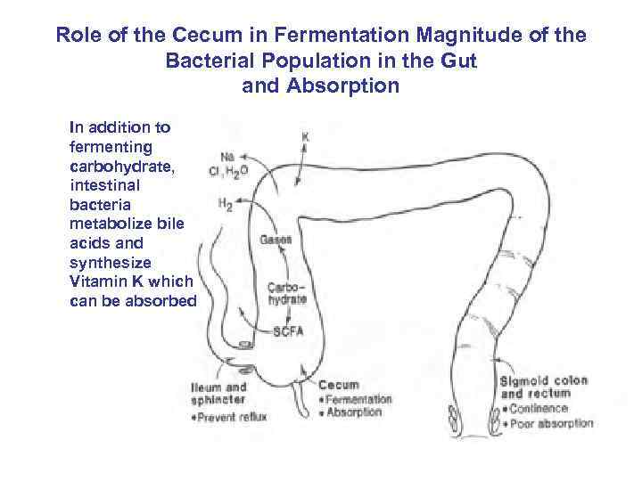 Role of the Cecum in Fermentation Magnitude of the Bacterial Population in the Gut and Absorption In addition to fermenting carbohydrate, intestinal bacteria metabolize bile acids and synthesize Vitamin K which can be absorbed
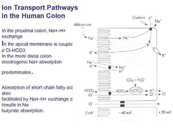 Ion Transport Pathways in the Human Colon In the proximal colon, Na+-H+ exchange In the apical membrane is coupled to a Cl-HCO 3 In the more distal colon electrogenic Na+ absorption predominates . Absorption of short chain fatty acids is also facilitated by Na+-H+ exchange and results in Na butyrate absorption.
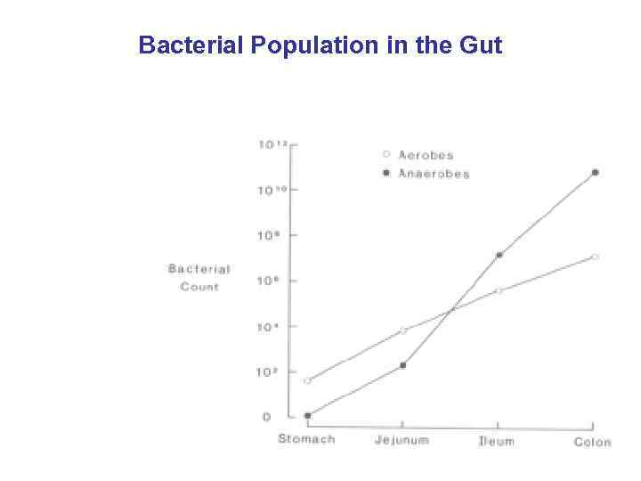 Bacterial Population in the Gut
 Intestinal bacteria • This bacterial flora is living in symbiosis with human and its effects are beneficial to the body. • Vitamin K and some B group vitamins as folic acid, biotin, thiamine and B 12 are synthesized by intestinal bacteria. • 1 - The bacteria-formed vitamin K is especially important because the amount of this vitamin in the daily ingested foods is normally insufficient to maintain adequate blood coagulation. • 2 - Bile salts are deconjugated and decarboxylated by intestinal bacteria. • 3 - Bile pigments are broken down in intestine to produce stercobilinogen which is excreted in stool giving it brown color.
 Intestinal bacteria • 4 - Intestinal bacteria decarboxylate some AA to produce amine and histamine. The amines are excreted in feces and are responsible for its smell. • 5 - Urea diffuses from blood to colon where it is broken down by its bacterial urease to ammonia. Most ammonia is absorbed and reconverted into urea by liver. In hepatic failure, accumulation of ammonia can cause hepatic encephalopathy. • 6 - Bacterial fermentation of undigested CHO may produce short chain FA and gases as CO 2.
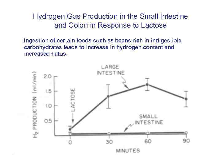 Hydrogen Gas Production in the Small Intestine and Colon in Response to Lactose Ingestion of certain foods such as beans rich in indigestible carbohydrates leads to increase in hydrogen content and increased flatus.
 The ileocaecal valve • It prevents backflow of contents from colon into small intestine. It remains closed and open only when an intestinal peristaltic wave reaches it. • * Distension of the cecum, Secretin, Ach, α adrenergic stimulation contract ileocaecal valve. • * Gastrin, CCk, β adrenergic stimulation relax ileocaecal valve.
 Response of the Ilecocecal Sphincter to Distension of the Ileum or Cecum
 Types of colonic movements • Mixing Movement (Haustrations) • ♦ In the same manner that segmentation movement occur in the small intestine, large circular constrictions also occur in the large intestine. • ♦ At each of these constrictions points, about 2. 5 cm of the circular muscle contracts, sometimes constricting the lumen of the colon to almost complete occlusion. • ♦ At the same time the longitudinal strips contract. • ♦ These combined contractions cause the unstimulated portion of the large intestine to bulge outward into baglike sacs called haustrations. • ♦
 Types of colonic movements • ♦ The haustral contraction once initiated usually reach peak intensity in 30 seconds & then disappear during the next 60 seconds. • ♦ They also at times move slowly analward during their period of contraction especially in the cecum & ascending colon. • ♦ After another few minute new haustral contractions occur in other areas nearby. • ♦ In this way all fecal material is gradually exposed to the surface of the large intestine & fluid is progressively absorbed until only 80 -150 ml of 1500 ml daily load of chyme is lost in feces.
 Types of colonic movements • Initiation of Mass Movement: • ♦ Gastrocolic & duodenocolic reflexes after meals. They result from distension of the stomach & duodenum. • ♦ Irritation of the colon e. g. , castor oil. • ♦ Intense stimulation of the parasympathetic nervous system. • ♦ Over distension of a segment of the colon.
 Antiperistalsis • It starts at the junction of ascending and transverse colon and traveling towards the cecum. • It mixes contents and help water absorption.
 Propulsive (mass) Movement • ♦ Peristaltic waves of the type seen in small intestine are rare. • Most of the propulsion in the cecum & ascending colon results from the slow but persistent haustral contractions requiring as many as 8 -15 hours to move the chyme only from the ileocecal valve to the transverse colon while the chyme itself becomes fecal in quality & also semisolid instead of semifluid. • ♦ From the transverse colon to the sigmoid, mass movement mainly take over the propulsive role.
 • ♦ These movements usually occur only few times each day, most abundantly for 15 min during the first hour after eating breakfast. • ♦ A mass movement is a modified type of peristalsis characterized by a constrictive ring occurs at a distended or irritated point in the colon. • Then rapidly the 20 or more cm of the colon distal to the constriction contract almost as a unit forcing the fecal material en mass down the colon
 Control of Colonic Motility • The intramural plexuses directly control the contractile behavior of the colon. • Stimulatory enteric motor neurons use acetylcholine & substance P as neurotransmitters. • Inhibitory enteric motor neurons release VIP & NO onto colonic smooth muscle cells. • The extrinsic autonomic nerves to the colon modulate the control of the colonic motility by the enteric nervous system.
 Defecation • During this process the haustrations disappear completely. • ♦ The initiation of contraction is complete in about 30 seconds, during the next 2 to 3 min before another mass movement occurs, this time perhaps farther along the colon. • ♦ But the whole series of mass movement will usually persist for only 10 min to half an hour. They will then return after a half day or even a day later. • ♦ Mass movement can occur in any part of the colon, though most often they occur in the transverse or descending colon. • ♦ When they have forced a mass of feces into the rectum the desire for defecation is felt.
 Defecation • It is a spinal reflex which is influenced by higher center. • Most of the time the rectum is empty and both internal and external sphincters are reflexly maintained in a state of tonic contraction. • Gastric or intestinal filling initiate a mass movement in the colon that pushes feces into rectum (gastrocolic & dudenocolic reflexes). • The rectum is distended and sends signals to cerebral cortex producing the desire to defecate.
 If the surrounding circumstances are suitable • • • Defecation reflex will be allowed. Stretch of the rectal wall is signaled to SC by pelvic nerve. Efferent pelvic impulses cause reflex contraction of the rectum and relaxation of IAS. This is followed by reduction in tonic impulses to EAS, so it relaxes and feces leave the rectum assisted by voluntary straining and contraction of pelvic floor muscle. Defecation Reflex 1. Distension of the rectum. 2. Stimulation of the stretch receptors in the rectum. 3. A. Short reflex: Stimulation of myenteric plexus in sigmoid colon and rectum. B. long reflex: stimulation of parasympathetic motor neurons in sacral spinal cord. C. stimulation of somatic motor neurons. 4. Increased local peristalsis, relaxation of internal anal sphincter and contraction of external anal sphincter.
 Defecation Reflex • 1. Distension of the rectum. • 2. Stimulation of the stretch receptors in the rectum. • 3. A. Short reflex: Stimulation of myenteric plexus in sigmoid colon and rectum. • B. long reflex: stimulation of parasympathetic motor neurons in sacral spinal cord. • C. stimulation of somatic motor neurons. • 4. Increased local peristalsis, relaxation of internal anal sphincter and contraction of external anal sphincter.
 If situation is not suitable for defecation • ◙ The reflex is inhibited by the cerebral cortex. Tonic contraction of EAS is voluntary maintained which leads to accommodation of the rectum to distension and return of tonic contraction of the IAS. • ◙ In infants( and in spinal cord lesion) the spinal reflex of defecation operates without interference from higher centers (fecal incontinence).
 Other Defecation Reflexes • Apart from the two main defecation reflexes mentioned above, other reflexes can also influence the defecation process. • Gastrocolic reflex – distention of the stomach while eating or immediately after a meal triggers mass movements in the colon. • Gastroileal reflex – distention of the stomach while eating or immediately after eating triggers the relaxation of the ileocecal sphincter and speeds up peristalsis in the ileum (end portion of the small intestine). This causes the contents of the ileum to rapidly empty into the colon. • Enterogastric reflex – distention and/or acidic chyme in the duodenum slows stomach emptying and reduces peristalsis. • Duodenocolic reflex – distention of the duodenum a short while after eating triggers mass movements in the colon
 Defecation Reflex • The defecation reflex is an involuntary response of the lower bowels to various stimuli thereby promoting or even inhibiting a bowel movement. These reflexes are under the control of the autonomic system and play an integral role in the defecation process along with the somatic system that is responsible for voluntary control of defecation. The two main defecation reflexes are known as the intrinsic myenteric defecation reflex and parasympathetic defecation reflex.
 Intrinsic Myenteric Defecation Reflex • The entry of feces into the rectum causes the distention of the rectal wall. • This stretching triggers signals to the descending and sigmoid colon via the myenteric plexus to increase peristalsis. • The myenteric plexus is part of the enteric nervous system which is the gut’s own internal neural network as discussed under stomach nerves. • The peristaltic waves extend all the way to the rectum an anus. In this manner, fecal matter is moved closer to the anus. • When the wave reaches the anus, it causes the internal anal sphincter, which is always constricted, to relax. • This is achieved by inhibitory signals via the myenteric plexus to reduce sphincter constriction. • Defecation may occur at this point if the external anal sphincter also relaxes. However, without the parasympathetic defecation reflex, defecation solely dependent on the intrinsic reflex would be weak.
 Parasympathetic Defecation Reflex • The parasympathetic defecation reflex works in essentially the same way as the intrinsic myenteric defecation reflex but involves parasympathetic nerve fibers in the pelvic nerves. I • t triggers peristaltic waves in the descending and sigmoid colon as well as the rectum. • It also causes relaxation of the external anal sphincter. The difference is that the parasympathetic defecation reflex enhances this process and makes the intrinsic reflex much more powerful. • If sufficiently stimulated, it may even cause the sigmoid colon to completely empty all of its contents in the rectum rapidly.
 The end!


