fibrous connective tissue.ppt
- Количество слайдов: 46


THE HISTOLOGY OF THE FIBROUSE CONNECTIVE TISSUE

The plan of the lecture 1. The general features of the fibrouse connective tissue. 2. Classification and the specific features of each one. 3. Micro- and submicroscopic organisation, histochemical features and the functions of the loose irregular connective tissue. 4. Dense connective tissue, it’s histological features and localization at the human organism.

FIBROUS CONNECTIVE TISSUE Loose irregular connective tissue Dense regular connective tissue

Loose irregular connective tissue Features: too many cells, small volume of the ground substance and small number of the fibers. The fibers are disordered Localization: walls of internal organs, the vascular adventitia, the proper connective tissue plate of the mucous coats, submucous tela.

Рыхлая irregular connective tissue Loose волокнистая неоформленная

CELLS HISTIOGENIC: ØFIBROBLASTS ØADIPOCYTES HEMATOGENIC: ØMACROPHAGES ØPLASMA CELLS ØMAST CELLS ØLYMPHOCYTES ØGRANULOCYTES FIBERS ØCOLLAGEN ØELASTIC ØRETICULAR
 Macrophages (monocytes). Functions – endocytosis, expression of the")
Fibroblasts (junior, mature, fibrocytes, myofibrocytes, fibroclasts) Macrophages (monocytes). Functions – endocytosis, expression of the BAS. Mast cells. Granules– heparin, serotonin, histamine, himasa, tripasa. Functions – releasing of the above noted enzymes, the synthesis of the DAS. Adventitial cells, pericytes, endotheliocytes, pigmentocytes, adipocytes, leucocytes (from vessels). Plasma cells (В-lymphocytes) – expression of antibodies.

FIBROBLASTS LOW DIFFERENTIATED JUNIOR MATURE FIBROCYTES MYOFIBROBLASTS FIBROCLASTS

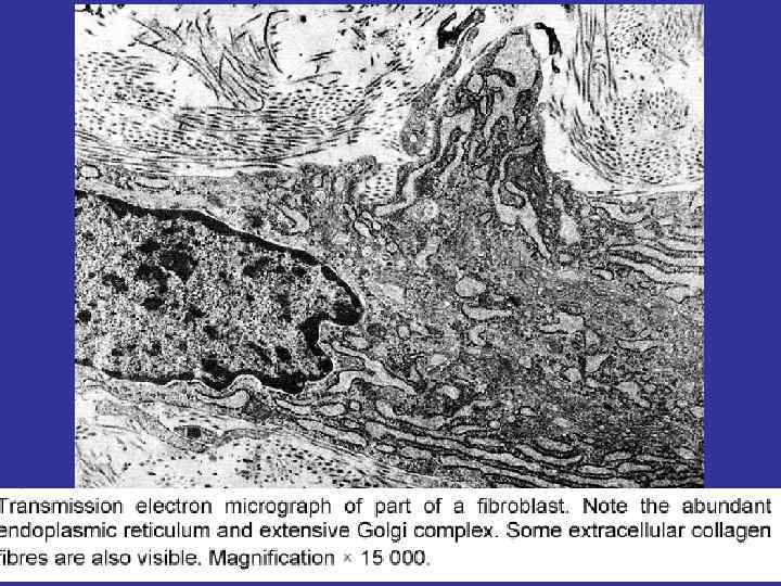
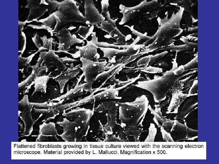
SOURCE FIBROBLASTS – ELEMENTS OF THE DIFFERON, SOURCED OF THE MESENCHIMA. THE PRECURSOR IS THE LOW-DIFFERENTIATED FIBROBLSTS WITH THE HIGH MITOTIC ACTIVITY. FUNCTION MATURE – PRODUCE THE COMPONENTS OF THE GROUND SUBSTANCE: PROTEINS (COLLAGEN & ELASTIN FOR FIBER’S SYNTESIS), PROTEOGLICANS AND GLICOPROTEINS. NUCLEI THERE ARE CELLS WITH THE HIGH LEVEL OF THE SYNTESIS – THIS IS WHY AN EUCHROMATIN MORE IN VOLUME AT THEIR NUCLEI WHICH ONE USUALLY PALE AND OVAL IN SHAPE CYTOPLASMA R-EPR. THE CELLS ARE SPIDER-SHAPED. AND THE CELL’S SHAPE MOTIONS CAN MOVE ALONG FIBERS USING FIBRONECTIN

FIBROCYTES NARROW, ELONGATED. NOT TOO MANY PROCESSUSES AND CYTOPLASMA. NARROW AND PICNIC NUCLEUS. FIBROCLASTS CAN BE PRESENT AT THE ORGAN IN THE CASE OF IT’S INVOLUTION. ENDOCITOSIS OF THE GROUND SUBSTANCE AND IT’S HYDROLISIS (INCLUDING COLLAQGEN FIBERS) AT THE NUMEROUSE LYSOSOMES. NUCLEI ARE OVAL AND PALE. THIS CELLS ARE BIGGER THEN FIBROBLASTS. MYO FIBROBLASTS CAN BE PRESENT AT THE ORGAN IN THE CASE OF IT’S REGENERATION AFTER VIOLATION. HAVE GOT A LOT OF MYOFILAMENTS AT THEIR CITOPLASMA.

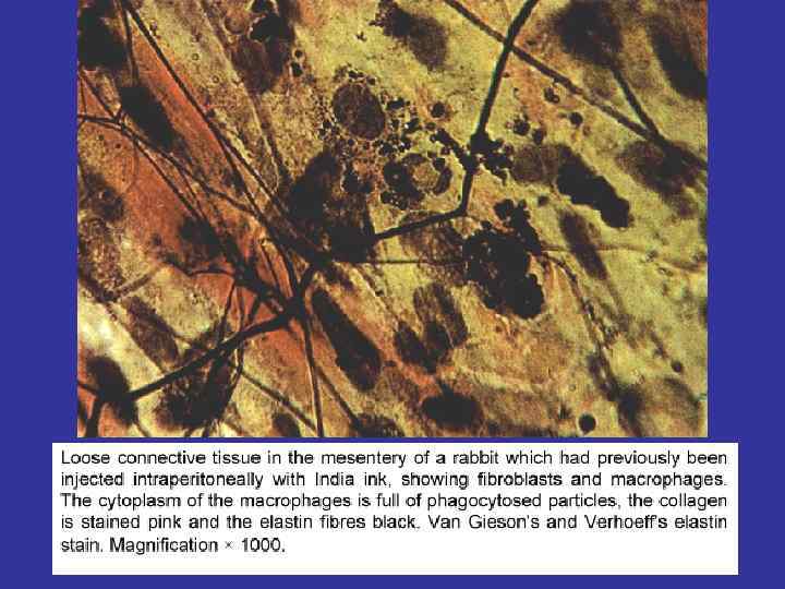
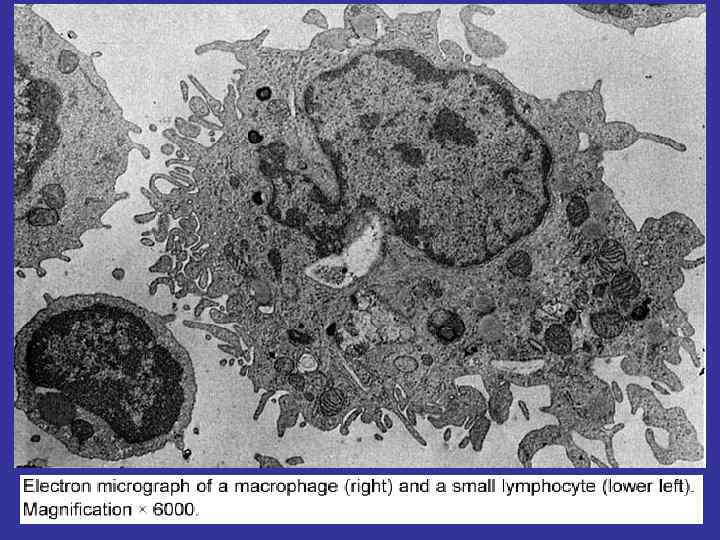
MACROPHAGES FUNCTIONS: ØFAGOCITOSIS, ØANTIGEN-PRESENTING, ØEXPRESSION OF THE CYTOCINES

MORPHOLOGY IRREGULAR SHAPED AND PICNIC NUCLEUS. AT THE CITOPLASMA – NUMEROUS VACUOLES AND GRANULES. FUNCTIONS ANTIGEN-PRESENTING, EXCRETION OF THE INTERLEUKINS, PIROGENES (STIMULATING LEUCOCYTORY MIGRATION AND ACTIVITIES), INTERFERON (ACTING AT THE VIRUSES), LISOCIM (ACTING AT THE BACTERIAL GLYCOCALIX), CYTOLITIC FACTORS(ACTING AT THE TUMOR CELLS). FAGOCITOSIS (DEAD CELL AND APOPTOTIC BODIES)
: ØHEPARIN, ØHISTAMIN, ØSEROTONIN, ØCYTOKINES, ØPROSTAGLANDINES")
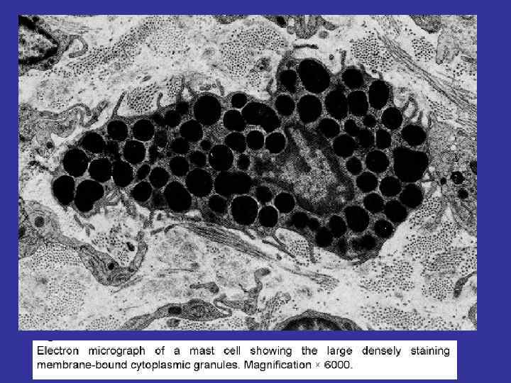
MAST CELLS FUNCTIONS: DEGRANULATION (EXCRETION OF THE NUMEROUSE BAS): ØHEPARIN, ØHISTAMIN, ØSEROTONIN, ØCYTOKINES, ØPROSTAGLANDINES N & LEUCOTRIENS
")
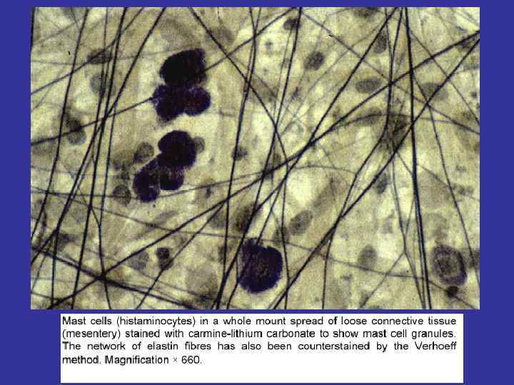
Main feature The presence of the big violet granules (full of heparin and serotonin) localization Near the blood vessels Nuclei Not too big, blue and place at the cell’s centre Granules Non-specific (too small in volume, invisible) Specific – big un volume, well visible. metachromasia The staining of the toluidin blue in 2 colors:
 Function They")
Adipocytes localization Near the blood vessels, but grouped (not like mast cells) Function They store the fat up to it’s utilization
")
PLASMACYTES THE DERIVATIVES OF THE B-CELLS LIFE-TIME ( 2 -3 TILL 10 -30 DAYS) MAIN PRODUCERS OF THE ANTIBODIES

Main feature This cell produce the immunoglobulins on the R-EPR. This is why the pyronine stain the plasm cells in purple-violet color (RNA-staining). Morphology Nuclei – decentralized. At the paranuclear area – not stained field (“light halo”). The Golgi complex is here.
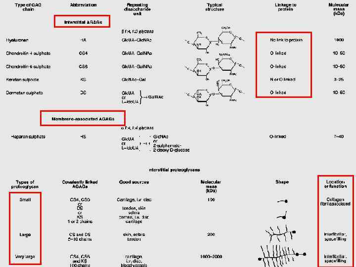
 ADHESIVE PRONEINS FIBRILLAR PROTEINS FIBRONECTIN, LAMININ GLICOSAMINEGLICANES HEPARIN,")
GRAUND SUBSTANCE FIBERS AMORPH SUBSTANCE (MATRIX) ADHESIVE PRONEINS FIBRILLAR PROTEINS FIBRONECTIN, LAMININ GLICOSAMINEGLICANES HEPARIN, HYALURON ACID, HONDROITINCULPHATES, DERMATANSULPHATES, CERATANSULPHATES

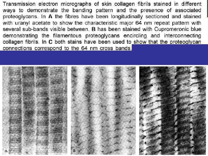
COLLAGEN FIBRES

THE SYNTESIS OF THE COLLAGEN AND COLLAGEN FIBRES TYROPOCOLLAGEN, PROTOFIBRILLS, FIBRILLS FIBERS.
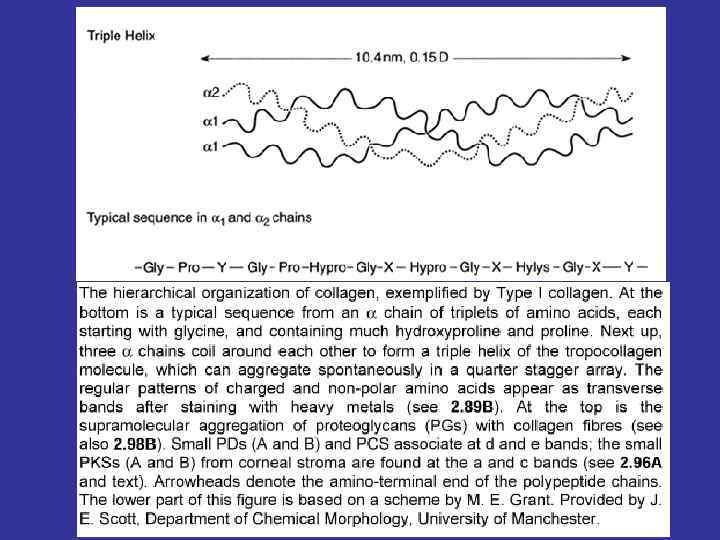

In the relationship to the amino acid content there are known 15 types of the collagen fibers^ 1 -st type – skin, bones, tendons; 2 -nd type – cartilage; 3 -rd type – reticular fibers; 4 -th type – basement membranes. Spiral part Globular part Acid glucosaminoglycans

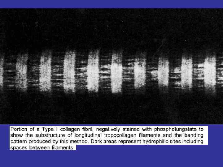
striation The reason is the special packing of the tropocollagen corpuscles. There are spaces in between newborn molecules. The corpuscles of newborn protofibrils are moved. Physical characteristics Too strong, resistant and hydrophilic fibers.

THE STAGES OF THE COLLAGEN FIBERS CREATION INTRACELLULAR PROCESS ORGANELLS GENE TRANSCRIPTION IN M-RNA SYNTESIS NUCLEUS THE ALFA-CHAIN SYNTESIS R-EPR PROLINE AND LISINE HYDROLISATION R-EPR PROCOLLAGEN SYNTESIS R-EPR PPACKAGE TO THE TRANSPORT VESICLES AND EXOCITOSIS Golgi complex EXTRACELLULAR PROCOLLAGEN - TROPOCOLLAGEN – FIBRILLS – FIBERS PLASMALEMMA

Elastic Thin, some time – bifid and creating compound mesh. fibers Elastin – globular protein (one polypeptide chain). That molecules are joined at protofibrils by lysine. Joining protofibrils produce the microfibrils, which joint them around glycoprotein amorphous component at the fibre. Thus: at the centre of the elastic fibre – amorphous component which is surrounded by numerous microfibrils.
. Proteoglycans: AGAG+protein Glicoproteins – fibronectin, laminin.")
Ground substance : glucosaminoglycans (interstitial and membrane assotiated). Proteoglycans: AGAG+protein Glicoproteins – fibronectin, laminin. Proteins, coming from the blood (60% of all albumins + globulins + ions). The consistence of the ground substance – jell. The degree of polymerization.

Плотная irregular connective tissue Dense волокнистая неоформленная Features: small number of cells, too many the fibers. The fibers are disordered Localization: the proper layer of the skin, the periosteum, the perichondrium

Main feature At the ground substance fibers much more then amorph substance The main aim of classification Direction of the fibers – this is why 2 types are known: regular and irregular. NB! – the orientating factor – an acting power. In relationship to the content, the dense regular connective tissue is classified in to parts – elastic and collagen.

localization There are 2 types of the fibrous tissue at the skin: loose irregular (at the papillary layer) and the dense irregular (at the reticular layer) Loose irregular connective tissue Collagen fibers (oxyfilic) and elastic fibers. Both types are thin and disordered. In between them there are numerous cell’s nuclei and not stained amorph matter (matrix).

Dense irregular Collagen fibers are grouped at the connective numerous thick tissue bundles. The bundles are disorientated, this is why this tissue termed as irregular. Because here more thick collagen bundles, the amorph matrix lightly oxyfilic as well. Elastic fibers are not stained. Cells – numerous fibroblasts.

Dense regular connective tissue Features: small number of cells, too many the fibers. The fibers are well ordered (grouped at bundles) Localization: the tendo, articular capsule, ligaments, fascia.
")
Tendo Endotenonium Peritenonium Articular capsule Fibrous layer Sinovial layer Subsinovial layer (main joints)

LONGITUDINAL AND CROSS SECTIONS OF THE TENDO

Reticular tissue components Reticulocytes + reticular fibers retuculocytes Like fibroblasts: big in square; have got contacting processuses, attached to the reticular fibers; the round shaped nucleus is at the centre of the cytoplasm. reticular fibers Collagen of the 3 -rd type; high concentration of the sulphates at the collagen, this is why so high argirophylia and a lot of anastomoses in between reticular fibers. Hydrophilic fibers.

Adipose tissue White adipose tissue Brown adipose tissue Every where Interscapular area, around the kidney, thyroid gland. The newborn organism most reach in this type of the adipose tissue Cells At the cytoplasm – one big droplet of the fat. The nucleus and the organells are decentralized. In between fat lobules – the loose irregular connective tissue At the cytoplasm – several small droplets of the fat. The nucleus and the organells are placed at the center. Too many big mitochondria. But an energy utilizing for the warming of the organism, and not stored as ATP. The color caused by numerous cytochromes.

ADIPOSE TISSUE WHITE BROWN
fibrous connective tissue.ppt