

3a391f179a2edb6d064c93aa41d0f705.ppt
- Количество слайдов: 77
 The Circulatory System
 • Need stethoscopes and straws/plastecine
 on their desk and •") Activity 1 • lay your hand (palm side up) on their desk and • count how many times you can open and close their hand for one minute. • Ready…. set…. go! • Timer
Activity 1 • lay your hand (palm side up) on their desk and • count how many times you can open and close their hand for one minute. • Ready…. set…. go! • Timer
 Activity 2 Discussion: • Over 170 years ago, a man named Laennec invented the first stethoscope. It was a wooden tube about 1 inch in diameter and about 10 inches long. Materials: • stethescopes Procedure: • pair up and listen to your partner's heartbeat by placing the tube over the partner's heart. • Count the number of beats per 30 seconds. Add this number together twice to find out how many times each minute the person's heart beats.
 Parts of the Circulatory System 1. Blood vessels 2. Blood 3. Heart Lets take a closer look…
 1. Blood Vessels Blood vessel = a tube for carrying blood • These tubes are how the heart pumps the blood all over the body. We call this CIRCULATION Blood moves through 3 kinds of blood vessels: a) Arteries b) Capillaries c) Veins
 Arteries • Carry blood AWAY from the heart TO the body or lungs") a) Arteries • Carry blood AWAY from the heart TO the body or lungs • Common misconception… • The largest artery = the aorta – This is the main branch off the heart, off which blood flows to everywhere except to the lungs – Stenosis of the aorta = … • Small arteries are called arterioles
a) Arteries • Carry blood AWAY from the heart TO the body or lungs • Common misconception… • The largest artery = the aorta – This is the main branch off the heart, off which blood flows to everywhere except to the lungs – Stenosis of the aorta = … • Small arteries are called arterioles
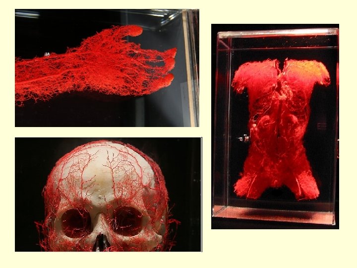
 Capillaries • As blood vessels branch smaller and smaller, they eventually become only") b) Capillaries • As blood vessels branch smaller and smaller, they eventually become only one cell thick = capillaries • This is how every cell in our body gets blood • Capillaries are the ONLY site of molecular exchange!!! • • – Gas, nutrients, water, nitrogenous, wastes, hormones, etc. http: //www. youtube. com/watch? v=Ul. AIHmv_0 p 8 http: //www. youtube. com/watch? v=2 Wh. Fm. GYt. T 0 M
b) Capillaries • As blood vessels branch smaller and smaller, they eventually become only one cell thick = capillaries • This is how every cell in our body gets blood • Capillaries are the ONLY site of molecular exchange!!! • • – Gas, nutrients, water, nitrogenous, wastes, hormones, etc. http: //www. youtube. com/watch? v=Ul. AIHmv_0 p 8 http: //www. youtube. com/watch? v=2 Wh. Fm. GYt. T 0 M
 Movement of blood – 2. Capillaries Nutrients cell Oxygen Wastes oxide i Carbon d
 Veins • Carry blood back TO the heart FROM the body or lungs") c) Veins • Carry blood back TO the heart FROM the body or lungs • Common misconception… • The biggest vein is the vena cava • Small veins are called venules
c) Veins • Carry blood back TO the heart FROM the body or lungs • Common misconception… • The biggest vein is the vena cava • Small veins are called venules
 Which way is the blood flowing?
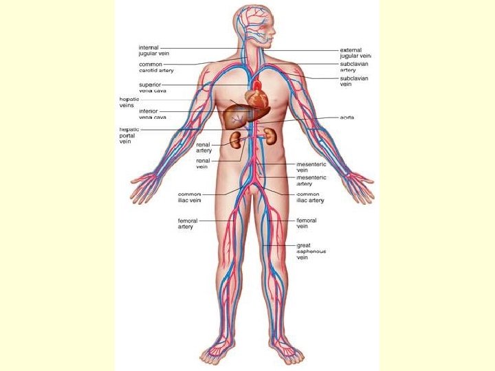
 Heart Aorta") Flow of blood through blood vessels in the body Heart Capillaries (exchange) Heart Aorta Venules Arteries Veins Arterioles Vena cava
Flow of blood through blood vessels in the body Heart Capillaries (exchange) Heart Aorta Venules Arteries Veins Arterioles Vena cava
 Comparison of Veins & Arteries
 • Discuss accidental amputations http: //www. youtube. com/watch? v=I 1 v 6 Yek MV_c – people fainting • http: //www. youtube. com/watch? v=8 r 9 LOap o. YRM • http: //www. youtube. com/watch? v=n 8 QHOUWECU • Nova article on Stapp http: //www. pbs. org/wgbh/nova/space/gravi ty-forces. html
 Comparison of Veins & Arteries 1. Have a pulse 2. Transport blood away from the heart; 3. Carry Oxygenated Blood (except the Pulmonary Artery); 4. Have relatively narrow lumens (see diagram); 5. Have more muscle / elastic tissue; 6. Transports blood under high pressure; 7. Do not have valves (except for the semi-lunar valves of the pulmonary artery and the aorta). Veins 1. Do not have a pulse 2. Transport blood towards the heart; 3. Carry De-oxygenated Blood (except the Pulmonary Vein); 4. Have relatively wide lumens (see diagram); 5. Have less muscle / elastic tissue; 6. Transports blood under low pressure; 7. Have valves to prevent blood flowing in the wrong direction
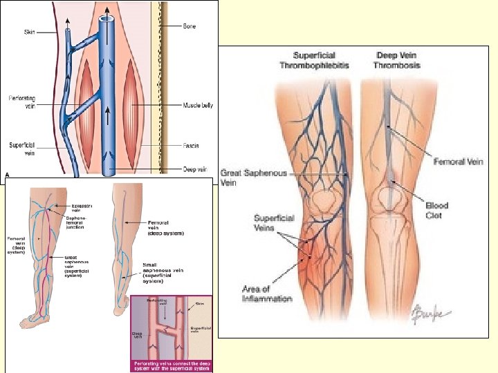
 Varicose veins • http: //www. youtube. com/w atch? v=49 j. Uy. Bu 4 M 1 Q • http: //www. youtube. com/w atch? v=VPe. XRCc 8 w 24 • See handout • Squeezing vein http: //www. youtube. com/w atch? v=XRs. NSSM 7 Pi. I
 Parts of the Circulatory System 1. Blood vessels 2. Blood 3. Heart Lets take a closer look…
 2. Blood • Average adult 4 -6 liters of blood
 2. Blood 1. Plasma • – • Clear/straw colored liquid portion of blood Can look cloudy after a fatty meal, gross!!! Components of plasma: – water, dissolved gases, proteins, hormones, electrolytes, sugar, vitamins, and waste products • 55% of blood volume • Function - Transport CO 2, nutrients from gut, hormones, and distributes heat
 • Aka erythrocytes • 45% of blood volume •") 2. Red Blood Cells (RBCs) • Aka erythrocytes • 45% of blood volume • primary function: carry O 2 lungs body • DO NOT have a nucleus • Contain iron and hemoglobin – – That’s why they’re red in color. Hemoglobin is the substance to which O 2 molecules bind for transport through the bloodstream. • 30 trillion RBCs • life span = 120 days • about 2 million die & are replaced every second • worn out RBC’s are filtered out by the liver and spleen
2. Red Blood Cells (RBCs) • Aka erythrocytes • 45% of blood volume • primary function: carry O 2 lungs body • DO NOT have a nucleus • Contain iron and hemoglobin – – That’s why they’re red in color. Hemoglobin is the substance to which O 2 molecules bind for transport through the bloodstream. • 30 trillion RBCs • life span = 120 days • about 2 million die & are replaced every second • worn out RBC’s are filtered out by the liver and spleen
 • • Aka leukocytes <1% of blood volume are") 3. White blood cells (WBCs) • • Aka leukocytes <1% of blood volume are a part of the immune system and help our bodies fight infection HAVE a nucleus Circulate in the blood so that they can be transported to an area where an infection has developed. If WBC # es = a sign of an infection somewhere in body. http: //www. youtube. com/watch? v=VAh. M 9 Ox. ZDk. U&NR=1 https: //www. youtube. com/watch? v=t. Rgq 3 v 1 W 61 w
3. White blood cells (WBCs) • • Aka leukocytes <1% of blood volume are a part of the immune system and help our bodies fight infection HAVE a nucleus Circulate in the blood so that they can be transported to an area where an infection has developed. If WBC # es = a sign of an infection somewhere in body. http: //www. youtube. com/watch? v=VAh. M 9 Ox. ZDk. U&NR=1 https: //www. youtube. com/watch? v=t. Rgq 3 v 1 W 61 w
 Macrophages") 3. White blood cell’s continued… • 2 main types of WBCs: WBCs (leucocytes) Macrophages Lymphocytes Are non-phagocytic Literally eat invaders = phagocytosis Can squeeze through walls of blood vessels to attack disease in tissue Play a role in the body’s “acquired immune system” When body recognizes invader, it lodges a massive attack against this specific invader
3. White blood cell’s continued… • 2 main types of WBCs: WBCs (leucocytes) Macrophages Lymphocytes Are non-phagocytic Literally eat invaders = phagocytosis Can squeeze through walls of blood vessels to attack disease in tissue Play a role in the body’s “acquired immune system” When body recognizes invader, it lodges a massive attack against this specific invader
 4. Platelets • Aka thrombocytes • <1% of blood volume • Platelets are sticky, irregularly-shaped cell fragments (NOT CELLS) How a clot is formed (the good kind): • Platelets detect air/injury and become sticky also begin to break apart these react with stuff in plasma to make thromboplastin Thromboplastin + prothrombin (+ Ca+ & Vit K) = Thrombin + Fibrinogen => Fibrin (tiny threads) fibrin threads form a web-like mesh traps the blood cells within it This hardens as it dries, forming a clot, or "scab/bruise“ • What do you think happens if Calcium and vitamin K are low/missing? • • http: //science. howstuffworks. com/environmental/life/human-biology/blood 3. htm http: //www. youtube. com/watch? v=--b. ZUeb 83 u. U&feature=related
 • blood vessel injured Platelets detect Air Ruptured platelets Thromboplastinogen") Blood clotting process (Steps) • blood vessel injured Platelets detect Air Ruptured platelets Thromboplastinogen Thromboplastin (active) (inactive) Prothrombin (inactive) Fibrinogen (inactive) (Ca, vit K) Thrombin (active) Fibrin (active)
Blood clotting process (Steps) • blood vessel injured Platelets detect Air Ruptured platelets Thromboplastinogen Thromboplastin (active) (inactive) Prothrombin (inactive) Fibrinogen (inactive) (Ca, vit K) Thrombin (active) Fibrin (active)
 What is the role of blood? TO HELP MAINTAIN HOMEOSTASIS by: 1. Transport • O 2, CO 2, food, nitrogenous wastes, hormones, etc 2. Protection – through immune system • Guards against microbial invasion (immune system) 3. Circulate clotting factors to prevent blood loss in case of injury 4. Regulation • controls levels of hormones, enzymes, temperature, p. H
 Blood Groups Antigens: • these are proteins that are presented on the surface of cells (even ours) and act as a name tag • They identify self vs. non-self • Are able to trigger the production of antibodies Antibodies • Proteins used by the immune system to identify invaders to our body which begins an immune response (an attack) to destroy the invader
 are present") Blood Types humans fall into 4 basic groups, based on which ANTIGEN(s) are present on their RBCs Type A - 41% Type B - 10% Type O - 45% Type AB - 4% The Rh (Rhesus) factor is the 2 nd grouping system based on if the Rh ANTIGEN is present / absent: 85% Rh+ 15% Rh –
Blood Types humans fall into 4 basic groups, based on which ANTIGEN(s) are present on their RBCs Type A - 41% Type B - 10% Type O - 45% Type AB - 4% The Rh (Rhesus) factor is the 2 nd grouping system based on if the Rh ANTIGEN is present / absent: 85% Rh+ 15% Rh –
 ABO Blood Groups Blood Type A B AB O Rh+ Rh- Antigen on RBC’s A B AB None Rh none Antibodies in Plasma Anti – B Anti – A None Anti A & B none Anti - Rh • Remember, antibodies are there to help destroy foreign (non-self) cells which are flagged by antigens • Why is this IMPORTANT ? – This chart indicates that people must know their blood types for safe transfusions because we are born with antibodies in our blood that will attack antigens of other blood groups.
 Transfusion Chart Donating A → A, AB B → B, AB AB → AB O → O, A, B, AB RH+ → RH+ RH- → RH+, RHUniversal donor = O RHUniversal receiver = AB RH+ Receiving A ← A, O B ← B, O AB ← AB, A, B, O O←O RH+ ← RH+, RHRH- ← RH-
 Rule of thumb: Whatever blood type you are, your body ‘hates’ all the rest
 Blood Transfusions • if blood is mixed and it is foreign to the recipient it will cause an immune response and clumping will occur → Agglutination
 Blood Transfusions • in emergency transfusions, O RH-, or even plasma is used (missing RBC’s & antigens) so blood types do not have to match *A simple blood test is used to determine the Rh factor of an individual.
 Just blood Add A antibodies Add B antibodies Add AB antibodies
 Pregnancy & antibodies • Problem when the mother is Rh- and baby is Rh+ (from father) • Does not affect 1 st child, but mother begins to produce antibodies • These antibodies stay in her body and if 2 nd child is also Rh+ then complications can arise • In this case mother is given an injection (Rhogam) to neutralize her antibodies (2 nd child will be safe) • These injections continue for each child that has opposite Rh factors from mother • Rhogam binds to, and lead to the destruction of, fetal Rh+ rbcs that have passed from the fetal circulation to the maternal circulation. • Prevents mom from producing permanent antibodies
 RH factor and Pregnancy
 Blood Genotyping • Genotype = the genetic makeup of an individual • Punnett Square = a diagram that is used to predict an outcome of a particular cross or breeding experiment • Ex: Mother blood type A Father blood type AB Mom has 2 options for her genotype Mom A Dad A B AA AB o Ao Bo
 • More examples Mom A Dad B O AB AO O BO OO Child is either type AB, A, B, or O Mom B Dad B O BB BO OO What are the parent’s genotypes?
 Announcements • Lab reports are OVERDUE • Quiz on Monday – it will be difficult – Blood vessels – Blood components (rbc, wbc, plasma, platelets) – Blood clotting – Blood compatibilities • lmccrindle@retsd. mb. ca
 Parts of the Circulatory System 1. Blood vessels 2. Blood 3. Heart Lets take a closer look…
 A Closer Look at the Heart Cool facts • The heart begins beating at four weeks after conception and does not stop until death • ~ size of fist • 72 beats/pumps 5 liters – every minute – A kitchen faucet would need to be turned on all the way for at least 45 years to equal the amount of blood pumped by the heart in an average lifetime • The heart pumps oxygenated blood through the aorta (the largest artery) at about 1 mile (1. 6 km) per hour. By the time blood reaches the capillaries, it is moving at around 43 inches (109 cm) per hour. • Blood is actually a tissue. When the body is at rest, it takes only six seconds for the blood to go from the heart to the lungs and back, only eight seconds for it to go the brain and back, and only 16 seconds for it to reach the toes and travel all the way back to the heart • Grab a tennis ball and squeeze it tightly: that’s how hard the beating heart works to pump blood
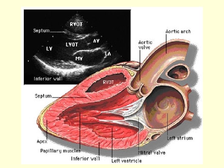
 3. Heart structure: • 1 heart = 2 side by side pumps – Each side has 2 chambers (= 4 chambers in total) 1 2 3 4
 3. Heart • Atrium = chamber that receives blood from the body / lungs • Ventricle = chamber that pushes blood out of the heart to the lungs / body – Have VERY strong muscles • Heart valves – flaps in the heart that open to allow blood to flow in only 1 direction – Will close to stop blood from back-flowing http: //www. kett 6. net/adulteducation/h eartanimations. html
 External Anatomy of the Heart
 Internal Anatomy of the Heart
 Superior Vena cava Right Pulm. Artery Right atrium Right Pulm. Veins Right Semilunar valve Right AV valve Ascending Aorta Left Pulm. Artery Pulmonary trunk Left Pulm. Veins Left Atrium Left AV Valve Left semilunar valve Right Ventricle Inferior Vena cava *AV = atrioventricular Left ventricle Septum Descending Aorta
 Blood Flow Through the Heart
 • http: //www. kett 6. net/adulteducation/h eartanimations. html • http: //highered. mcgrawhill. com/sites/0072495855/student_view 0/c hapter 22/animation__the_cardiac_cycle__qu iz_1_. html • http: //www. youtube. com/watch? v=rk. W 1 sm. Po XKA • http: //www. youtube. com/watch? v=Cs. Idppya. PQ
 The “Cardiac Cycle” Circulation of Blood • Circulation is divided into 2 major pathways: 1. Pulmonary circulation (heart ↔ lungs) 2. Systemic circulation (heart ↔ all body cells)
 Pulmonary Circulation • Pulmonary = lungs • Adds O 2 to blood & removes CO 2 • Heart ↔ lungs • Please follow along on your handout… Pathway: Right ventricle pulmonary trunk L&R pulmonary arteries L&R Lungs (arterioles capillaries O 2 ↔ CO 2 venules) L&R pulmonary veins Left Atrium
 • The pulmonary") NOTE: • The pulmonary ARTERY carries deoxygenated blood ( CO 2) • The pulmonary VEIN carries oxygenated blood • Also, remember that the L & R sides of the heart are reversed in ALL ALL! diagrams
NOTE: • The pulmonary ARTERY carries deoxygenated blood ( CO 2) • The pulmonary VEIN carries oxygenated blood • Also, remember that the L & R sides of the heart are reversed in ALL ALL! diagrams
 to") Systemic Circulation • Systemic = body • Delivers oxygenated blood, nutrients, (and hormones) to all cells and picks up waste products • heart ↔ body • Please follow along on your handout… Pathway: Left ventricle aorta body (arteries, arterioles, capillaries (exchange), venules, veins) inferior & superior vena cava right atria
Systemic Circulation • Systemic = body • Delivers oxygenated blood, nutrients, (and hormones) to all cells and picks up waste products • heart ↔ body • Please follow along on your handout… Pathway: Left ventricle aorta body (arteries, arterioles, capillaries (exchange), venules, veins) inferior & superior vena cava right atria
 The Cardiac Cycle cont’d… The cardiac cycle has 2 main phases: 1. Systole = When the heart muscles contract – Includes atrial systole & ventricular systole – The heart beats in this order: • Atrial systole ventricular systole diastole • (or squeeze, rest) a. Atrial systole • The atria both contract • The atrioventricular valves (av valves) open • Blood is pumped into the ventricles
 b. Ventricular systole: – Semilunar valves open – Atrioventricular valves shut • If didn’t when ventricles contract, we’d get backwashing into the atria = BAD – Contraction pushes blood into aorta (from L ventricle) and Pulm. Arteries (from R ventricle) – Makes “Lub” sound as AV valves close (in unison) – When ventricles contract, it sends a huge gush of blood through the body this is a period of increased pressure in the arteries • = SYSTOLIC PRESSURE
 The Cardiac Cycle cont’d… 2. Diastole = when heart muscles relax • L&R semilunar valves shut • “DUB” sound as semilunar valves close • The atria (L&R) now fill with blood – Eventually, leading into atrial systole, the weight of the blood causes the L&R atrioventricular valves to open, and blood pumps from atria to ventricles • Because blood is not being shoved into the arteries, there is LESS pressure in them – = DIASTOLIC PRESSURE – http: //www. youtube. com/watch? v=H_3 V 9 xl. DMA 0 heart valves on echocardiogram (ultrasound)
 • the “lub-DUB” sound heard during a heartbeat is created by the opening and closing of the 2 sets of heart valves • Sphygmomanometer = blood pressure monitor – I. e. – the cuff thingy Systolic arteriole pressure • Optimal BP at rest = 90 - 120 60 - 80 Diastolic arteriole pressure
 Factors that Affect Blood Pressure • Effect on BP?
 Factors that Affect Blood Pressure • Effect on BP? *Temporarily, but longterm effects are unclear
 Factors that Affect Blood Pressure • Effect on BP? http: //science. howstuffworks. com/nicotin e 3. htm video https: //www. youtube. com/watch? v=Dj 64 0 y. HTNM 4
 Factors that Affect Blood Pressure • Effect on BP? Shock
 Factors that Affect Blood Pressure • they diminish the effects of adrenaline and other stress hormones • Used particularly for the management of cardiac arrhythmias, treatment following a heart attack, and hypertension • Effect on BP? “Beta Blockers”
 Factors that Affect Blood Pressure • Effect on BP? Stress
 How to measure blood pressure • https: //www. youtube. com/watch? v=Gmi c 13 mvsgo - polyfit manual cuff measurement • https: //www. youtube. com/watch? v=S 64 8 x. ZDK 7 b 0 better video – 4 yourcna. com
 • Thrombosis = a blood clot") What is a heart attack? (aka coronary thrombosis) • Thrombosis = a blood clot • Atherosclerosis = is a hardening of an artery specifically due to an atheromatous plaque (white blood cells, calcium, lipids) – Decreases or stops blood flow in that area • Stenosis = the narrowing of a blood vessel, especially arteries, due to plaque buildup • Coronary arteries = The arteries that branch off of the aorta and bring a blood supply TO THE HEART TISSUE itself • Coronary veins = the veins that collect blood back from the heart tissue and bring it to the vena cava
What is a heart attack? (aka coronary thrombosis) • Thrombosis = a blood clot • Atherosclerosis = is a hardening of an artery specifically due to an atheromatous plaque (white blood cells, calcium, lipids) – Decreases or stops blood flow in that area • Stenosis = the narrowing of a blood vessel, especially arteries, due to plaque buildup • Coronary arteries = The arteries that branch off of the aorta and bring a blood supply TO THE HEART TISSUE itself • Coronary veins = the veins that collect blood back from the heart tissue and bring it to the vena cava
 • Coronary arteries = the arteries that branch off of the aorta and bring a blood supply to the heart itself • Coronary veins = collect blood from the heart tissue and bring it back to the vena cava
 Heart attack aka coronary thrombosis • https: //www. youtube. com/watch? v=di. G 519 d. FVNs – Hypertension • http: //www. youtube. com/watch? v=H_Vs. Hmo. RQKk – having a heart attack moment to moment • http: //www. youtube. com/watch? v=w 8 w. Xdto. W-HQ bursting clot • http: //www. youtube. com/watch? v=BL 92 vtuj 9 nc – heart attack • angina https: //www. youtube. com/watch? v=f. Bn 9 munof. Vs • Angioplasty http: //www. youtube. com/watch? v=N 7 nghr 9 Tp. SU • Bypass surgery http: //www. youtube. com/watch? v=3 Nf 6 Q 2 sk. GOM – Bypass surgery p 328, figure 9. 29
 Strokes • http: //www. youtube. com/watch? v=442 s 9 a_TEL 8
 Control of Heartbeat The heartbeat is controlled in 2 ways 1. Nervous control (pacemaker/nervous system) 2. Chemical control (medulla oblongata in brain)
 1. Nervous Control of Heartbeat • Basic rate of heartbeat is controlled by electrical impulses • Heart has a built in biological pacemaker – called sinoatrial (SA) node • Produces impulses all by itself that cause atria to contract • Then impulse spreads to the AV (atrioventricular) node • After a slight delay it’s passed into nerve fibers known as the bundle of His, then the Purkinje fibers which contour the ventricles • This causes the ventricles to contract
 http: //highered. mc grawhill. com/sites/0072 507470/student_vi ew 0/chapter 20/ani mation__conductin g_system_of_the_ heart. html
 What is cellular respiration • Cellular respiration is when our CELLS take in OXYGEN and food (GLUCOSE) and uses it to create ATP, a molecule which the cell uses for energy. Carbon dioxide is released from the cells as a waste product. • The simplified formula is: – C 6 H 12 O 6 + 6 O 2 → 6 CO 2 + 6 H 2 O + Energy (as ATP) • The word equation for this is: – Glucose + Oxygen → Carbon dioxide + Water + Energy (as ATP)
 2. Chemical Control of the Heartbeat • When you physically exert yourself in any way, the pacemaker (i. e. nervous control) can no longer keep up with gas exchange demand need someway to pump heart faster • Chemical control now kicks in (more complete story on p. 318) • Exercise causes ed CO 2 production in the cells due to cellular respiration • CO 2 goes into the blood • Medulla oblongata in brain picks up on ed CO 2 levels (via chemoreceptors) • Brain (medulla oblongata) now causes adrenaline to be released tells heart to beat faster • When exercise stops, CO 2 levels go es, therefore Medulla oblongata ‘shuts off’ its control (stoping the release of adrenaline from adrenal glands) pacemaker back in charge • Other ways of initiating chemical control of heartbeat: • Adrenalin in the fight or flight response