
The Autonomic Nervous System The Autonomic Nervous

lecture_15_-_autonomic_nervous_system.ppt
- Размер: 1.6 Mегабайта
- Количество слайдов: 26
Описание презентации The Autonomic Nervous System The Autonomic Nervous по слайдам
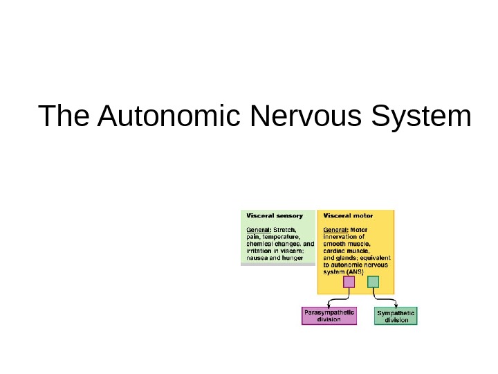
The Autonomic Nervous System
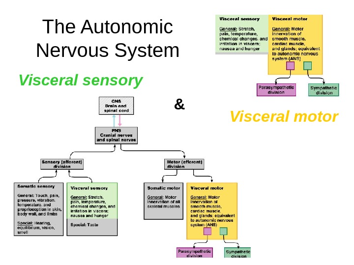
The Autonomic Nervous System Visceral sensory Visceral motor&
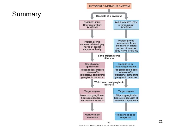
3 Autonomic nervous system The autonomic nervous system is the subdivision of the peripheral nervous system that regulates body activities that are generally not under conscious control Visceral motor innervates non-skeletal (non-somatic) muscles Visceral sensory will be covered later
4 ANS is the subdivision of the peripheral nervous system that regulates body activities that are generally not under conscious control Visceral motor innervates non-skeletal ( non-somatic) muscles Composed of a special group of neurons serving: Cardiac muscle (the heart) Smooth muscle (walls of viscera and blood vessels) Internal organs Skin. To repeat…
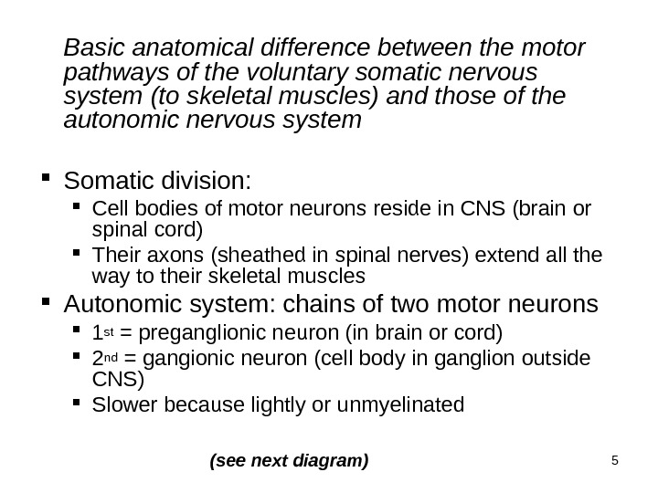
5 Basic anatomical difference between the motor pathways of the voluntary somatic nervous system (to skeletal muscles) and those of the autonomic nervous system Somatic division: Cell bodies of motor neurons reside in CNS (brain or spinal cord) Their axons (sheathed in spinal nerves) extend all the way to their skeletal muscles Autonomic system: chains of two motor neurons 1 st = preganglionic neuron (in brain or cord) 2 nd = gangionic neuron (cell body in ganglion outside CNS) Slower because lightly or unmyelinated (see next diagram)
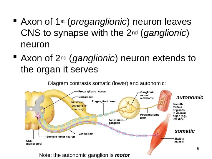
6 Axon of 1 st ( preganglionic ) neuron leaves CNS to synapse with the 2 nd ( ganglionic ) neuron Axon of 2 nd ( ganglionic ) neuron extends to the organ it serves Diagram contrasts somatic (lower) and autonomic: autonomic somatic Note: the autonomic ganglion is motorthis dorsal root ganglion is sensory
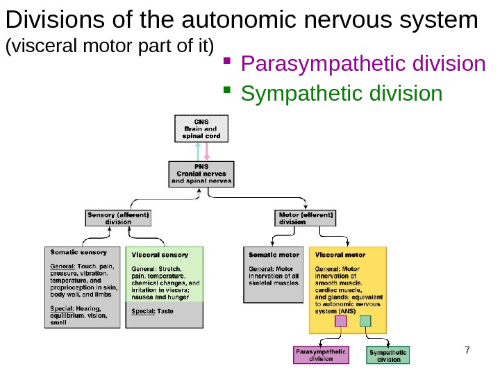
7 Divisions of the autonomic nervous system (visceral motor part of it) Parasympathetic division Sympathetic division
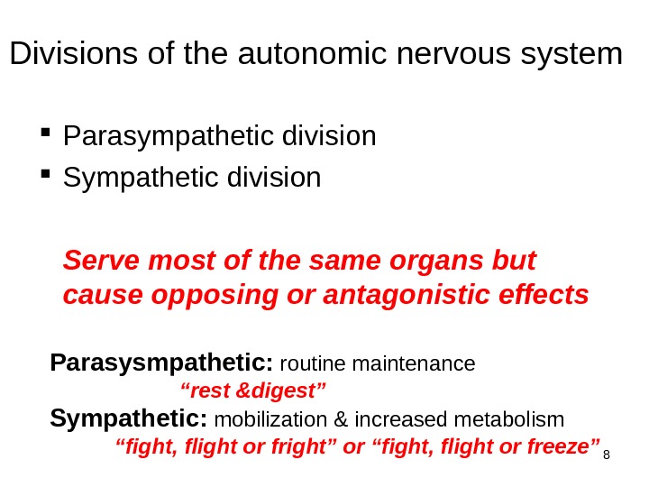
8 Divisions of the autonomic nervous system Parasympathetic division Serve most of the same organs but cause opposing or antagonistic effects Parasysmpathetic: routine maintenance “ rest &digest” Sympathetic: mobilization & increased metabolism “fight, flight or fright” or “fight, flight or freeze”
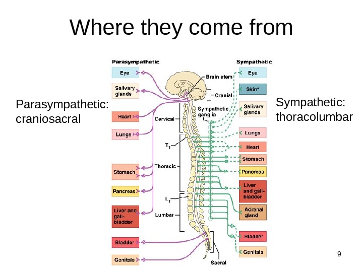
9 Where they come from Parasympathetic: craniosacral Sympathetic: thoracolumbar
10 Parasympathetic nervous system “rest & digest” Also called the craniosacral system because all its preganglionic neurons are in the brain stem or sacral levels of the spinal cord Cranial nerves III, VII, IX and X In lateral horn of gray matter from S 2 -S 4 Only innervate internal organs (not skin) Acetylcholine is neurotransmitter at end organ as well as at preganglionic synapse: “cholinergic”
11 Parasympathetic continued Cranial outflow III — pupils constrict VII — tears, nasal mucus, saliva IX – parotid salivary gland X (Vagus n) – visceral organs of thorax & abdomen: Stimulates digestive glands Increases motility of smooth muscle of digestive tract Decreases heart rate Causes bronchial constriction Sacral outflow (S 2 -4): form pelvic splanchnic nerves Supply 2 nd half of large intestine Supply all the pelvic (genitourinary) organs
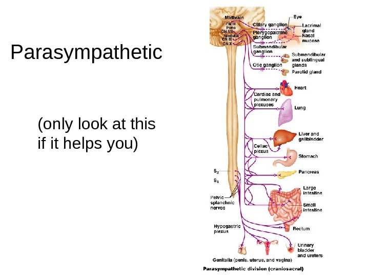
12 Parasympathetic (only look at this if it helps you)
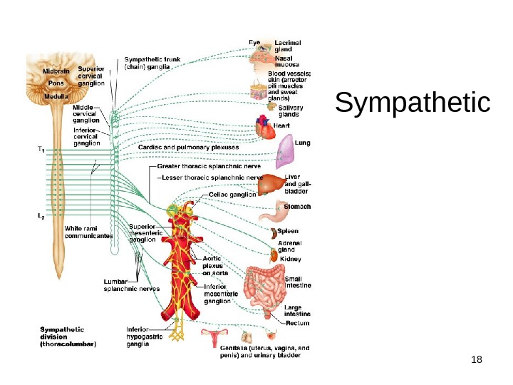
13 Sympathetic nervous system “fight, flight or fright” Also called thoracolumbar system: all its neurons are in lateral horn of gray matter from T 1 -L 2 Lead to every part of the body (unlike parasymp. ) Easy to remember that when nervous, you sweat; when afraid, hair stands on end; when excited blood pressure rises (vasoconstriction): these sympathetic only Also causes: dry mouth, pupils to dilate, increased heart & respiratory rates to increase O 2 to skeletal muscles, and liver to release glucose Norepinephrine (aka noradrenaline) is neurotransmitter released by most postganglionic fibers (acetylcholine in preganglionic): “adrenergic”
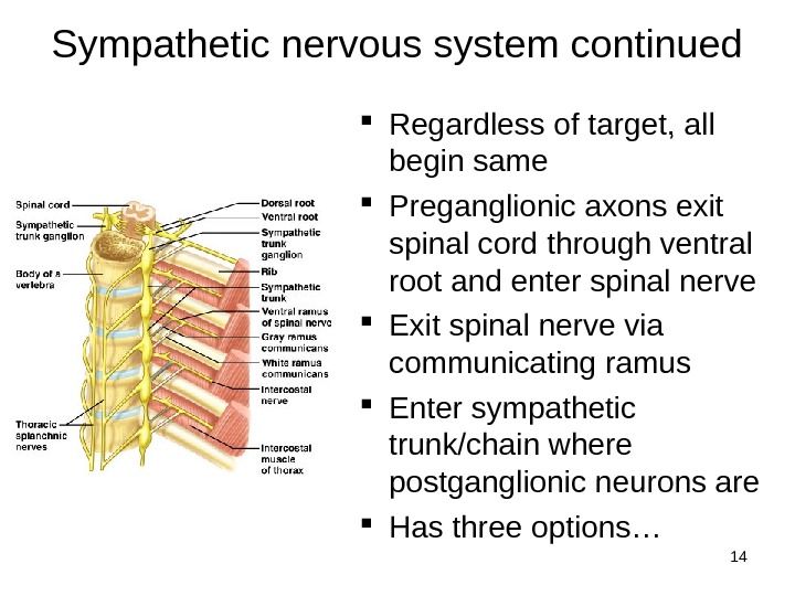
14 Sympathetic nervous system continued Regardless of target, all begin same Preganglionic axons exit spinal cord through ventral root and enter spinal nerve Exit spinal nerve via communicating ramus Enter sympathetic trunk/chain where postganglionic neurons are Has three options…
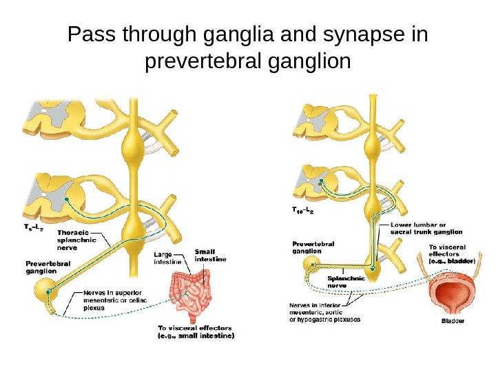
15 Options of preganglionic axons in sympathetic trunk 1. Synapse on postganglionic neuron in chain ganglion then return to spinal nerve and follow its branch to the skin 2. Ascend or descend within sympathetic trunk, synapse with a posganglionic neuron within a chain ganglion, and return to spinal nerve at that level and follow branches to skin 3. Enter sympathetic chain, pass through without synapsing, form a splanchnic nerve that passes toward thoracic or abdominal organs These synapse in prevertebral ganglion in front of aorta Postganglionic axons follow arteries to organs (see next slides for drawing examples)
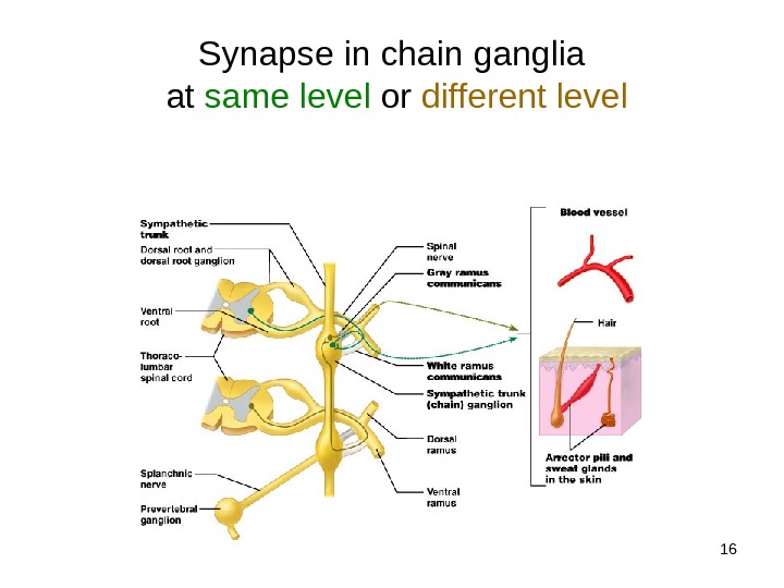
16 Synapse in chain ganglia at same level or different level
17 Pass through ganglia and synapse in prevertebral ganglion
18 Sympathetic
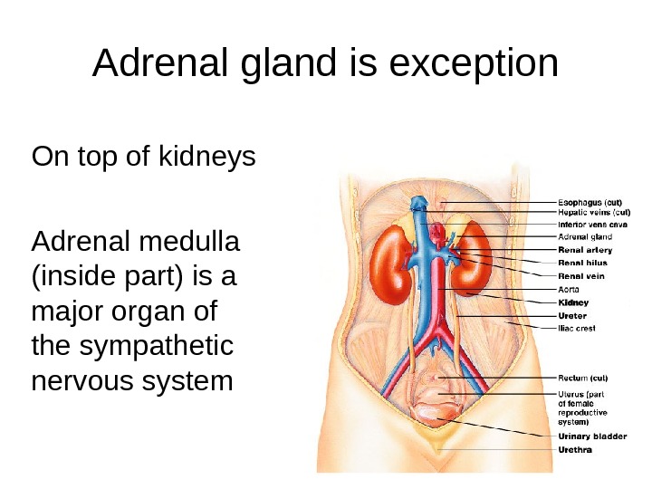
19 Adrenal gland is exception On top of kidneys Adrenal medulla (inside part) is a major organ of the sympathetic nervous system
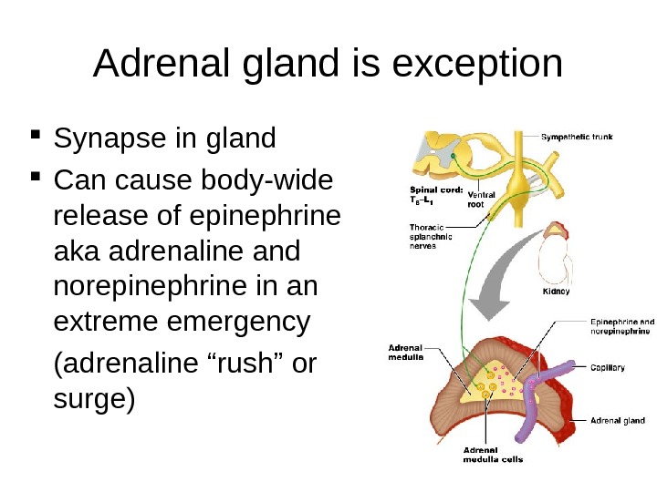
20 Adrenal gland is exception Synapse in gland Can cause body-wide release of epinephrine aka adrenaline and norepinephrine in an extreme emergency (adrenaline “rush” or surge)
21 Summary
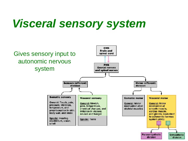
22 Visceral sensory system Gives sensory input to autonomic nervous system
23 Visceral sensory neurons Monitor temperature, pain, irritation, chemical changes and stretch in the visceral organs Brain interprets as hunger, fullness, pain, nausea, well-being Receptors widely scattered – localization poor (e. g. which part is giving you the gas pain? ) Visceral sensory fibers run within autonomic nerves, especially vagus and sympathetic nerves Sympathetic nerves carry most pain fibers from visceral organs of body trunk Simplified pathway: sensory neurons to spinothalamic tract to thalamus to cerebral cortex Visceral pain is induced by stretching, infection and cramping of internal organs but seldom by cutting (e. g. cutting off a colon polyp) or scraping them
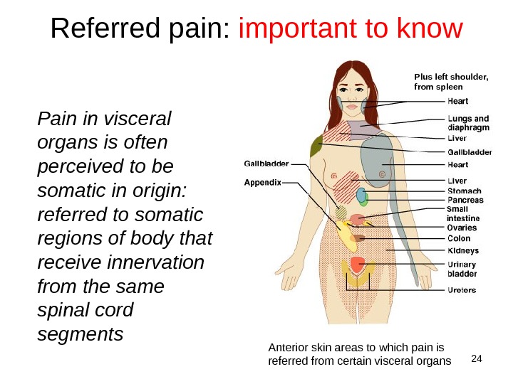
24 Referred pain: important to know Pain in visceral organs is often perceived to be somatic in origin: referred to somatic regions of body that receive innervation from the same spinal cord segments Plus left shoulder, from spleen Anterior skin areas to which pain is referred from certain visceral organs
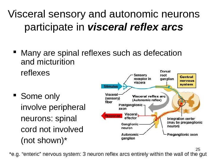
25 Visceral sensory and autonomic neurons participate in visceral reflex arcs Many are spinal reflexes such as defecation and micturition reflexes Some only involve peripheral neurons: spinal cord not involved (not shown)* *e. g. “enteric” nervous system: 3 neuron reflex arcs entirely within the wall of the gut
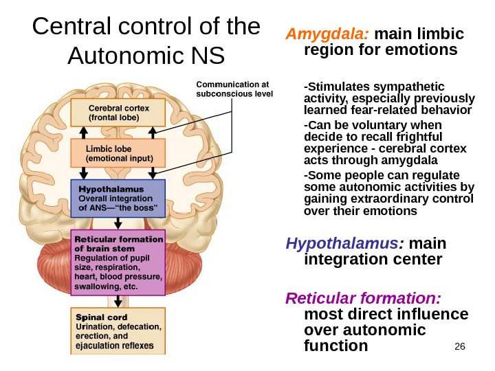
26 Central control of the Autonomic NS Amygdala: main limbic region for emotions -Stimulates sympathetic activity, especially previously learned fear-related behavior -Can be voluntary when decide to recall frightful experience — cerebral cortex acts through amygdala -Some people can regulate some autonomic activities by gaining extraordinary control over their emotions Hypothalamus : main integration center Reticular formation: most direct influence over autonomic function