56ad99ed89313cd3124bc4099a755d20.ppt
- Количество слайдов: 161
 The 4 functions of muscles: 1. Name all 4
 The 4 functions of muscles: 1. Movement: a. Skeletal muscles pull against bones as levers. b. Visceral organs do their “work” such as digestion c. The heart moves blood 2. Maintain posture: skeletal muscles function constantly, always making tiny adjustments
 Functions (cont. ): 3. Stabilize joints: tendons wrap over and around joints and the tautness of skeletal muscles helps stabilize the joint and keep it from being injured during stress. 4. Generate heat: As muscles contract and use energy; they emit heat.
 A quick review: Define: ? ? ? : ? ? ? ? : The relatively immovable bone the skeletal muscle is attached to The relatively movable bone that skeletal muscle is attached to
 A quick review: Define: Muscle origin: Muscle insertion: The relatively immovable bone the skeletal muscle is attached to The relatively movable bone that skeletal muscle is attached to Origin Insertion
 Muscle fibers • Is a long, cylindrical cell, also called a muscle fiber(1). • Is huge compared to other cells – are very thin, but can be up to a foot long! • Is formed by the fusion (connecting together) of many cells during the embryo stage of development.
 Muscle fibers EACH FIBER CONTAINS: • ? ? – the connective tissue “overcoat” for the cell. Protects and gives it more strength. Also separates it from other cells. Is very permeable (porous).
 Muscle fibers EACH FIBER CONTAINS: • Endomysium – the connective tissue “overcoat” for the cell. Protects and gives it more strength. Also separates it from other cells. Is very permeable (porous). endomysium
 EACH FIBER CONTAINS: Contains Name these? ? ? – rod-like organelles that run the length of the fiber and are divided into the contractile segments of the muscle cell. • Account for 80% of the cell volume, and take up most of the space in the sarcoplasm.
 EACH FIBER CONTAINS: Contains myofibrils – rod-like organelles that run the length of the fiber and are divided into the contractile segments of the muscle cell. • Account for 80% of the cell volume, and take up most of the space in the sarcoplasm.
 Muscle fibers EACH FIBER CONTAINS: • Contain thick and thin “stringy” protein myofilaments called m? ? (thicker one) and a? ? ? (thinner one). • Are divided into segments called s? ? which contain bands due to the arrangement of the myofilaments.
 Muscle fibers EACH FIBER CONTAINS: • Contain thick and thin “stringy” protein myofilaments called myosin (thicker one) and actin (thinner one). • Are divided into segments called sarcomeres which contain bands due to the arrangement of the myofilaments.


 Muscle Fiber Stimulation Cause Contraction • Name the ? ? ? theory shows how the muscle mechanically contracts…but how is it told to contract? • All sarcomeres within the fibers of a muscle must get the message at the same time and contract together. • The following shows how the muscle is told to contract…
 Muscle Fiber Stimulation Cause Contraction • The sliding filament theory shows how the muscle mechanically contracts…but how is it told to contract? • All sarcomeres within the fibers of a muscle must get the message at the same time and contract together. • The following shows how the muscle is told to contract…
 Neuromuscular Junction
 Name this condition • Lack of oxygen causes ATP deficit • Lactic acid builds up from anaerobic respiration (or as the latest research indicates calcium ions leak across cell membranes losing the ability of muscles to contract)
 Muscle Fatique • Lack of oxygen causes ATP deficit • Lactic acid builds up from anaerobic respiration
 Muscle A? • Weakening and shrinking of a muscle • May be caused – Immobilization – Loss of neural stimulation Multiple Sclerosis Ted Talk
 Muscle Atrophy • Weakening and shrinking of a muscle • May be caused – Immobilization – Loss of neural stimulation Multiple Sclerosis Ted Talk
 Muscle Hypertrophy • Enlargement of a muscle • More capillaries • More mitochondria • Caused by – Strenuous exercise – Steroid hormones
 Name this type of Contraction • Produces no movement • Used in – Standing – Sitting – Posture
 Isometric Contraction • Produces no movement • Used in – Standing – Sitting – Posture
 ? ? Contraction • Produces movement • Used in – Walking – Moving any part of the body
 Isotonic Contraction • Produces movement • Used in – Walking – Moving any part of the body
 Name that Muscle!
 Muscle #1 & 2 #1 #2
 Trapezius Action: l Upper fibers: they extend the head and neck (both); laterally flex the head and neck to the same side, rotate the head and neck to the opposite side (singly), elevate the scapula, and upwardly rotate the scapula. l Middle fibers: adduct the scapula, stabilize the scapula. l Lower fibers: depress the scapula, upwardly rotate the scapula. l Origin: External occipital protuberance, medial portion of superior nuchal line, ligamentum nuchae, and spinous processes of C-7 to T-12. l Insertion: Lateral 1/3 of clavicle, acromion and spine of scapula. l http: //www. preventdisease. com/home/muscleatlas/shtraplat. jpg
 Latissimus Dorsi A: extends shoulder, adducts shoulder, medially rotates shoulder (same as teres major) l O: spinous processes of last 6 thoracic vertebrae, last 3 or 4 ribs, and posterior iliac crest l I: Crest of lesser tubercle of humerus (anterior aspect; same insertion as teres major) l http: //www. octc. kctcs. edu/gcaplan/anat/images/Image 359. gif http: //courses. washington. edu/hubio 553/atlas/images/shtraplat. jpg http: //www. trans health. com/images/latspread. jpg
 Muscle #3
 Deltoid A: abduct shoulder (all); flex shoulder, medially rotate shoulder, horizontally adduct shoulder (anterior fibers); extend shoulder, laterally rotate shoulder, horizontally abduct shoulder (posterior fibers) l O: lateral 1/3 of clavicle, acromion process and spine of scapula l I: deltoid tuberosity (of humerus) l http: //www. gpc. edu/~jaliff/deltoid. gif http: //courses. washington. edu/hubio 553/atlas/images/108. jpg
 Muscle #4 #4
 Serratus Anterior l l A: abduct, depress scapula; hold medial border of scapula O: upper 8 -9 ribs I: anterior surface of medial border of scapula Note: well developed on superheroes http: //www. okpatents. com/phosita/images/superhero. jpg http: //courses. washington. edu/hubio 553/atlas/images/shserant. jpg https: //secure. contactdesigns. net/disabilityproducts. com/ht docs/contactcommerce/images/items/Big%20 Serrated. JPG
 http: //www. aurelia art. com/art/cinema/daredevil. jpg
 Muscle #5
 Coracobrachialis A: Flexes the shoulder; adducts the shoulder l O: Coracoid process of the scapula l I: Medial surface of the mid-humeral shaft l l "Armpit" muscle. In anatomical position, it is deep to pectoralis major and anterior deltoid. http: //www. gpc. e du/~jaliff/hubibo 24. gif http: //www. rad. washington. edu/staticpix/atlas/co racobrachialisant. jpg
 Muscle #6 #6
 External Intercostals l Action: assist w/inhalation by pulling ribs up l Origin: inferior border of rib above l Insertion: superior border of rib below http: //www. gpc. edu/~jaliff/serratus. gif http: //www. octc. kctcs. edu/gca plan/anat/images/Image 391. gif
 Muscle #7 #7
 Teres Major A: extend, adduct, medially rotate shoulder (same as latissimus dorsi). l O: lateral inferior angle and lower ½ of lateral border of scapula. l I: crest of lesser tubercle of humerus l Notes: "lat's little helper. " Synergist w/lat. dorsi. Antagonist to teres minor. l Back http: //www. rad. washington. edu/staticpix/atlas/teresmajorpost 2. jpg Front http: //www. rad. washington. edu/staticpix/atlas/teresmajorant 2. jpg
 Muscle #8
 Pectoralis Minor l A: depress, abduct, anteriorly tilt scapula, l O: 3 rd-5 th ribs l I: coracoid process of scapula http: //www. rad. washington. edu/staticpix/atlas/pectoralisminor. jpg
 Muscle #9
 Levator Scapulae A: elevate scapula, downwardly rotate scapula, laterally flex head & neck, rotate head & neck to same side (singly); extend head & neck (both) l O: transverse processes of 1 st 4 th cervical vertebrae l I: upper medial border & superior angle of scapula l http: //www. rad. washington. edu/staticpix/atlas/levatorscapulae 2. jpg http: //www. vancouveryoga. com/assets/photos/levator. jpg
 Muscle #10
 Internal Intercostals l Action: assist w/exhalation by drawing the ribs down l Origin: inferior border of rib above l Insertion: superior border of rib below http: //www. octc. kctcs. edu/gcaplan/anat/images/Image 392. gif
 Muscle #11
 Rotator Cuff Muscle 1 of 4: Supraspinatus l A: abducts shoulder stabilizes head of humerus in glenoid cavity (common function in all four rotator cuff muscles) O: supraspinous fossa of the scapula I: greater tubercle of the humerus Note: deep to trapezius, runs under the acromion process 1. 2. l l l http: //www. rad. washingt on. edu/staticpix/atlas/sup raspinatuspost. jpg http: //www. shoulderdoc. co. uk/img/should erdoc/cuff_tendons 1. jpg
 Muscle #12
 Rotator Cuff Muscle 4 of 4: Subscapularis l A: medially rotate shoulder 2. stabilize head of humerus in glenoid cavity O: subscapular fossa of scapula I: lesser tubercle of humerus Note: On anterior surface 1. l l l http: //www. orthop. washington. edu/_Rainbow/Album/10357 m 1 ef 96049 ad 8 d 4 e 97 a 88 a cdbe 200 be 928. jpg
 Rotator Cuff Muscles l l l Four Muscles: Supraspinatus Teres Minor Infraspinatus Subscapularis http: //www. fitstep. com/Advanced/Anatomy/Rot_cuff. htm
 Muscles #13 & 14 #13 #14
 Rhomboideus Major A: adduct scapula, elevate scapula, downwardly rotate scapula l O: Spinous processes of T-2 to T-5 l I: Medial border of scapula l http: //anatquest. nlm. nih. gov/Visible. Human/Image. Data/Rendered/jpg/DSR 110224363. jpg http: //www. ncpad. org/get/images/strengthexercisehandouts/latpulldown_RHOM_Major. Minor. jpg
 Rhomboid Minor l A: see rhomboid major. l O: Spinous processes of C-7 and T-1 l I: medial border of scapula http: //www. dartmouth. edu/~anatomy/assets/surface/shoulder/scsurface 1. gif http: //www. rad. washington. edu/staticpix/atlas/rhomboids 2. jpg
 Muscle #15
 Rotator Cuff Muscle 2 of 4: Teres Minor l A: laterally rotate, adducts, extends, and horizontally abducts shoulder 2. stabilizes head of humerus in glenoid cavity O: superior ½ of lateral border of scapula I: greater tubercle of the humerus Note: antagonist to teres major in rotation of humerus 1. l l l http: //www. rad. washington. edu/staticp ix/atlas/teresminorpost. jpg http: //www. fotosearch. com/comp/LIF 1 25/3 D 506030. jpg http: //www. chirotx. com/_derived/Shoulder_Pain_ Active_Release_Technique. html_txt_wpe 2. gif
 Muscle #16
 Pectoralis Major l A: adducts shoulder 2. medially rotates shoulder l O: medial ½ of clavicle, sternum, and cartilage of 1 st-6 th ribs l I: crest of greater tubercle of humerus 1. http: //www. rad. washington. edu/atlas/pectoralismajor. html http: //www. ironman israel. com/gallery/schwarzenegger_arno ld/schwarzenegger_arnold_004. jpg http: //www. getbodysmart. com/ap/muscularsy stem/armmuscles/menu/image. gif
 Muscle #17
 Rotator Cuff Muscle 3 of 4: Infraspinatus l l A: See teres minor O: infraspinatus fossa of scapula I: greater tubercle of the humerus Note: convergent muscle fibers. Underneath trapezius and deltoid. Synergist w/teres minor in rotation of shoulder http: //www. abcbodybuilding. com/anatomy/s houldersanatomy 1_files/image 006. gif http: //mywebpages. comcast. net/wnor/postshouldermus 1. jpg http: //www. weldoen. nl/schouder/img/rupturen_02. jpg
 Answers 1. 2. 3. 4. 5. 6. 7. 8. Trapezius Latissimus dorsi Deltoid Serratus anterior Coracobrachialis External intercostals Teres major Pectoralis minor 9. 10. 11. 12. 13. 14. 15. 16. 17. Levator scapulae Internal intercostals Supraspinatus Subscapularis Rhomboid minor Rhomboid major Teres minor Pectoralis major Infraspinatus
 Name this type of Muscle Tissue striated fibers multinucleated voluntary control
 Skeletal Muscle striated fibers multinucleated voluntary control
 Skeletal Muscle Name 3 Characteristics of This type of Muscle tissue
 Skeletal Muscle striated fibers multinucleated voluntary control long cylindrical cells attached to bones

 Name this type of Muscle Tissue • No striations • single nuclei • autonomic control
 Smooth Muscle • No striations • single nuclei • autonomic control
 Smooth Muscle • Name 3 characteristics of this type of muscle tissue
 Smooth Muscle • • • No striations single nuclei autonomic control Fusiform cells Viscera (guts), GI, lining internal organs
 Name this type of Muscle Tissue • • striations intercalated disks gap junctions autonomic control
 Cardiac Muscle • Name 3 characteristics
 Cardiac Muscle • • • striations intercalated disks gap junctions autonomic control Heart Branching cells
 Skeletal Muscle- CT wrappings • • Bundles are formed by: ? ? = upon ? ? = around ? ? = within
 Skeletal Muscle • • Bundles are formed by: epimysium epi = upon perimysium peri = around endomysium end = within
 Tendons • Band like • A? ? ? flat sheet –like attachment of tendon to bone
 Tendons • Band like • Aponeurosis flat sheet –like attachment of tendon to bone
 Every Muscle Has. . . • Name this attachment point that is – proximal – least movable • Insertion
 Every Muscle Has. . . • Origin – proximal – least movable • Insertion
 Grouping by Function 1 • • • 2 (1) (2) (3) (4) (5) 5 4 3
 Grouping by Function • • • Flexors Extensors Adductors Abductors Circumduction
 1 2
1 2

 1) 2) 3)
 Skeletal Muscle Bundles are formed by: • epimysium epi = upon • perimysium peri = ? ? ? • endomysium endo = within all continuous with the TENDON “mysium” = presence of muscle *Name this term = strong cord that connects muscle with the bone (periosteum)
 Skeletal Muscle Bundles are formed by: • epimysium epi = upon • perimysium peri = around • endomysium endo = within all continuous with the TENDON “mysium” = presence of muscle *Tendon = strong cord that connects muscle with the bone (periosteum)
 Epimysium • “upon” • Coarse sheath covers muscle and holds f? ? ? in place • Lubricating surface for muscles to rub
 Epimysium • “upon” • Coarse sheath covers muscle and holds fasicles in place • Lubricating surface for muscles to rub
 Endomysium • “within” the muscle – connective tissue membrane that covers muscle fibers • Maintains the chemical environment that muscle cells need to contract
 Name this term • “around” • Tougher connective envelope that binds groups of muscle fingers together (= fascicle) • Fascicle is under independent control of a motor neuron
 Perimysium • “around” • Tougher connective envelope that binds groups of muscle fingers together (= fascicle) • Fascicle is under independent control of a motor neuron
 : • What is the aponeurosis?
 Tendons • Band like Aponeurosis flat sheet –like attachment of tendon to bone CT of one muscle merges with another muscle
 Interactions of Skeletal Muscles in the Body • • Muscles usually work in g? ? P? ? M? ? (agonist), Antagonist, Synergist, Fixator Muscles can swap roles depending on the motion Muscles are usually arranged in antagonistic pairs – F? ? ? decreases the angle between bones bending motions – E? ? ? and hyperextension increases the angle between bones; straightening motions – A? ? ? move a part away from the median of the body – A? ? ? moves a part toward the median of the body
 Interactions of Skeletal Muscles in the Body • Muscles usually work in groups, i. e. perform “group actions” • Prime mover (agonist), Antagonist, Synergist, Fixator • Muscles can swap roles depending on the motion • Muscles are usually arranged in antagonistic pairs – Flexion decreases the angle between bones bending motions – Extension and hypertension increases the angle between bones; straightening motions – Abduction move a part away from the median of the body – Adduction moves a part toward the median of the body
 Interactions of Skeletal Muscles • Prime Mover (agonist): the muscle that does what? ? ? – ex: biceps brachii, flexion of forearm • Antagonist: the principle muscle that causes the opposite movement – ex: triceps brachii, extension of forearm
 Interactions of Skeletal Muscles • Prime Mover (agonist): the principle muscle that causes a movement – ex: biceps brachii, flexion of forearm • Antagonist: the principle muscle that causes the opposite movement – ex: triceps brachii, extension of forearm
 Interactions of Skeletal Muscles (1)? ? : muscles that assist the prime mover – ex: extensor carpi (wrist) muscles are these for the flexor digitorum muscles when you clench your fist (2)? ? : these that stabilize the origin of a prime over – ex: several back muscles that stabilize scapula when the deltoid flexes the arm
 Interactions of Skeletal Muscles (1) Synergists: muscles that assist the prime mover – ex: extensor carpi (wrist) muscles are synergists for the flexor digitorum muscles when you clench your fist (2) Fixators: synergists that stabilize the origin of a prime over – ex: several back muscles that stabilize scapula when the deltoid flexes the arm
 • What muscle(s) assists with mastication?
 What muscle assists with mastication? Masseter, temporalis, pterygoids
 Muscles of Mastication Masseter • Action: closes jaw • Origin: zygomatic arch • Insertion: mandibular ramus • Notes: strongest muscle in body (per in 2) 150 lbs of pressure http: //face-andemotion. com/dataface/anatomy/media/ Hand. Atlas_masseter-fascia. jpg http: //instruct. westvalley. edu/g ranieri/headmuscles. jpg
 Muscles of Facial Expression Temporalis • Action: closes jaw • Origin: temporal line of skull • Insertion: coronoid process of mandible • Note: tight band around head while chewing… http: //sungag. buddhism. org/tech 22 pe/board/jungyun/upfile/temporalis. gif http: //face-and-emotion. com/dataface/anatomy/media/Hand. Atlas_buccinator-temporalis. jpg
 Muscles of Facial Expression Pterygoids (Medial& Lateral) • Action: grating, "chewing the cud" muscle; protraction of mandible • Origin: sphenoid bone • Insertion: medial surface of ramus (m. pterygoid); TMJ & neck of mandible (l. pterygoid) http: //www. wetcanvas. com/Community/i mages/06 -Mar-2005/30792 -pterygoids. jpg http: //www. med. mun. ca/anatomy/media/ov erview/Protract. Image 292. jpg
 What muscle draws the eyebrows together?
 Muscles of Facial Expression Corrugator supercilii • Action: pulls eyebrows together • Origin: frontal bone, near nasal bone, above orbit • Insertion: skin of eyebrow • Other notes: deep to frontalis • Frowning http: //www. bartleby. com/107/Images/large/image 379. gif http: //waddell. ci. manchester. ct. us/g arden_images/h_corrugated. jpg http: //www. uned. es/psico-2 -psicologia-general. II/links/FACS/index_archivos/image 006. jpg
 • Of the abdominal muscles, which one has fibers that are horizontal? Which one compresses the abdomen?
 Muscles of the Abdominal Wall Transversus Abdominis • Action: compress abdominal contents • Origin: inguinal ligament, iliac crest & internal surface of lower 6 ribs • Insertion: linea alba http: //www. aerobicmania. cz/images/core/ta. gif http: //www. getbodysmart. com/ap/muscular system/abdominalmuscles/menu/image. gif
 Muscles of the Abdominal Wall Internal Oblique • Action: laterally flex and rotate vertebral column to the same side; flex vertebral column & compress abdomen • Origin: lateral inguinal ligament, iliac crest • Insertion: internal surface of lower 3 ribs, linea alba http: //www. octc. kctcs. edu/gca plan/anat/images/Image 341. gif http: //www. getbodysmart. com/ap/muscul arsystem/backabdomen/menu/image. gif
 Muscles of the Abdominal Wall Rectus Abdominis • Action: flex vertebral column • Origin: pubic symphysis • Insertion: cartilage of 5 th, 6 th, and 7 th ribs and xiphoid process • Note: “six pack” muscle http: //www. octc. kctcs. edu/gcaplan/anat/images/Image 339. gif
 What posterior arm muscle extends the forearm?
 • What posterior arm muscle extends the forearm? Triceps brachii
 Extensor of Upper Arm: Triceps Brachii Action: Extend elbow; long head – extend & adduct shoulder. • O: – Long head: Infraglenoid tubercle of scapula. – Lateral head: Proximal ½ of humerus. – Medial head: Distal ½ of humerus. • I: Olecranon process of ulna. • Only muscle on posterior upper arm. • Antagonist to biceps brachii. http: //www. rad. washington. e du/atlas/tricepsbrachii. html http: //en. wikipedia. org/wiki/Image: Triceps_brachii. png
 • What does the brachialis muscle do?
 • What does the brachialis muscle do? Flexion of the elbow Synergist w/biceps brachii

 • What muscles are of the quadriceps group?
 Muscles of the Hips & Legs: Quadriceps • A: Extend the knee. • O: – RF: AIIS – VL, VM, VI: Femur • I: Tibial tuberosity via patellar tendon. • Four heads: – – Rectus femoris Vastus lateralis Vastus medialis Vastus intermedius http: //www. aleve. com/images/Aleve_Sketches/Knee_Extension. jpg http: //www. preventdisease. com/home/muscleatlas/203. jpg
 • Where are the hamstrings located?
 Muscles of the Hips & Legs: Hamstrings Group • A: Flex knee, extend hip. • O: All at ischial tuberosity • I: – ST: Proximal, medial shaft of tibia. – SM: Posterior aspect of medial condyle of tibia. – BF: Head of fibula. • Palpate distal tendons around back of knee. • Three muscles (medial to lateral): – Semitendinosus (superficial), “half tendon”; distal half is a classically shaped cylindrical tendon – Semimembranosus (deeper, wider), “half membrane”, its prox. half is a flat (or membranous) aponeurotic tendon – Biceps femoris (long, superficial & short, deep), “two headed of the thigh”, ALWAYS SPECIFY WHICH HEAD http: //www. exrx. net/Muscles/Hamstrings. html
 Posterior Femoral hamstrings • Biceps femoris long head O: ischial tuberosity short head O: linea aspera Insertion for both fibular head; “pin” head; both fibula and perone = pin
 Muscles of the Lower Leg: Extensor Digitorum Longus • A: Extend 2 nd 5 th toes, dorsiflexion of ankle, evert foot. • O: Proximal fibula. • I: Middle & distal phalanges of 2 nd 5 th toes. • Between peroneus muscles and tibialis anterior. • Four tendons are palpable on dorsal surface of foot.
 • What are the common tendons of the gastrocnemius and the soleus?
 Some Important Muscles of the Lower Leg • Posterior: – Gastrocnemius (calf) [“belly of the leg”, gaster = stomach/belly; kneme = leg] – Soleus (deep to gastrocnemius) [“sandal shaped” alias filet of Soleus ala le Tramp] – Flexor digitorum longus – Flexor hallucis longus “long flexor of big toe” – Tibialis posterior “back of tibia” • Anterior: – Tibialis anterior – Extensor digitorum longus – Extensor hallucis longus • Lateral: – Peroneus longus – Peroneus brevis
 Muscles of the Lower Leg
 Muscles of the Lower Leg: Gastrocnemius • A: Flex knee, plantar flex ankle • O: Femur • I: Calcaneus via calcaneal tendon • Crosses two joints. • Has medial & lateral heads. http: //www. octc. kctcs. edu/gcaplan/anat/images/Image 413. gif http: //www. therunningdoctor. com/achilles_fig. jpg
 Tendon vs. Ligament http: //mywebpages. comcas t. net/wnor/livepalmofhandf lexretinac. jpg http: //health. allrefer. com/pictures images/tendon vs ligament. html
 • Approximately how many muscles are in the body?
 More than 600
 • When a muscle contracts, which bone moves, the origin or the insertion?
 Insertion
 • Is the direction of the muscle fibers important to its functions?
 Question 23 The direction of muscle fibers influences the line of action made when the muscle contracts.
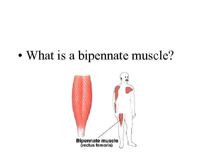 • What is a bipennate muscle?
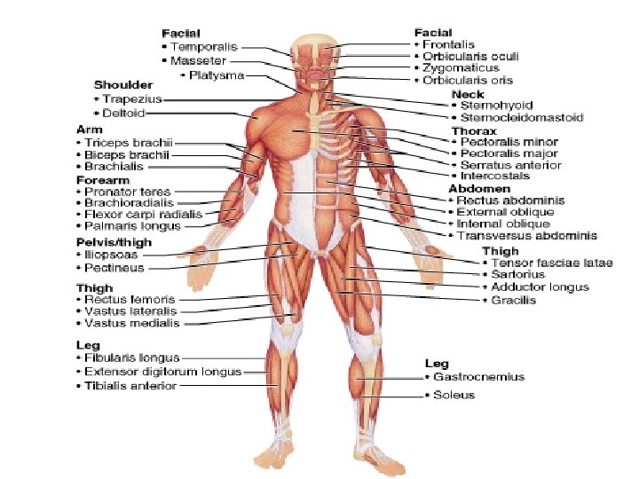
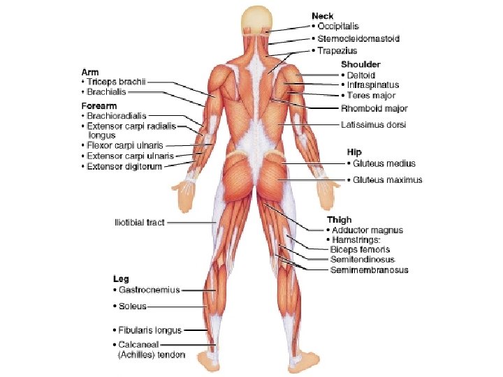
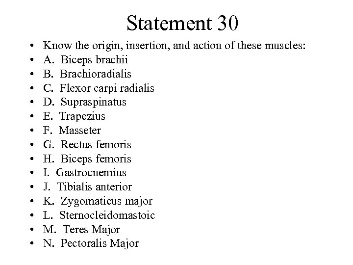 Statement 30 • • • • Know the origin, insertion, and action of these muscles: A. Biceps brachii B. Brachioradialis C. Flexor carpi radialis D. Supraspinatus E. Trapezius F. Masseter G. Rectus femoris H. Biceps femoris I. Gastrocnemius J. Tibialis anterior K. Zygomaticus major L. Sternocleidomastoic M. Teres Major N. Pectoralis Major
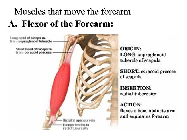 Muscles that move the forearm A. Flexor of the Forearm: http: //home 1. gte. net/imagine/biceps%20 brachii. jpg
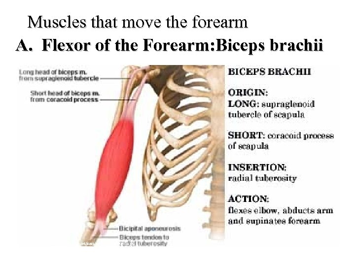 Muscles that move the forearm A. Flexor of the Forearm: Biceps brachii http: //home 1. gte. net/imagine/biceps%20 brachii. jpg
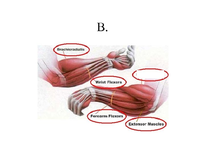 B.
B.
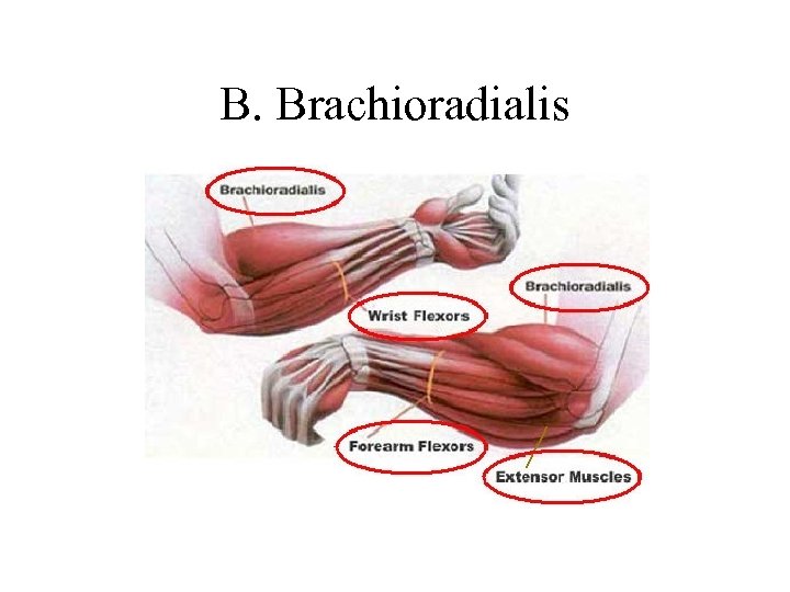 B. Brachioradialis
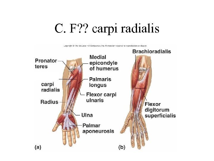 C. F? ? carpi radialis
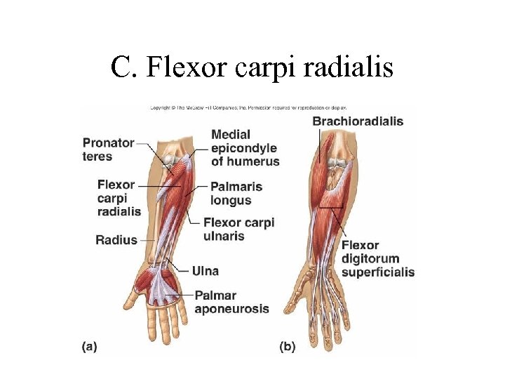 C. Flexor carpi radialis
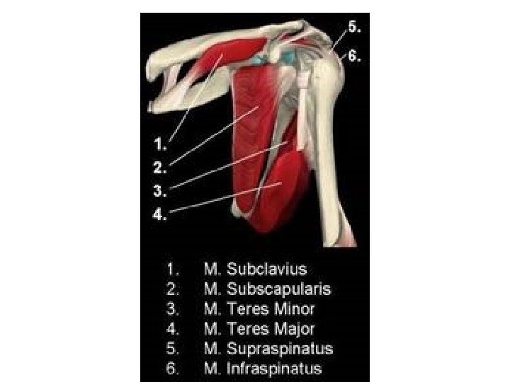
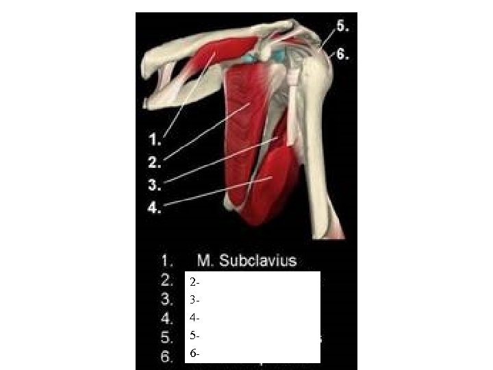 2 3 4 5 6
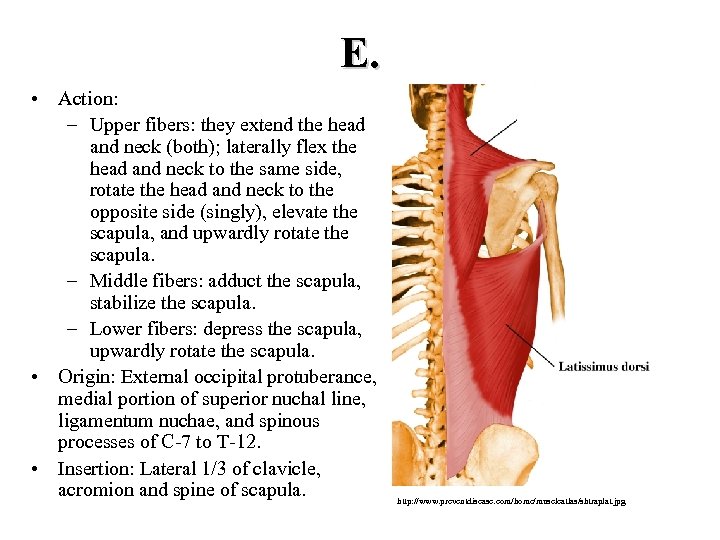 E. • Action: – Upper fibers: they extend the head and neck (both); laterally flex the head and neck to the same side, rotate the head and neck to the opposite side (singly), elevate the scapula, and upwardly rotate the scapula. – Middle fibers: adduct the scapula, stabilize the scapula. – Lower fibers: depress the scapula, upwardly rotate the scapula. • Origin: External occipital protuberance, medial portion of superior nuchal line, ligamentum nuchae, and spinous processes of C 7 to T 12. • Insertion: Lateral 1/3 of clavicle, acromion and spine of scapula. http: //www. preventdisease. com/home/muscleatlas/shtraplat. jpg
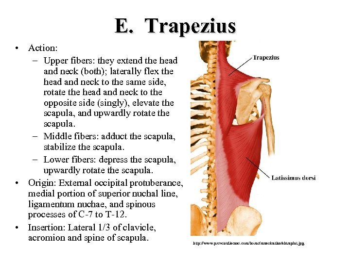 E. Trapezius • Action: – Upper fibers: they extend the head and neck (both); laterally flex the head and neck to the same side, rotate the head and neck to the opposite side (singly), elevate the scapula, and upwardly rotate the scapula. – Middle fibers: adduct the scapula, stabilize the scapula. – Lower fibers: depress the scapula, upwardly rotate the scapula. • Origin: External occipital protuberance, medial portion of superior nuchal line, ligamentum nuchae, and spinous processes of C 7 to T 12. • Insertion: Lateral 1/3 of clavicle, acromion and spine of scapula. http: //www. preventdisease. com/home/muscleatlas/shtraplat. jpg
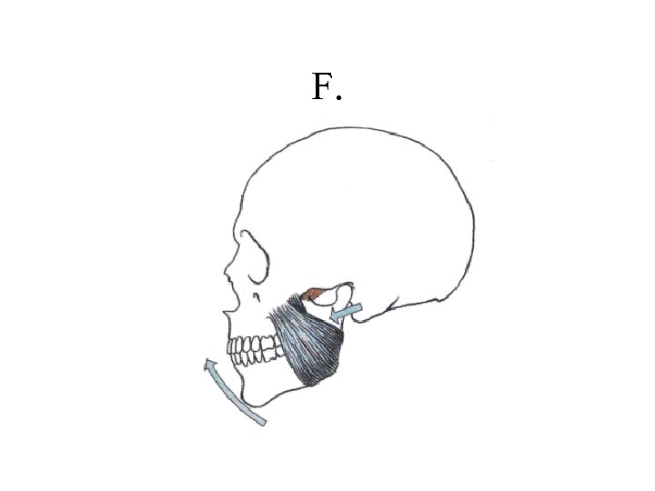 F.
F.
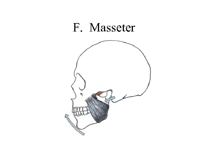 F. Masseter
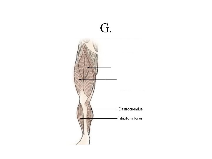 G.
G.
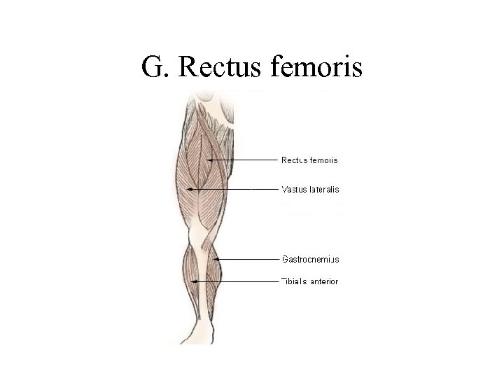 G. Rectus femoris
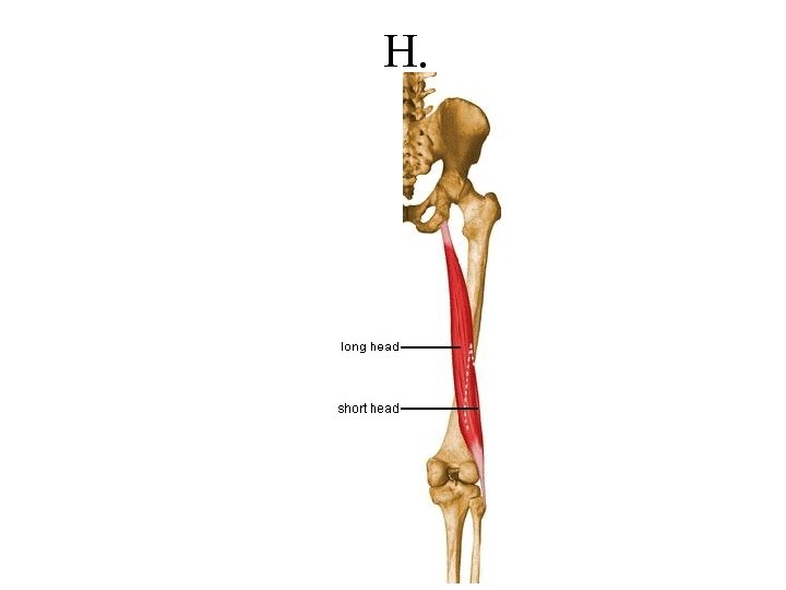 H.
H.
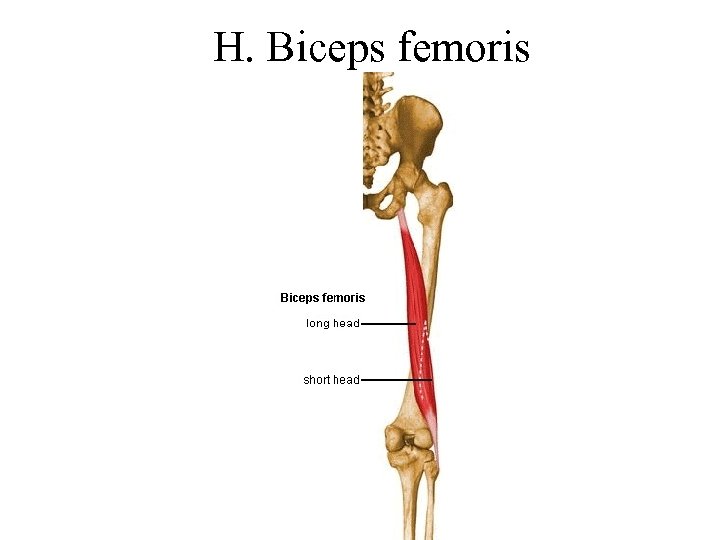 H. Biceps femoris
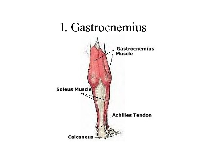 I. Gastrocnemius
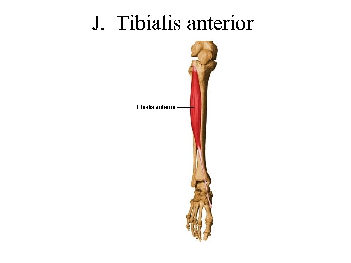 J. Tibialis anterior
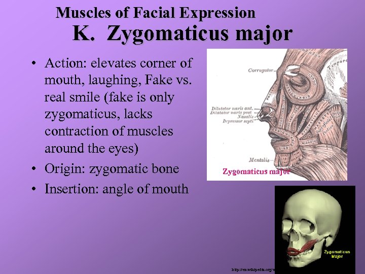 Muscles of Facial Expression K. Zygomaticus major • Action: elevates corner of mouth, laughing, Fake vs. real smile (fake is only zygomaticus, lacks contraction of muscles around the eyes) • Origin: zygomatic bone • Insertion: angle of mouth http: //www. gamasutra. com/features/20000414/lander_03. gif Zygomaticus major http: //en. wikipedia. org/wiki/Image: Gray 378. png
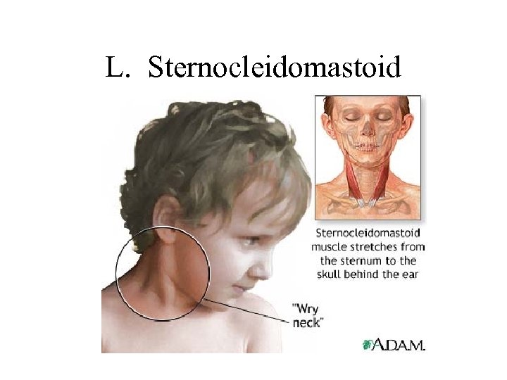 L. Sternocleidomastoid
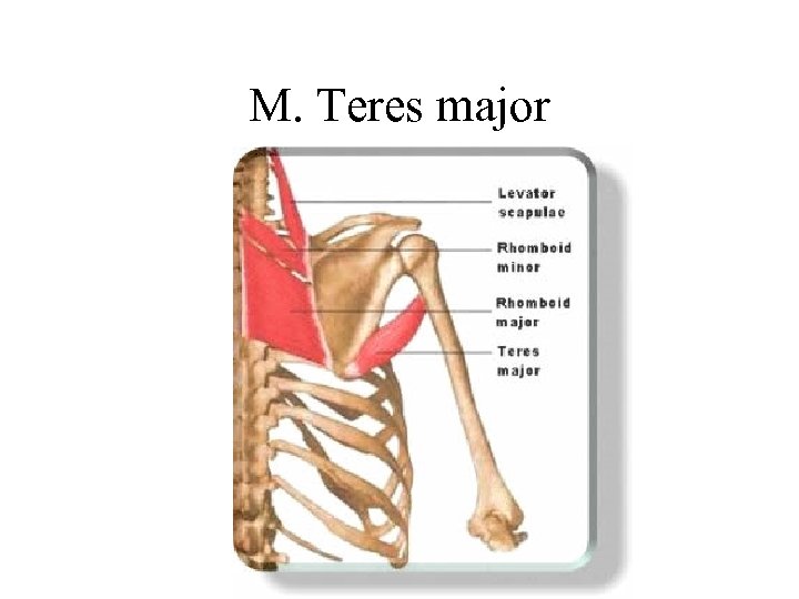 M. Teres major
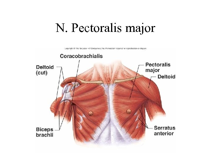 N. Pectoralis major
 The End


