1 лекция INTRODUCTION.ppt
- Количество слайдов: 42


 of anatomy. A general characteristic of fabrics, bodies, systems of")
Subject and problems (tasks) of anatomy. A general characteristic of fabrics, bodies, systems of an organism. Initial stages эмбриогенеза the person.
 of anatomy. 3.")
The plan of lecture: 1. A subject of anatomy. 2. Problems(Tasks) of anatomy. 3. Kinds of anatomy. 4. A place of anatomy among other biological disciplines. 5. Initial stages эмбриогенеза the person. 6. A general characteristic of fabrics, bodies of systems. 7. Types of a constitution. Concept of norm and variants of norm

INTRODUCTION Human anatomy is a science about (he genesis, development and struc ture of the human body. It studies external forms and proportions of the human body and its parts, separate organs, their constitution and micro scopic structure. Anatomy also studies the main stages of human develop ment, particularities of structure of the body and separate organs at differ ent age periods. Anatomy studies the structure of the human body taking into consid eration biological patterns, natural for all living organisms. At the same time human beings differ very much from animals due to their living in society. Mankind was formed by labor and social needs, an increase in which led to changes in structure, to biological progress. The environ ment has an influence on the human organism as well. The human body consists of many organs and a tremendous amount of cells, however it is not a sum of separate parts but a whole coordinated living organism. This is why it is impossible to examine organs without considering at their relationship with each other and without taking into account the integrative role of the nervous and vascular systems and the endocrine apparatus. The main methods of anatomical research are the following: observation, examination of the body, dissection and studying of separate organs and groups of organs (macroscopic, or gross anatomy), including their internal fine structure (microscopic anatomy). With the appearance of microscopes anatomy gave start to histology (the study of tissues) and cytology (the science of structure and functions of the cell).

The structure of the skeleton, internal organs, the disposition of blood and lymphatic vessels are examined with the help of rays. In the clinic the internal surface of many internal organs is studied by means of endo scopy. Antropometrical methods are used for studying external forms and proportions of the body. The study of the human body by the systems (skeletal, muscular, diges tive, etc. ) is called systematic (descriptive) anatomy. Systematic anatomy studies the structure of «normal» that is, healthy human beings, whose organs have not been altered by disease or distur bance of development. Topographic (surgical) anatomy studies the structure of the human body by regions, as well as the location of organs and their interrelation ■ with each other and with the skeleton. External forms of the body and its proportions are studied by surface anatomy. Modern anatomy is called functional, as it examines the structure of the body in correlation with its functions.

Anatomy examines the structure and functions of organs taking into account the developments the human being goes through from birth to death (during ontogenesis). Developments that take place before birth (the prenatal period) are examined by embryology. After birth (the postnatal period) the human being is studied by developmental anatomy. Peculiarities of body structure of elderly and senile people are studied by gerontology — the science of aging. In accordance with the length and width of the body and other anthro pometric features anatomy distinguishes the dolichomorphic, brachymor phic and mesomorphic types of constitution. The dolichomorphic constitu tional type is defined by a narrow long trunk and long extremities (astheme); the brachymorphic type is characterized by a short wide trunk and short extremities (hypersthenic). The intermediate type of constitution is called mesomorphic. This type is close to an «ideal» (normal) human being (normosthenk; ). While analyzing the peculiarities of structure of the human body (an alytical approach), anatomy studies the organism as a whole, using the synthetic approach. That is why anatomy is not only an analytical science, but also a synthetic one.

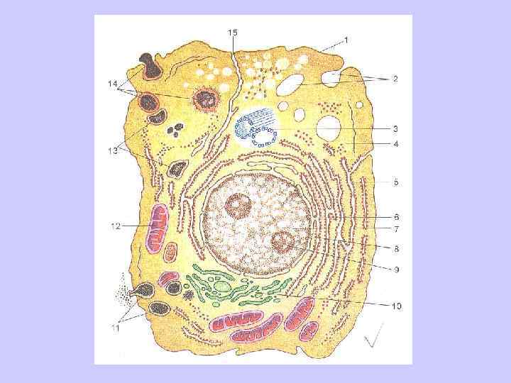
The human organism is a unified, complexly organized system. It consists of cells, tissues and organs. Organs, which are made of tissues, make up systems and apparatuses, which in combination form an entire human organism. CELLS The cell (cellula), as an elementary well organized unit, carries out the functions of reproduction, growth, m tabolism, adaptation to changes jn. the environment and regeneration. Cells vary in shape, structure and ■' chemical composition. In the human body there are flat, spherical, ovoid, f cuboid, prismatic, pyramidal and stellate cells. The size of cells varies from several micrometers (small lymphocyte) to 200 micrometers (ooeyte). All cells have a plasma membrane, or cytolemma (which consists of hyaloplasm, containing organelles and inclusions), and a nucleus.

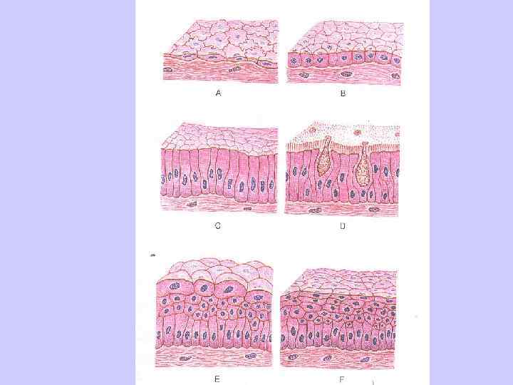
TISSUES Cells and their derivatives combine to form tissues. A tissue is a com bination of cells and extracellular matrix united by a common embryolog ical derivation, structure and function. The tissues in the human organism are subdivided into four types: epithelial, connective, muscular and ner vous. Each type of tissue develops from a specific germ layer. Epithelial 1 tissue derives from the endo , ecto and mesoderm. Nervous tissue devel / ops from the ectoderm. EPITHELIAL TISSUE Epithelial tissue covers the surface of the body and mueosas. Epi thelial tissue also forms glands (glandular epithelium). Furthermore, there is sensory epithelium in the hearing, vestibular, smell and taste organs, the cells of which can perceive specific stimuli.
 portion glands")
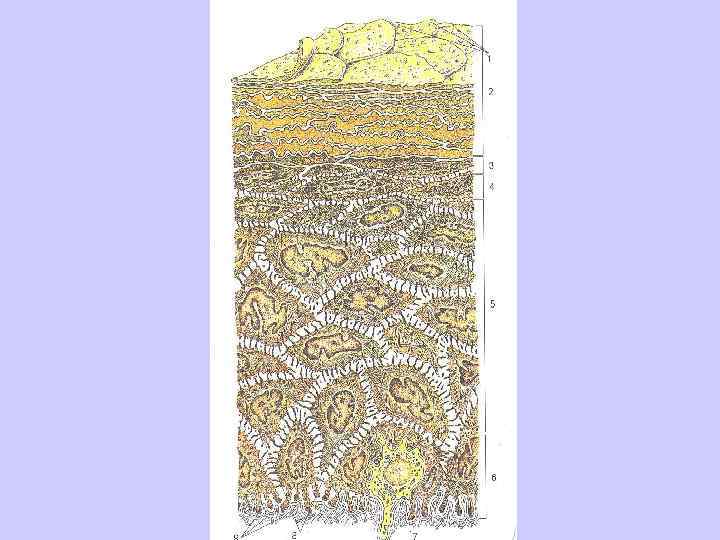
Classifications of multicellular glands. According to the structure of the initial (secretory) portion glands are subdivided into tubular (in the form of a tube), acinar (shaped like a pear or an oblong grape) and alveo lar (rounded, spherical). There also tubuloacinar and tubuloalvcolar glands.

Types of exocrine glands (ace. to 4 — simple tubular gland with noncompound termir loncompound terminal region; С — simple tubular glai Depending on the structure of their duct glands can be simple, with ! only one duct, and compound. In compound glands the main duct collects t many duct branches, each of which collects several secretory portions. I Glands produce different types of secretion: protein (secretory glands). ' mucus (mucous glands) or mixed.
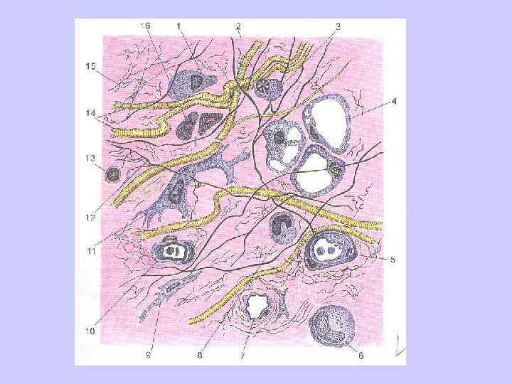

CONNECTIVE TISSUE Connective tissues make up a large group, which includes fibrous connective tissues {loose and dense), specialized tissue (reticular, pigmen tal adipose), rigid , (bones and cartilage) and liquid (blood and lymph) ones. Connective tissues perform many functions. For example, fibrous connective tissues, cartilage and bones provide support; blood has a trophic(nourishing) and protection (e. g. phagocytosis) functions. Connective tis sues are made up of cells andextracellular matrix, which consists of gly cosaminoglycans, proteoglycans and various fibers (collagen, elastic, re ticular). Extracellular matrix is solid in bone tissue and liquid in blood. Fibrous connective tissues include loose (areolar) and dense (collag enous) types dense connective tissue in turn subdivides into irregular and regular. Loose fibrous connective tissue (Fig. 12) (LFCT) is located mainly around blood and lymph vessels and nerves; it covers muscles and forms the stroma of most internal organs, lamina propria mucosae, submucosa, subserosa and adventitia. Loose connective tissue contains many cells such as fibroblasts, fibrocytes, reticular cells, as well as wandering macrophag es, mast cells (tissue basophils), plasma cells, adipocytes, pigment cells, lymphocytes and leukocytes. All these cells are situated in the extracellu larmatrix. The matrix is secreted by fibroblasts and consists of collagen, . elastic and reticular fibers and a ground (amorphous) substance. I Fibroblasts are the main fixed cells, the quantity of which may I vary in different types of connective tissue. Particularly large numbers of I fibroblasts are found in loose fibrous connective tissue.

Skeletal tissues Connective tissue also includes bone and cartilage tissues. The func tions of these are support, movement and defense. They also participate in metabolism of mineral substances. Cartilage tissue forms joint cartilages, vertebral disks, cartilages of |he larynx, trachea, bronchi and external nose. Cartilage consists of spe cialized cells (chondroblasts and chondrocytes) and a dense elastic extra cellular matrix (Fig. 15). Cartilage contains approx imately 70 80% water, 10 15% organic substanc es and 4 7% salts. About 50 70% of the dry sub stance in cartilage is col lagen. The extracellular substance (matrix), which is produced by cartilage cells, consists of complex compounds, which include proteoglycans, hyaluronic acid and glycosaminogly cans. Proteoglycan mole cules bind most of the wa ter in the cartilage. Chondroblasts are young cells round or ovoid in shape, which are able to divide and synthe size components of theex tracellularmatrix. Their cy tolemm forms a large number of villi. The cytoplasm is rich in RNA, rough and smooth endoplasmic reticulum, Golgi complex, mitochondria, lysos omes and granules of glycogen. The nucleus is rich in chromatin and can have one or two nucleoli.
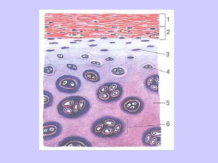

Chondrocytes are mature large cells in oval, rounded or polygo nal in shape, cytoplasmic projections and well developed organelles. They are situated in cavities called lacunae, which are surrounded by extracellu larmatrix. If a lacuna contains only one cell, it is called primary. More often cells lie in a secondary lacuna in isogenic groups (2 3 cells). Walls of the lacunae are made up of collagen fibers, and aggregates of proteoglycans. The structural and functional unit of cartilage is a chondron, which includes a cell or group of isogenic cells, extracellular matrix and a lacuna capsule. / Depending on its structure, cartilage is divided into three types: hya ^__line, fibrocartilage and elastic. / Hyaline cartilage has a bluish color. It consists of ground sub / stance and fibers of collagen. Hyaline cartilage forms joint and costal car ! tilage and most cartilage plates in the larynx. Fibrocartilage contains a large number of collagen fibers in its ground substance and is highly durable. Elongated cells, which are found between collagen fibers, contain oblong rod shaped nuclei and have thin bands of basophilic cytoplasm. This type of cartilage forms fibrous rings in vertebral disks, articular disks and menisci, covers the articular surfac es of the temporomandibular and sternoclavicular joints. Elastic cartilage is resilient and flexible. Besides collagen, its matrix contains a large number of complexly interwoven elastic fibers. Elastic cartilage forms the epiglottis, the cuneiform and corniculate carti lages of the larynx, processus vocalis of the arytenoid cartilage, the outer ear and the cartilaginous part of the auditory tube.

Bone tissue is characterized by special mechanical properties. It con sists of bone cells, bricked up in bone ground substance, which contains collagen fibers and is saturated with inorganic compounds. There are two types of bone cells: osteoblasts and osteocytes. Another category of cells found in bones is osteoclasts, which are not bone cells. They have a monocyte origin and pertain to the system of macrophages.

Osteoblasts are young process bearing cells polygonal in shape. They contain rough endoplasmic reticulum, ribosomes, well developed Golgi complex and highly basophilic cytoplasm. Osteoblasts are situated in su perficial layers of bones. They have a round or oval nucleus, which contains one large nucleolus usually in the peripheral zone. There are microfibrils surrounding the osteoblasts. Osteocytes are mature fusiform cells with a large round nucleus, which contains a clearly visible nucleolus, and many cytoplasmic pro cesses. They lie in lacunae, where they are surrounded by a thin layer of so called bone fluid (tissue fluid). Osteocytes are not in direct contact with the calcified matrix. Their long cytoplasmic processes lie inside bone canaliculi. They are also separated from the calcified matrix by space con taining tissue fluid, which feeds theosteocytes. The distance between an osteocyte and the nearest capillary does not exceed 100 200 mm. Osteoclastsare large multinuclear cells of size 190 mm. The have a monocyte origin and are able to destroy bone and cartilage and reabsorb bone tissue during its physiological and reparative regeneration. Their cytoplasm contains a large number of mitochondria, elements of granular endoplasmic reticulum and Golgi complex, free ribosomes and lysosomes. Osteoclasts have numerous cytoplasmic projections, usually found on the surface, that is, facing the deteriorating bone. This brush border increases the surface area of the osteoclast in contact with the bone. The projections are covered with microvilli, spaces between which contain hydroxyapatite crystals. These crystals can be found inside osteoclasts in phagolysosomes, where they are dissolved.

Bone tissue can be subdivided into two varieties: reticulofibrous and lamellar. Reticulofibrous bone tissue is found in zones where ten dons attach to bones and in sutures of the skull after their fusion. This type of bone contains thick bundles of collagen, spaces between which are filled with an amorphous substance. Reticulofibrous tissue is covered on the outside by periosteum. Lamellar bone tissue is formed by bone lamellae 4 20 mm thick, which consist of osteocytes and fibrous ground substance, hi each lamella collagen fibers lie parallel to each other. Fibers in neighboring lamella are oriented differently, which provides for high durability of the bone.

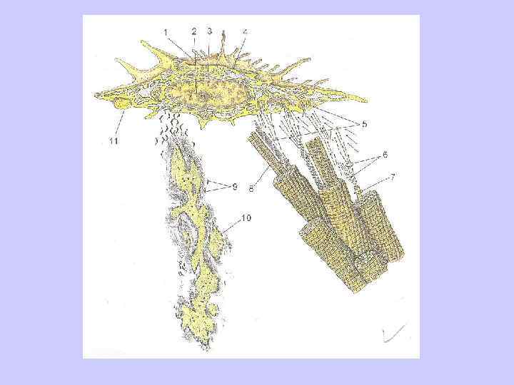
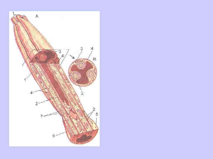
MUSCLE TISSUE Muscle tissue includes skeletal, smooth and cardiac muscles, all of which have different embryological derivation and structure. A common characteristic of these tissues is their ability to contract or change their length. The human organism also contains muscle tissue of ectodermal derivation (myoepithelial cells of glands and myocytes of the iris). Skeletal {striated) muscle tissue is made up of muscle fibers 4 cm or more in length and 0. 1 mm thick. Each fiber consists of a myosymplast and myosatellitocytes, covered with a common membrane called sarco lemma. The sarcolemma consists of a basement membrane with interwoven collagen and reticular fibers. The myosymplast, which lies beneath the sarcolemma is also called the sarcoplasm. It contains a large number of ellipsoid nuclei (up to 100), myofibrils and cytoplasm. The sarcoplasm is

rich in granular endoplasmic retic ulum. A considerable part of the muscle fiber is occupied by myo fibrils. Between the muscle fibers there are many granules of glyco gen and mitochondria with well developed cristae. The sarcoplasm contains a lot of the protein myo globin, which can bind with oxy gen like hemoglobin. Depending on thickness and myoglobin con tent, striated muscle fibers are di vided into red and white types. Red fibers are rich in sarcoplasm, myoglobin and mitochondria but contain fewer myofibrils. These fibers contract slowly and can re main in a contracted /working/ condition for a long time. White muscle fibers contain little sarco plasmand myoglobin and few mi tochondria, but have a lot of myo fibrils. White fibers contract fast er than red fibers, but tire quickly. Combinations of slow ( «red» ) and fast ( «white» ) muscle fibers pro vides quickness of muscle reac tion (contraction) and long lasting work capability.
. Each")
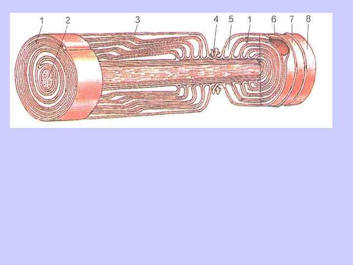
The main part of the sarco plasmis made up of myofibrils (Fig. 18). Each myofibril consists of interchanging segments of dark anisotropic A bands and light iso tropic I bands. In the middle of each a band there is a light strip called the H zone. The middle of the H zone is marked by the M line, or mesophragm, and in the middle of the I band is the Z line, or the so called telophragm. The alter nating of the light and dark bands in neighboring myofibrils creates thestriated appearance. The dark bands are formed by thick myosin strands 10 15 nm in diameter. These strands are made up of a high molecular protein myosin. The light bands consist of thin actin fila ments 5 8 nm in diameter and about 1 mm in length.

These thin filaments are formed by a low molecular protein actin, and two other low molecular proteins troponin and tropo myosin. The section between two telophragms (Z lines) is called the sarcomere and is consid ered to be the functional unit of the myofibril. Asarcomere is about 2. 5 urn long and in cludes a dark A band the adjoining half of a light I band at each side. Thin actin fila ments are situated in the inter vals between the heavy myo sin filaments. During contrac tions of the muscle theactin and myosin filaments slide towards each other; during muscle relax ation they move in opposite directions. On borders between A and I bands the sarcolemma of the muscle fiber invaginates, forming transverse tubules (T tubules). These play an important role in fast conduction of the action potential to each myofibril. The action potential spreads along T tubules, passes onto the sarcoplas micreticulum and between myofibrils. Myosatellitocytes are situated directly beneath the sarcolemma. These are flattened cells with large nuclei. Each myosatellitocyte has a centrosome and a small number of organelles and lacks myofibrils. Myosatellitocytes are precursor cells of striated (skeletal) muscle tissue. They are able to synthesize DNA and undergo mitosis.
 muscle tissue consists of muscle cells called myocytes, which are found in")
Smooth (non-striated) muscle tissue consists of muscle cells called myocytes, which are found in the walls of circulatory and lymph vessels and hollow organs, forming their contractile apparatus. Smooth myocytes are fusiform cells 20 500 xm long and 5 15 цт thick (Fig 19). There is no striation in these cells. Myocytes are situated in groups with their pointed ends in between two neighboring cells. Each smooth myocyte has a base ment membrane, which is absent at sites of cell junctions. These cells contain elongated rod shaped nuclei, which can reach 10 25 xm in length and during contraction acquire a corkscrew shape. Adjoining the plasma membrane from within are dense (adhesive) corpuscles.

Smooth myocytes contain thin and thick myofilaments. During con tractionactin and myosin filaments move towards each other and the myocyte shortens. Nerve impulses are passed from one myocyte to anoth er through cell junctions at a speed of approximately 8 10 cm/s. Smooth myocytes contract considerably slower (by 1000 times) than striated muscle fibers. Smooth muscles carry out prolonged tonic con tractions and relatively slow movements. Cardiac muscle tissue differs by structure and functions from skeletal muscles. It consists of cardiac myocytes (cardiomyocytes), which form interconnected complexes. Cardiac muscle resembles skeletal muscle tis sue by its microscopic structure (transverse striation), but it contracts in voluntarily like smooth muscle tissue. Cardiomyocytes have an irregular cylindrical shape; cells are 100 150 mm long and 10 20 mm in diameter. Each cardiomyocyte contains 1 or 2 elongated oval nuclei, which lie in the center of the cell and are surrounded by longitudinally posi tioned myofibrils. Between neighboringcardiomyocytes there are spe cialized contacts in the form of intercalated disks, which actively take part in passing excitation from one cell to another. The structure of myofibrils in cardiomyocytes is analogous to that in skeletal muscles. Beneath the cytolemma and between mitochondria there are granules of glycogen and elements of smooth endoplasmic reticulum. Cardiomyocytes contain many large mitochondria with well developed cristae, which are situated in groups between myofibrils. The cytolemma of cardiomyocytes also forms T tubules, near which there accumulations of smooth endoplasmic reticulum cisterns.

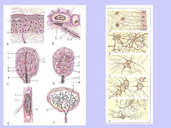
NERVOUS TISSUE Nervous tissue is the main structural element of organs of the ner vous system, namely, the brains and spinal chord, ganglia, nerves and nerve endings. Nervous tissue consists of nerve cells (neurocytes or neu rons) and accessory neuroglial cells, which are anatomically and func tionally associated with them.

MAIN STAGES OF DEVELOPMENT IN ONTOGENESIS Each human being possesses individual traits of outer appearance and internal organ structure, which are determined by hereditary factors and 1 influences of the environment. V Individual development of a human organism lasts throughout all ' periods of life — from conception until death. Ontogenesis of a person is • divided into the intrauterine (prenatal) and the «after birth» (postnatal) | periods. During the intrauterine period, which lasts*from conception until / birth, the embryo develops inside the mothers womb. In the course of the first 8 weeks of embryogenesis the main processes of organ and body part ; formation take place. This period is called the embryonic period, and the future human organism — the embryo. Starting on the 9 th week, when main external features have begun to designate, the organism enters the fetal period of development and begins to be called a fetus. After conception, which usually takes place in a uterine tube, fused v sex cells (ovum and spermatozoid) form a unicellular 'germ' called a zygote, which possesses all the attributes of both sex cells. From this mo ment begins the development of a new daughter organism.

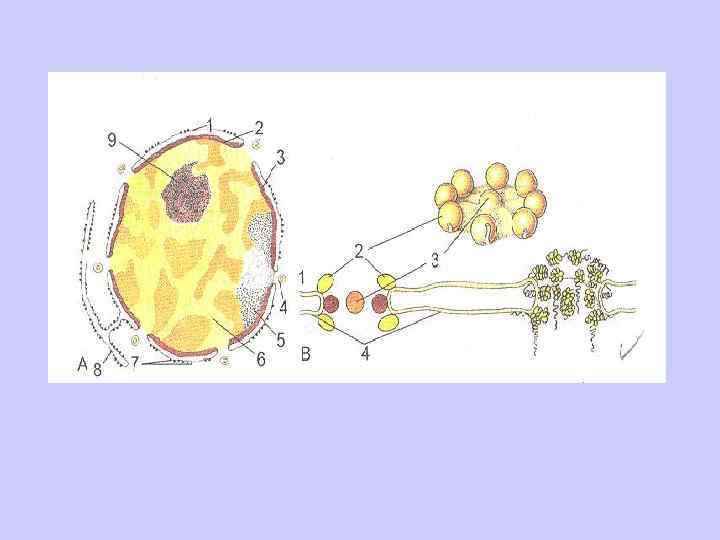
During its first week of development the zygote divides into • daughter cells (cleavage stage). In the first 3 4 days the zygote is si imultaneously dividing and moving along the uterine tube towards the /uterus. As a result of its divisions the zygote transforms into a multi | cellular vesicle (blastula) with a cavity inside (Fig. 29). The walls of | this vesicle are made up partially of larger and partially of smaller / cells. The smaller cells form the outside layer of the blastula wall and / is called the trophoblast. Later on the trophoblast cells form the outer j layer of the extraembryonic membranes. The larger cells (blastomer I es) form a cell mass called an embryoblast, which is situated to the inside of the trophoblast. This inner cells mass /embryoblast/ later de velops into the embryo. Between the trophoblast and the embryoblast accumulates a small amount of fluid. The second week of embryonic development is a stage when cells of the embryoblast divide into two layers (discs), which form into two vesi cles. The outside cell layer, which adjoins the trophoblast, forms the ecto blastic vesicle, which becomes filled with amniotic fluid. The inside layer forms an endoblastic vesicle (yolk sac). The embryonic disk ( «body» ) is situated where the amniotic cavity connects to the yolk sac. During this stage the embryo has the shape of a bilaminar disk made up of an external (ectoderm) and internal (endoderm) embryonic sheets. The ectoderm fac es the amniotic cavity, and the endoderm adjoins the yolk sac. At this stage it is possible to define surfaces of the embryo.

The dorsal surface adjoins the amniotic cavity and the ventral surface contacts the yolk sac. The trophoblast cavity around the amniotic and yolk vesicles is loosely filled with cells of extraembryonic mesenchyme. By the end of the 2 nd week the length of the embryo is approximately 1. 5 mm. During this stage the bilaminar embryonic disk thickens in its posterior (caudal) part. This is where the axial organs (chorda, neural tube) will later develop. During the third week of development the formation of the three layered embryo takes place. Some cells of the ectoderm migrate toward its posterior end, forming a cell cord called the primitive streak. In the anterior (head) section cells of the primitive streak grow and divide faster, which results in the formation of the primary nodule (Hensel's nodule). The prim itive streak determines the bilateral symmetry of the body. The location of ; the primary nodule marks the cranial (head) end of the embryo. Later on, the cells of the primitive streak and primary nodule grow in between the ectoderm and endoderm. This creates the middle embryonic layer — the mesoderm. Cells of the mesoderm, which stay between the two layers of the embryonic disk are called intraembryonic mesoderm; cells that have migrated outside the disc make up the extraembryonic mesoderm. Part of the mesoderm cells within the primary nodule grows particu larly actively, forming the cranial (chordal) process.

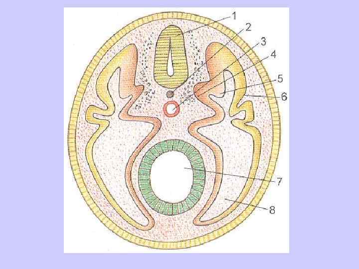
This process pene trates between the external and internal layers up to the caudal end, form ing thechorda (notochord). The cranial part of the embryo grows faster than the caudal end. At the end of the 3 rd week a longitudinal strip of actively dividing cells (neural plate) appears within the ectoderm in front of the primary nodule. This plate then forms a longitudinal fold — the neural sulcus. As the sulcus deepens, its edges thicken, converge and grow together, forming the neural tube. The ectoderm closes over the neural tube and 'detaches' from it. Later on, the entire nervous system will de velop out of the neural tube. The endoderm inside the embryo rolls up into a tube, forming the embryonic rudiment of the future gut. A narrow opening, which connects the embryonic gut with the yolk sac, later turns into the umbilical ring. The endoderm forms the epithelium and glands of the gastrointestinal tract. The ectoderm forms the nervous system, the dermal epithelium and its derivatives, the epithelial tegument of the mouth, the anal part of the rec tum and the vagina. The mesoderm develops into internal organs (except ing the derivatives of the endoderm), the cardiovascular system, organs of the locomotive apparatus (bones, joints, muscles) and the dermis.

After birth the child grows quickly, his mass, length and surface area iof tfle body increase likewise. A human being grows during the first / 20 years of life. For men growth of body length ends at approximately 18 22 years; for women — at 18 20 years. Up to 60 65 years of age. body length stays almost constant. During old age (after 60 70 years), howev er, the length of the body decreases by 1 1. 5 mm each year due to the increase of curving in the vertebral column, thinning of intervertebral disks and flattening of foot arcs. In the course of the first year of life the height of a child increases by 21 25 cm. In the beginning of the second childhood period (8 12 years) height increases at a rate of 4. 5 5. 5 cm per year and later speeds up. Dur ing adolescence (12 16 years) body length increases yearly by 5. 8 cm for boys and 5. 7 cm for girls. The most intensive growth period for girls is between 10 13 years and for boys — during adolescence (13 16 years). After that growth slows down. The mass of the human body at 5 6 months after birth doubles, and by two years of age increases by 4 times. Themaximum yearly mass increase for girls is during the thirteenth year, and for boys during the fifteenth year. Weight continues to increase up to 20 25 years and then remains stable up to age 40 46. It is considered impor tant and physically justified to keep body mass within limits of its values t at age 19 20. During the recent 100 150 years a speeding up (acceleration) of mor phological and functional development of the whole body has been noted in children and adolescents. Thus, the body mass for newborns has in creased over the century by 100 300 grams, and for one year olds by 1500 2000 grams. Body length during the second childhood period and adolescence increased by 10 15 cm, and for adult males — by 6 8 cm.

The time period during which height increases shortened. In the end of the XIX century growth continued until 23 26 years. In the end of the XX century height increases in men until age 20 22, and in women until age 18 20. Dentition of both milk and permanent teeth has accelerated. Men tal development and sexual maturation proceed faster. In the end of the XX century, compared to its beginning, the average of menarche for girls lowered from 16. 5 to 12 13 years. The age of menopause for an adult shifted from 43 45 to 48 50 years. A newborn has a relatively large round head, short neck and chest, a long abdomen, short legs and long arms. The cerebral part of the skull is comparatively larger than the facial part. The shape of the thorax is barrel like. The vertebral column does not have curves. The internal organs are relatively bigger than those of an adult. Thus, the mass of the liver for a newborn makes up 1/20 of total body mass, while in adults it is 1/50. The length of the intestine is twice as long as body length, whereas in adults it is 4 4. 5 times as long. The mass of the brain in a newborn makes up 13 14% of the body mass, whereas in an adult it is only approximately 2%. The thymus and adrenal glands are especially large. During the suckling age (10 days — 1 year) the baby's body grows rapidly. At about 6 months teething of milk teeth begins. During the first year the size of some organs reaches adult size (the eye, inner ear, central nervous system). During the first year of life there is a quick development of organs of the locomotion apparatus, the digestive system and the respi ratory system.
 primary dentition ends. Psychic de velopment, speech and")
In early childhood (1 -3 year) primary dentition ends. Psychic de velopment, speech and memory abilities progress quickly. The child starts to become oriented in space. In the end of this period secondary /perma nent/ dentition begins. Because of fast development of the brain, the mass of which by now reaches 1100 1200 g, mental capabilities develop quickly, as well as long term ability for recognition and orientation in time and days of the week. During the second childhood period (3 -12 years) growth in width predominates again, although growth in length, which in this period is greater for girls, also increases. The psychic development of children progresses. Orientation in months and days of the calendar develops. Sexual maturation starts, beginning earlier for girls, due to an increase in female hormone secretion. In girls at the age of 8 9 the pelvis and hips begin to widen, sebaceous gland secretion increases and hair appears on the pubis. In boys of 10 11 year the larynx, testes and penis begin to grow. In adolescence (12 -16 years) sex organs develop quickly and sec ondary sexual characteristics strengthen. For girls the amount of hair on the pubis increases and hair appears in the axillary fossae. Sexual organs and gonads increase in size. The basic p. H of vaginal secretions becomesacidic, menstruation appears and the size of the pelvis increases. For boys the testes and penis grow quickly. At first, pilosis of the pubis develops as in females and the mammary glands swell. By the end of adolescence (15 16 years) hair growth begins on the face, body, axillar fossae and pubis (male type).
 occur. Mechanical")
The skin of the scrotum becomes pigmented and first ejaculations (involuntary ejaculations) occur. Mechanical and verbo log ical memory develops during adolescence. Juvenile age (16 -21 years) coincides with the period of maturation. During this period growth and development of the organism comes to a conclusion, all apparatuses and organ systems reach a morphological and functional maturity. The body structure during mature /adult/ age (22 -60) changes lit tle. Duringold age (61 -74) and senile period (75 -90) certain changes take place that are characteristic of these ages and which are studied by a specialized science— gerontology. Time frontiers of aging vary within a wide range for different individuals. During old age the ability of the or ganism foradaption decreases.
1 лекция INTRODUCTION.ppt