d3e2a0920e65b68d7e01aa141f24a139.ppt
- Количество слайдов: 181
 Stroke In The Young Adult Victoria E. Judd MD, MBA
 What Is A Stroke ? n n A stroke occurs when blood flow to the brain is interrupted by a blocked or a ruptured blood vessel. A brain attack.
 Stroke n Acute stroke is typically characterized by the sudden onset of a focal neurologic deficit, though some patients have a stepwise or gradual progression of symptoms.
 Dysarthria (difficulty speaking) Hemianopia (difficulty") Stroke n n Common deficits include: Dysphasia (difficulty swallowing) Dysarthria (difficulty speaking) Hemianopia (difficulty with sight) Weakness
Stroke n n Common deficits include: Dysphasia (difficulty swallowing) Dysarthria (difficulty speaking) Hemianopia (difficulty with sight) Weakness
 Stroke Common Deficits n n Ataxia Sensory loss Neglect Consciousness is generally normal but maybe impaired
 Stroke Warning Signs n n n Sudden weakness or numbness of the face, arm or leg, especially on one side of the body Sudden confusion, trouble speaking or understanding Sudden trouble seeing in one or both eyes
 Stroke Warning Signs n n Sudden trouble walking, dizziness, loss of balance or coordination Sudden, severe headaches with no known cause (for hemorrhagic stroke)
 Stroke Warning Signs n Acute loss of focal cerebral function • Abrupt onset • Symptoms occur in all affected areas at the same time • Symptoms resolve gradually • Symptoms are “negative”
 Nature of Symptoms n n Positive symptoms indicate active discharge from central nervous system neurons. Typical positive symptoms can be visual (e. g. , bright lines, shapes, objects), auditory (e. g. , tinnitus, noises, music), somatosensory (e. g. , burning, pain, paresthesias), or motor (e. g. , jerking or repetitive rhythmic movements). Negative symptoms indicate an absence or loss of function, such as loss of vision, hearing, feeling, or ability to move a part of the body.
") Annual Incidence of Ischemic Stroke n n n In young adults (15– 45 years) has been estimated at approximately 2– 11 per 100, 000 in Caucasians, 22. 8 per 100, 000 in African Americans 10/100, 000 in a Mayo Clinic study of women ages 15 to 29 About 2– 12% of cerebral infarcts occur in young adult patients, with a higher frequency between 31 and 45 years
Annual Incidence of Ischemic Stroke n n n In young adults (15– 45 years) has been estimated at approximately 2– 11 per 100, 000 in Caucasians, 22. 8 per 100, 000 in African Americans 10/100, 000 in a Mayo Clinic study of women ages 15 to 29 About 2– 12% of cerebral infarcts occur in young adult patients, with a higher frequency between 31 and 45 years
 Annual Incidence of Ischemic Stroke n Stroke ranks second after ischemic heart disease as a cause of lost disability-adjusted life-years in highincome countries
 Mortality of Strokes n n Mortality in the first month after stroke has been reported to range from 2. 5% in patients with lacunar infarcts to 78% in patients with space-occupying hemispheric infarction. Lacunar stroke or lacunar infarct (LACI) is a type of stroke that results from occlusion of one of the penetrating arteries that provides blood to the brain's deep structures.
 Stroke n n Stroke in young adults is surprisingly common. The differential diagnosis for potential etiologies is broader than that for older adults.
 Stroke n n n In children and young adults; Congenital and acquired heart problems, Hematologic conditions, Vasculopathies, Metabolic disorders, Drug ingestion are more common.
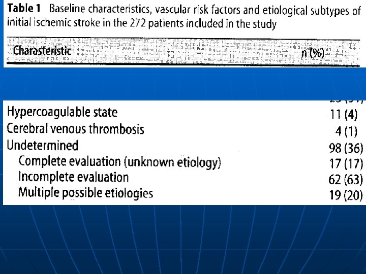
 Causes of Stroke n n The largest series studies of young adults with ischemic stroke cite undetermined as the most frequent etiology (up to 35% of patients) Ischemic stroke is much more common than hemorrhagic
 Causes of Stroke n n Up to 45% of strokes in young adults are due to spontaneous intracerebral hemorrhage. Vascular malformations, aneurysms, hypertension, and illicit drug use are the main causes.
 • Causes of Ischemic Stroke in Young Adults Eur Neurol 2007; 57: 212– 218
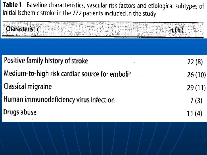
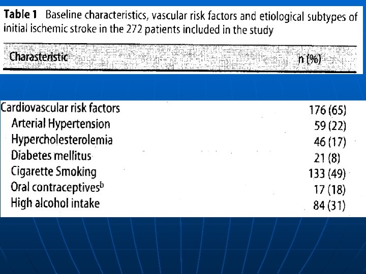
 n n n Major cardiovascular") Cardiovascular Risk Factors in 272 Young Patients % (n) n n n Major cardiovascular risk factors 35 (96) Arterial hypertension 22 (59) Diabetes mellitus 8 (21) Hypercholesterolemia 17 (46) Atherosclerosis 5 (14) Causes of Ischemic Stroke in Young Adults Eur Neurol 2007; 57: 212– 218
Cardiovascular Risk Factors in 272 Young Patients % (n) n n n Major cardiovascular risk factors 35 (96) Arterial hypertension 22 (59) Diabetes mellitus 8 (21) Hypercholesterolemia 17 (46) Atherosclerosis 5 (14) Causes of Ischemic Stroke in Young Adults Eur Neurol 2007; 57: 212– 218
 n n Minor cardiovascular risk") Cardiovascular Risk Factors in 272 Young Patients % (n) n n Minor cardiovascular risk factors 63 (172) Cigarette smoking 49 (133) Oral contraceptives 18 (17) High alcohol intake 31 (84) Causes of Ischemic Stroke in Young Adults Eur Neurol 2007; 57: 212– 218
Cardiovascular Risk Factors in 272 Young Patients % (n) n n Minor cardiovascular risk factors 63 (172) Cigarette smoking 49 (133) Oral contraceptives 18 (17) High alcohol intake 31 (84) Causes of Ischemic Stroke in Young Adults Eur Neurol 2007; 57: 212– 218
 Prevention of Stroke n n n Control high blood pressure Prevent heart disease Stop cigarette smoking Recognize signs of TIA Reduce blood cholesterol levels
 Stroke Risk Factors That Can Be Treated n Hypertension/High Blood Pressure n Heart Disease n Cigarette Smoking n Transient Ischemic Attacks
 Stroke Risk Factors That Can Be Treated n Diabetes n Elevated Blood Cholesterol/Lipids n Asymptomatic Carotid Bruits
 Stroke Risk Factors That Cannot Be Treated n n n Age Gender Race Prior stroke Family history
 Stroke Risk Factors Less Well. Documented n Geographical Location n Socioeconomic Factors n Excessive Alcohol Intake n Certain Kinds of Drug Abuse
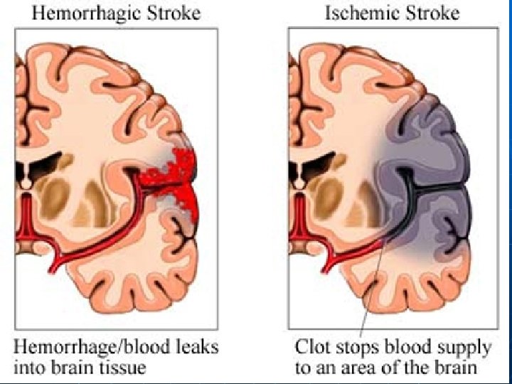
 • Caused by a") What Are the Types of Stroke ? Ischemic Stroke (Blockage) • Caused by a blockage in blood vessels in brain Hemorrhagic Stroke (Bleeding) • Caused by ruptured or leaking blood vessels in brain
What Are the Types of Stroke ? Ischemic Stroke (Blockage) • Caused by a blockage in blood vessels in brain Hemorrhagic Stroke (Bleeding) • Caused by ruptured or leaking blood vessels in brain
 Stroke Background • Inadequate blood flow n Ischemic stroke • Focal – thrombotic or embolic occlusion of major artery • Global – inadequate cerebral perfusion • Hemorrhage Parenchymal – into brain tissue n Subarachnoid – surrounding subarachnoid space n
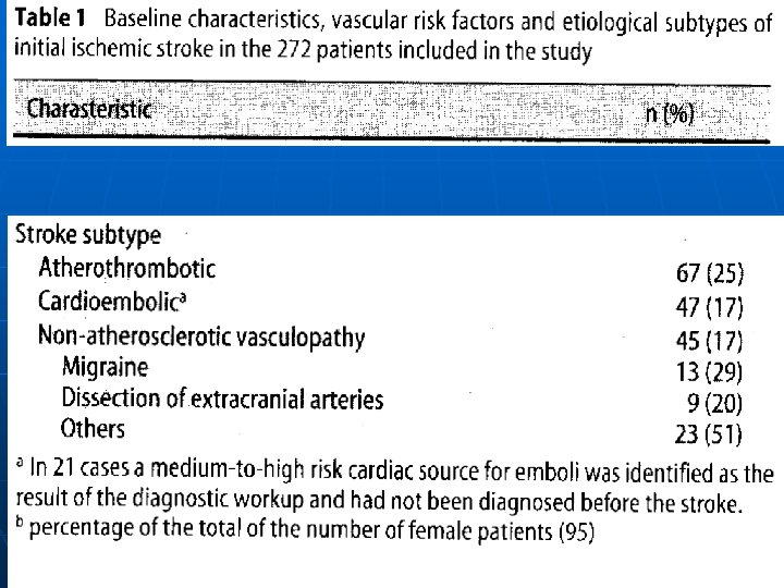
 Causes of Stroke n n n Some of the most common causes of ischemia in the young Cardioembolism (20– 35%), Dissection of extracranial arteries (6– 25%), Migraine with aura (1– 20%) Drugs (10%) Hypercoagulable states (5– 10%) Premature atherosclerosis (20 -25%)
 Ischemic Stroke n In patients younger than 55 years, only about 10% of strokes are caused by large-vessel atherosclerotic disease.
 What Are the Causes of Ischemic Stroke? Large vessel disease n n n n n Premature atherosclerosis Dissection (spontaneous or traumatic) Inherited metabolic diseases (homocystinuria, Fabry’s, pseudoxanthoma elasticum, MELAS syndrome) Fibromuscular dysplasia Infection (bacterial, fungal, tuberculosis, syphilis, Lyme) Vasculitis (collagen vascular diseases — systemic lupus erythematosus, rheumatoid arthritis, Sjögren’s syndrome, polyarteritis nodosa; Takayasu’s disease, Wegener’s syndrome, cryoglobulinemia, sarcoidosis, inflammatory bowel disease, isolated central nervous system angiitis) Moyamoya disease: (Japanese, "puff of cigar smoke") is an inherited disease in which certain arteries in the brain are constricted Radiation Toxic (illicit drugs — cocaine, heroin, phencyclidine; therapeutic drugs — L -asparaginase, cytosine arabinoside, ephedra, phenylephrine)
 What Are the Causes of Ischemic Stroke? n Cardiac disease (including congenital, rheumatic valve disease, mitral valve prolapse, patent foramen ovale, endocarditis, atrial myxoma, arrhythmias, cardiac surgery)
 What Are the Causes of Ischemic Stroke? n n Small vessel disease Vasculopathy (infectious, noninfectious, microangiopathy) Independent predictors of arteriopathy are sickle cell disease and recent upper respiratory infection.
 What Are the Causes of Ischemic Stroke? n n n n Hematologic disease Sickle-cell disease Leukemia Hypercoagulable states (antiphospholipid antibody syndrome, deficiency of antithrombin III or protein S or C, resistance to activated protein C, increased factor VIII) Disseminated intravascular coagulation Thrombocytosis Polycythemia vera Thrombotic thrombocytopenic purpura Venous occlusion (dehydration, parameningeal infection, meningitis, neoplasm, polycythemia, leukemia, inflammatory bowel disease)
 Hematologic Disorders n n n Many hematologic disorders are associated with ischemic stroke. The disorders most likely to cause ischemic stroke in patients younger than 45 years are: Antiphospholipid antibody syndrome Sickle cell anemia Heparin induced thrombocytopenia
 or antiphospholipid antibody syndrome is a disorder") APS n Antiphospholipid syndrome (APS or APLS) or antiphospholipid antibody syndrome is a disorder of coagulation that causes blood clots (thrombosis) in both arteries and veins as well as pregnancy-related complications such as miscarriage, stillbirth, preterm delivery, or severe preeclampsia. The syndrome occurs due to the autoimmune production of antibodies against phospholipid (a. PL), a cell membrane substance. In particular, the disease is characterized by antibodies against cardiolipin (anti-cardiolipin antibodies) and β 2 glycoprotein.
APS n Antiphospholipid syndrome (APS or APLS) or antiphospholipid antibody syndrome is a disorder of coagulation that causes blood clots (thrombosis) in both arteries and veins as well as pregnancy-related complications such as miscarriage, stillbirth, preterm delivery, or severe preeclampsia. The syndrome occurs due to the autoimmune production of antibodies against phospholipid (a. PL), a cell membrane substance. In particular, the disease is characterized by antibodies against cardiolipin (anti-cardiolipin antibodies) and β 2 glycoprotein.
 Hematologic disorders n Most of the common hereditary hypercoagulable disorders, such as factor V Leiden/activated protein C resistance, the prothrombin gene mutation (G 20210 A), antithrombin III deficiency, protein C deficiency, and protein S deficiency, typically cause venous thrombosis much more often than they cause arterial thrombosis.
 What Are the Causes of Ischemic Stroke? Migraine: especially with aura
 • Artery-to-artery • Fat") Embolism: • Cardiogenic (atrial fibrillation, mural thrombus, myxoma, valvular vegetations) • Artery-to-artery • Fat • Air • Paradoxical (emboli of venous origin passing through a patent foramen ovale)
Embolism: • Cardiogenic (atrial fibrillation, mural thrombus, myxoma, valvular vegetations) • Artery-to-artery • Fat • Air • Paradoxical (emboli of venous origin passing through a patent foramen ovale)
 Cardiogenic Embolism n Major risk factors: Anticoagulation Indicated • Atrial fibrillation • Mitral stenosis • Prosthetic cardiac valve • Recent MI • Thrombus in LV or LA appendage • Atrial myxoma • Infective endocarditis (No anticoagulation) • Dilated cardiomyopathy
 Cardiogenic Embolism n Minor risk factors: Best treatment unclear • Pathologic Mitral valve prolapse (2% of population) • Mitral annular calcification • Patent foramen ovale (25% 0 f population) • Atrial septal aneurysm • Calcific aortic stenosis • LV regional wall motion abnormality • Aortic arch atheromatous plaques • Spontaneous echocardiographic contrast
 Cardiogenic Embolism n One-fifth to one-third of strokes in the young may be caused by cardioembolic phenomena.
 Cardiogenic Embolism n n Paradoxical embolization from the right heart to the left is believed to occur via a patent foramen ovale or atrial septal defect (which can be found on autopsy in up to one fourth of all people. Atherosclerosis of the aorta or carotid arteries can be a source of both atheroemboli and thromboemboli
 Cardiogenic Embolism n Left atrial thrombi account for nearly half of cardiac thromboemboli. The most common cause is atrial fibrillation; other causes are dilated cardiomyoapthy, mitral valve stenosis, and some hypercoagulable states.
 Left atrium Right atrium Left atrium Valsalva Figure 1. Transesophageal Echocardiograms of a Patent Foramen Ovale. In Panel A, a transesophageal echocardiogram in the Right atrium longitudinal plane shows a separation between the primum septum (arrowhead) and the secundum septum — a finding consistent with the presence of patent foramen ovale. Panel B shows a transesophageal echocardiogram, also in the longitudinal plane, obtained during the injection of agitated-saline contrast material through an antecubital vein with use of the Valsalva maneuver. There is complete opacification of the right atrium, and passage of a cloud of bubbles between the primum and secundum septa into the left atrium is visible.
 passing from right atrium") RA LA Figure 2. Transesophageal echocardiogram showing a thrombus (arrows) passing from right atrium (RA) to left atrium (LA) through a patent foramen ovale.
RA LA Figure 2. Transesophageal echocardiogram showing a thrombus (arrows) passing from right atrium (RA) to left atrium (LA) through a patent foramen ovale.
 Right atrium Left ventricle Right atrium Right ventricle Figure 2. Transesophageal Echocardiograms of an Atrial Septal Aneurysm. In Panel A, a transesophageal echocardiogram (in the horizontal plane) shows an atrial septal aneurysm protruding into the right atrium (arrow). Atrial septal aneurysm is defined as either sustained bowing of a 15 -mm segment of interatrial septal membrane in the fossa ovalis of at least 11 mm (or at least 15 mm by a more conservative definition) beyond the plane of the interatrial septum or as phasic excursion to either side totaling the same distance. Panel B shows a transesophageal echocardiogram showing the same atrial septal aneurysm (arrow) viewed in the longitudinal plane.
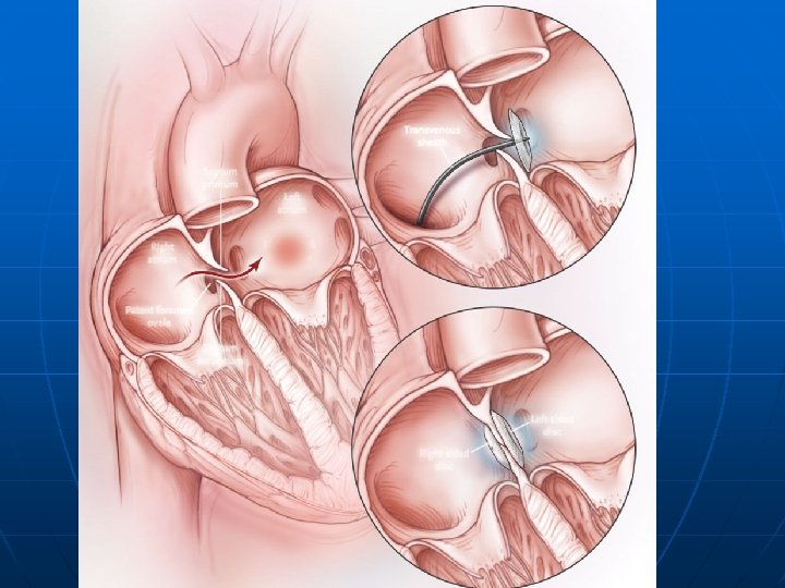
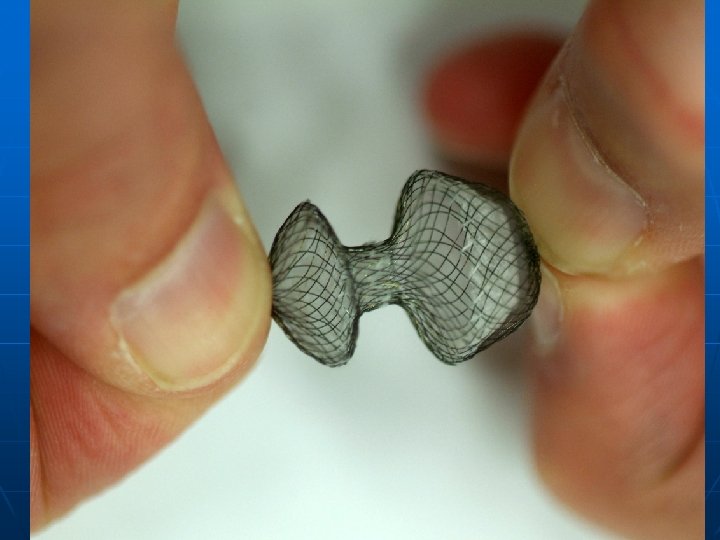
 PFO n n Figure 3. Percutaneous Closure of a Patent Foramen Ovale. With use of a femoral approach, a transvenous sheath is advanced across the foramen into the left atrium, where a folded disk is expanded and pulled back, apposing the primum and secundum septa closed. This step is followed by deployment of a right-sided disk, at which time the twodisk device is released. Clopidogrel and aspirin are recommended for a period of three months to prevent thrombus formation on the device, with aspirin therapy continued for an additional three months, when endothelialization is complete. Antibiotic prophylaxis for six months is recommended. Complete late closure of the foramen has been reported in 80 to 95 percent of patients.
 Cocaine Abuse n Another important cause of ischemic stroke is the use of sympathomimetic drugs such as cocaine amphetamines, ephedra, or phenylephrine. The strongest association is with cocaine, which has been seen in case series to cause cerebral vasoconstriction in a dose-dependent manner. Vasoconstriction is also related to a longer duration of cocaine use. Several case-control studies have found that the risk of stroke is 4. 5 to 6. 5 times higher in drug abusers than in controls, and that use of catecholamines or cocaine alone was associated with a significantly increased risk of stroke.
 What Are the Causes of Hemorrhagic Stroke? Occurs when a weakened blood vessel ruptures • Aneurysms: Ballooning of a weakened region of a blood vessel • Arteriovenous Malformations (AVMs): Cluster of abnormal blood vessels
 Arteriovenous Malformations n n Cerebral AVMs are most commonly discovered in young adults aged 20 -40 years. These lesions are usually detected in patients as the result of a seizure or hemorrhage. AVMs hemorrhage at a rate of 4% per year. Approximately half of these hemorrhages will carry significant morbidity or mortality.
 Hematologic") HEMORRHAGIC n n n Arteriovenous malformation Neoplasm (primary central nervous system, metastatic, leukemia) Hematologic (sickle-cell disease, neoplasm, thrombocytopenia) Moyamoya disease Drug use (warfarin, amphetamines, cocaine, phenypropanolamine) Iatrogenic (peri-procedural)
HEMORRHAGIC n n n Arteriovenous malformation Neoplasm (primary central nervous system, metastatic, leukemia) Hematologic (sickle-cell disease, neoplasm, thrombocytopenia) Moyamoya disease Drug use (warfarin, amphetamines, cocaine, phenypropanolamine) Iatrogenic (peri-procedural)
 Moyamoya n n Moyamoya syndrome is characterized by progressive stenosis of the internal carotid arteries and formation of collateral vessels that give a "puff of smoke" appearance on angiography. Moyamoya disease occurs mainly in Japanese and other Asian populations and may have a genetic basis. Secondary moya syndrome is seen in association with neurofibromatosis, Down syndrome, Williams syndrome, sickle cell disease, and as a sequela of cranial irradiation. Intracranial hemorrhage is common in young adults. Dissection — Arterial dissection is the most common vascular abnormality in some young adult series
 Intracerebral Hemorrhage n n n Diffuse – subarachnoid hemorrhage Focal – intraparenchymal Accounts for 20% of all strokes Acute rise in intracranial pressure from arterial rupture frequently results in loss of consciousness at outset Some patients die from herniation
 n Intraparenchymal hemorrhage • Trauma • Hypertension •") Causes of Spontaneous Intracerebral Hemorrhage (ICH) n Intraparenchymal hemorrhage • Trauma • Hypertension • Amyloid angiopathy • Arteriovenous malformation • Bleeding diathesis (anticoagulants, thrombolytics) • Drugs (amphetamines, cocaine)
Causes of Spontaneous Intracerebral Hemorrhage (ICH) n Intraparenchymal hemorrhage • Trauma • Hypertension • Amyloid angiopathy • Arteriovenous malformation • Bleeding diathesis (anticoagulants, thrombolytics) • Drugs (amphetamines, cocaine)
 n n Cervical arterial dissection causes up to") Causes of Spontaneous Intracerebral Hemorrhage (ICH) n n Cervical arterial dissection causes up to 20% of strokes in patients younger than 45 years. Dissections usually involve the extracranial portion of the vessel, and involve the internal carotid arteries at least three times as often as the vertebral arteries.
Causes of Spontaneous Intracerebral Hemorrhage (ICH) n n Cervical arterial dissection causes up to 20% of strokes in patients younger than 45 years. Dissections usually involve the extracranial portion of the vessel, and involve the internal carotid arteries at least three times as often as the vertebral arteries.
 n n In many cases the dissection is") Causes of Spontaneous Intracerebral Hemorrhage (ICH) n n In many cases the dissection is preceded by mild neck trauma, which may be as minor as a vigorous cough or turning of the head. Typical features of dissection include: Neck pain, headache, and Horner syndrome, followed minutes to hours later by symptoms of ocular or cerebral ischemia, usually a transient ischemic attack rather than a stroke.
Causes of Spontaneous Intracerebral Hemorrhage (ICH) n n In many cases the dissection is preceded by mild neck trauma, which may be as minor as a vigorous cough or turning of the head. Typical features of dissection include: Neck pain, headache, and Horner syndrome, followed minutes to hours later by symptoms of ocular or cerebral ischemia, usually a transient ischemic attack rather than a stroke.
 n n n Inherited disorders that are associated") Causes of Spontaneous Intracerebral Hemorrhage (ICH) n n n Inherited disorders that are associated with increased risk of cervical arterial dissection include: Ehlers-Danlos syndrome type IV Marfan syndrome Autosomal dominant polycystic kidney disease Osteogenesis imperfecta type I Fibromuscular dysplasia
Causes of Spontaneous Intracerebral Hemorrhage (ICH) n n n Inherited disorders that are associated with increased risk of cervical arterial dissection include: Ehlers-Danlos syndrome type IV Marfan syndrome Autosomal dominant polycystic kidney disease Osteogenesis imperfecta type I Fibromuscular dysplasia
 Diagnosis, Management, and Prognosis of ICH n CT diagnostic test of choice • Hyperintense area with mass effect and later hypointense surrounding edema n MRI less sensitive in early stages
 Diagnosis, Management, and Prognosis of ICH n Management depends on size and location • In acute phase, mass effect far greater than in large cerebral infarction, so greater risk of herniation and death • In chronic phase, prognosis for surviving patients much better than with ischemic stroke
 Subarachnoid Hemorrhage n n n Aneurysms can rupture any time but more common during strenuous activity Most common manifestation is headache • “worst headache of my life” Neck pain and rigidity Loss of consciousness and vomiting common Seen on CT in 95% of cases – location may suggest site of rupture Normal CT does not rule out so do lumbar puncture – xanthochromia (develops after 6 hours)
 n Subarachnoid hemorrhage • Congenital saccular aneurysm (85%)") Causes of Spontaneous Intracerebral Hemorrhage (ICH) n Subarachnoid hemorrhage • Congenital saccular aneurysm (85%) • Unknown (15%)
Causes of Spontaneous Intracerebral Hemorrhage (ICH) n Subarachnoid hemorrhage • Congenital saccular aneurysm (85%) • Unknown (15%)
 What Parts of the Brain Are Affected by Stroke?
 Hemisphere Stroke: Common Pattern n n n n Neglect of left visual") Right (Non-dominant) Hemisphere Stroke: Common Pattern n n n n Neglect of left visual field Extinction of left-sided stimuli Left hemiparesis Left-sided sensory loss Left visual field defect Poor left conjugate gaze Dysarthria Spatial disorientation
Right (Non-dominant) Hemisphere Stroke: Common Pattern n n n n Neglect of left visual field Extinction of left-sided stimuli Left hemiparesis Left-sided sensory loss Left visual field defect Poor left conjugate gaze Dysarthria Spatial disorientation
 Hemisphere Stroke: Common Pattern n n n Aphasia Right hemiparesis Right-sided sensory") Left (Dominant) Hemisphere Stroke: Common Pattern n n n Aphasia Right hemiparesis Right-sided sensory loss Right visual field defect Poor right conjugate gaze Dysarthria Difficulty reading, writing, or calculating
Left (Dominant) Hemisphere Stroke: Common Pattern n n n Aphasia Right hemiparesis Right-sided sensory loss Right visual field defect Poor right conjugate gaze Dysarthria Difficulty reading, writing, or calculating
 Brain Stem / Cerebellum / Posterior Hemisphere Stroke: Common Pattern n n n n Motor or sensory loss in all four limbs Crossed signs Limb or gait ataxia Dysarthria Dysconjugate gaze Nystagmus Amnesia Bilateral visual field defects
 Small Subcortical Hemisphere or Brain Stem Stroke: Common Pattern n Pure Motor • Weakness of face and limbs on one side of the body without abnormalities of higher brain function, sensation, or vision n Pure Sensory • Decreased sensation of face and limbs on one side of the body without abnormalities of higher brain function, motor function, or vision
 Physical Exam • Neurologic Exam • Carotid Bruits • Cardiac Exam • Peripheral Pulses • Dermatologic • Ophthalmologic
 Xanthoma (hyperlipidemia) Café-au-lait spots") Dermatologic n n n Splinter hemorrhages and needle tracks (endocarditis) Xanthoma (hyperlipidemia) Café-au-lait spots Neurofibromas (neurofibromatosis) Purpura (coagulopathy) Capillary angiomata (cavernous malformation)
Dermatologic n n n Splinter hemorrhages and needle tracks (endocarditis) Xanthoma (hyperlipidemia) Café-au-lait spots Neurofibromas (neurofibromatosis) Purpura (coagulopathy) Capillary angiomata (cavernous malformation)
 Xanthoma, eruptive CAFÉ AU LAIT spots Neurofibromas splinter hemorrhages
 Corneal opacity (Fabry’s disease) Lisch") Opthalmlogic n n n n n Corneal arcus (hypercholesterolemia) Corneal opacity (Fabry’s disease) Lisch nodules Optic atrophy (neurofibromatosis) Lens subluxation (Marfan’s syndrome, homocystinuria) Retinal perivasculitis (sickle-cell disease, syphilis, connective tissue diseases, inflammatory bowel disease) Occlusions (emboli) Angioma (cavernous malformation) Hamartoma (tuberous sclerosis).
Opthalmlogic n n n n n Corneal arcus (hypercholesterolemia) Corneal opacity (Fabry’s disease) Lisch nodules Optic atrophy (neurofibromatosis) Lens subluxation (Marfan’s syndrome, homocystinuria) Retinal perivasculitis (sickle-cell disease, syphilis, connective tissue diseases, inflammatory bowel disease) Occlusions (emboli) Angioma (cavernous malformation) Hamartoma (tuberous sclerosis).
 Corneal arcus optic atrophy in tuberous sclerosis lens dislocation in marfan syndrome
 Diagnostic Testing for Patients With Stroke n n n Basic stroke evaluation Cranial computed tomography (CT) Carotid ultrasonography ± transcranial Doppler Transthoracic echocardiography EKG monitoring Routine blood studies (complete blood count with differential and platelet count, prothrombin time (international normalized ratio), activated partial thromboplastin time, glucose, chemistries, serology for syphilis, and an erythrocyte sedimentation rate)
 Diagnostic Testing for Patients With Stroke n n n Comprehensive stroke evaluation Cranial magnetic resonance imaging (MRI) Imaging of the intracranial arteries (MR, CT, or catheter angiography of the brain) Imaging of the extracranial arteries (MR, CT, or catheter angiography of the neck) Transesophageal echocardiography (TEE) Prolonged EKG monitoring with Holter or event loop recorder Urine toxicology screen (often productive) Urine pregnancy test Blood testing for a hypercoagulable state anticardiolipin antibodies, lupus anticoagulants, protein S, protein C, activated protein C resistance, antithrombin III) is requested in patients without a firmly identified cause of stroke or if the patient or family members have a history of thromboses. It is advantageous to send such a profile prior to initiating anticoagulation, as heparin can alter interpretation of some of those assays. In select cases, blood testing for rare genetic causes of stroke (CADASIL, Fabry disease, MELAS)
 How Are Strokes Treated? Ischemic Stroke • Clot-busters e. g. , t-PA • Anticoagulants – warfarin, aspirin n Carotid Endarterectomy n Angioplasty/Stents
 Treatment of Strokes • Antiplatelet therapy remains treatment of choice to prevent recurrent thromboembolism in majority of patients • Anticoagulation may be appropriate n Atrial fibrillation n Recent MI n Suspected propagation of thrombus or stroke in evolution
 Treatment of Strokes n n CT or MRI of the brain should be performed promptly; MRI is more sensitive for early ischemic changes, but either method can fully rule out hemorrhage. Treatment of TPA was associated with an increase of about 1. 2 with minimal or no disability for every 10 patients treated.
 Limitations of Imaging n n n CT will miss a minority of acute bleeds MRI with DWI (diffusion weighted imaging), quite sensitive for acute stroke, has an occasional false negative result (17 out of 782 patients in a recent study) MRA’s resolution is not yet on par with conventional angiography.
 Thrombolysis t-PA • Guidelines for treatment: n Present within 3 hours of onset of clearly defined stroke – frequency of symptomatic hemorrhage most likely increases after this time CT scan shows no evidence of intracranial hemorrhage • No anticoagulants or antiplatelet agents given for 24 hours • Avoid BP values > 185/110 n
 Guidelines Not to Treat Previous stroke or serious head trauma in preceding 3 months n History of intracranial hemorrhage n Repeated systolic BP’s > 185 mm Hg or diastolic BP’s > 110 mm Hg n
 Guidelines Not to Treat Requires aggressive treatment to reduce BP to specified limits n Taking anticoagulants or propensity to hemorrhage n Recent invasive surgical procedure n Rapidly improving neurological deficit or minor symptoms n
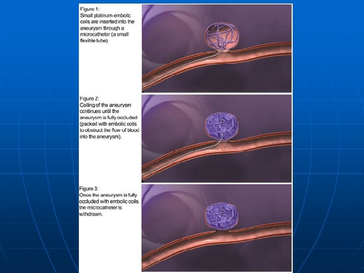
 How Are Strokes Treated Hemorrhagic Stroke n Surgical Intervention n Endovascular Procedures, e. g. , “coils”
 What Is the Impact of Stroke? Stroke is the third leading cause of death in the United States • On average, someone suffers a stroke every 40 seconds • About 795, 000 Americans suffer a stroke each year • About every 4 minutes, someone dies of a stroke
 What Is the Impact of Stroke? n n n Stroke is a leading cause of serious, long term disability About 6. 4 million Americans are stroke survivors Americans will pay about $73. 7 billion in 2010 for stroke-related medical costs and lost productivity long-term
 Rehabilitation n n After suffering a stroke, it’s important to begin a rehabilitation program as soon as possible. Types of rehabilitation programs: Hospital programs Extended care facilities Outpatient programs Home-based programs
 Rehabilitation Specialists n n n n Provider Rehabilitation specialist Physical therapist Speech therapist Occupational therapist Physiatrist Psychiatrist
 Family Relationships n n Overall, 13 studies reported consequences of stroke for family relationships and in those studies, 5% to 54% of the samples experienced family problems. Nine studies reported marital problems after stroke, including separation or divorce. Six of these reported that marital problems were a direct consequence of the stroke. One study reported that 5% of the sample had experienced deterioration in the spousal relationship, whereas another found that 38% of couples had experienced conflict since the stroke.
 Sexual Relationships n n Ten studies investigated the impact of stroke on sexual relationships, reporting problems in participants’ sexual relationships or frequency of sexual activities. Prevalence of deterioration in sexual relationships reported in 8 studies ranged from 5% to 76%.
 Social Activities n n Nine studies reported consequences of stroke on social or leisure activities with 4 of these reporting deterioration or decrease in these activities. Five studies quantified reported decrease in leisure activities ranging from 15% to 79%.
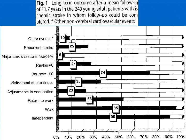
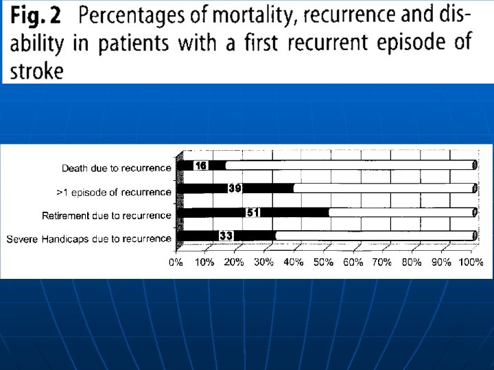
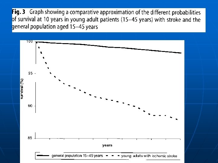
 Prognosis n n The outcome of stroke in young adults is better than that for older adults. In a recent study of 330 patients with first stroke or transient ischemic attack, followed for an average of 96 months, 8% died, 3% had another stroke, and 3% had a myocardial infarction. Approximately 16% were dependent, but 56% had returned to work. Unfortunately, only a minority of those who smoked at the time of their stroke subsequently stopped using tobacco. The overall annual recurrence rate is less than 1%. Prognosis is often closely associated with the underlying cause. A relatively good outcome may be found after many cases of arterial dissection. Risk of stroke recurrence is low (2% over 5 years) in women whose first stroke occurred in pregnancy.


 Stroke Chameleons n n n Strokes with atypical presentations that take on the appearance of other disease process may be termed stroke chameleons, for like the chameleon, these disguised strokes may change and evolve with time. The provider is left with the daunting problem of discovering the unusual manifestation of an uncommon clinical process. The presence of historical risk factors for cerebrovascular disease and the abrupt onset of symptoms may be the best clues available to the provider to detect these unusual stroke syndromes.
 Stroke Chameleons n n n In the majority of cases of stroke, making the diagnosis is straightforward. Especially in patients with unusual features (e. g. , Gradual onset, Seizure at the onset of symptoms Impaired consciousness The differential diagnosis should include migraine, postictal paresis, hypoglycemia, conversion disorder, subdural hematoma, and brain tumors.
 and cardioembolism") Stroke Chameleons n n n Atherosclerosis (leading to thromboembolism or local occlusion) and cardioembolism are the leading causes of brain ischemia. Unusual causes should be considered, especially if patients are younger (e. g. , below 50 years of age) and have no apparent cardiovascular risk factors. Some clinical clues that suggest alternative diagnoses are ptosis and miosis contralateral to the deficit (carotid-artery dissection), fever and a cardiac murmur (infective endocarditis), and headache and an elevated erythrocyte sedimentation rate.
Stroke Chameleons n n n Atherosclerosis (leading to thromboembolism or local occlusion) and cardioembolism are the leading causes of brain ischemia. Unusual causes should be considered, especially if patients are younger (e. g. , below 50 years of age) and have no apparent cardiovascular risk factors. Some clinical clues that suggest alternative diagnoses are ptosis and miosis contralateral to the deficit (carotid-artery dissection), fever and a cardiac murmur (infective endocarditis), and headache and an elevated erythrocyte sedimentation rate.
 begin with") Differential Diagnosis n n Seizures and migraine auras characteristically (but not always) begin with positive symptoms, while TIAs invariably are characterized by negative symptoms. Seizures occasionally cause paralytic attacks but, on close observation, there are usually features of the history and physical examination that suggest the presence of a seizure disorder such as minor twitching of a finger or toe or a tingling sensation in the affected limb.
Differential Diagnosis n n Seizures and migraine auras characteristically (but not always) begin with positive symptoms, while TIAs invariably are characterized by negative symptoms. Seizures occasionally cause paralytic attacks but, on close observation, there are usually features of the history and physical examination that suggest the presence of a seizure disorder such as minor twitching of a finger or toe or a tingling sensation in the affected limb.
 Differential Diagnosis of Stroke n n Intracranial mass: Tumor, Subdural hematoma Seizure with persistent neurological signs Migraine with persistent neurological signs Metabolic
 Hypoglycemia Post-cardiac arrest ischemia") Differential Diagnosis of Stroke n n Hyperglycemia (nonketotic hyperosmolar coma) Hypoglycemia Post-cardiac arrest ischemia Drug/narcotic overdose
Differential Diagnosis of Stroke n n Hyperglycemia (nonketotic hyperosmolar coma) Hypoglycemia Post-cardiac arrest ischemia Drug/narcotic overdose
 Focal symptom s Nonfocal symptoms Seizures ++ ++ TIAs ++++ occasionally Migraine ++++ Common disorders Syncope 0 0 Less common disorders Vestibulopathy ++ ++ Metabolic + +++ "Tumor attacks" +++ + Multiple sclerosis ++++ 0 Psychiatric ++ ++ Nerves and nerve root ++++ 0 Transient global amnesia ++++ 0
 Differential Diagnosis of Transient Neurological Diseases n n n TIA Seizure Migraine with aura Syncope Hypoglycemia
 Hypoglycemia n n n n That transient hypoglycemia may produce a stroke like picture with hemiplegia and aphasia has been known for years. These patients may be drowsy but are often alert and do not show the more common response to hypoglycemia of confusion, diminished level of consciousness, or coma. Aphasia may make the history of diabetes more difficult to discover. Syndrome has also been reported in alcoholics with hypoglycemia. The pathogenesis of this focal CNS dysfunction is unclear. Hypoglycemia is generally defined as a blood glucose level of less than 45 mg/dl in these studies. The wide use of bedside rapid laboratory testing for glucose now makes this easily detectable and treatable. The hemiplegia may resolve immediately with the administration of intravenous glucose but resolution over a hours is also reported.
 Mass Lesions n n n Subdural hematoma, cerebral abscess, primary CNS tumors, and metastatic tumors are among the clinical conditions simulating stroke in the studies cited above. The typical clinical presentation of a slowly increasing mass is a progressive syndrome; an abrupt onset of symptoms of these masses seems counter-intuitive. A review of patients with brain tumors presenting to an ED showed that 6% of patients had symptoms that were of less than one day’s duration; it was thought that these patients with brief symptom duration might reflect a subpopulation who suffer acute deterioration from hemorrhage into the tumor or who develop obstructive hydrocephalus. Secondary effects of mass or edema on cerebral vasculature have been identified as possible causes of abrupt onset of seizures as well. Chronic subdural hematoma has been frequently reported as a cause of stroke and TIA-like symptoms.
 Functional Hemiparesis n n n Little is written about a factitious or feigned stroke yet several studies discover rare patients initially thought to have cerebrovascular disease but later determined to have a functional cause of the hemiparesis or other stroke syndrome. Conversion disorder is the most commonly assigned psychiatric disorder. One study of emergency department presentations of conversion disorder noted that symptoms of paresis, paralysis, or movement disorders were common and were a presentation in almost 30% of patients. They noted significant comorbidity in this population, often other psychiatric disorders, and emphasized that conversion disorder is a diagnosis of exclusion. Patients often undergo multiple diagnostic tests before the diagnosis is assigned.
 Seizures Demographics n Any age, often younger
 Seizures CNS Symptoms n n Light-headed, dim vision, noises distant, decreased alertness Transient loss of consciousness
 Seizures CNS Symptoms n n Positive symptoms: limb jerking, head turning, loss of consciousness Negative symptoms may develop, remain postictally, and persist
 Seizures Timing n n 20 to 80 seconds Absence, atonic seizures and myoclonic jerks are shorter Postictal depression Spells occur over years
 Seizures Associated Symptoms n Tongue biting, incontinence, sore muscles, headache after attack
 seizure • Positive sensory or motor symptoms • Spread") Focal Seizure n Partial (focal) seizure • Positive sensory or motor symptoms • Spread quickly (60 seconds) • Negative symptoms afterward (Todd’s paresis) • Multiple attacks
Focal Seizure n Partial (focal) seizure • Positive sensory or motor symptoms • Spread quickly (60 seconds) • Negative symptoms afterward (Todd’s paresis) • Multiple attacks
 What is a TIA • Acute loss of focal cerebral function • Symptoms last less than 24 hours • Due to inadequate blood supply Thrombosis n Embolism n
 n n Warning strokes” that can happen before a major") Transient Ischemic Attacks (TIAs) n n Warning strokes” that can happen before a major stroke Occur when blood flow through a brain artery is blocked or reduced for a short time Symptoms are temporary (<24 hours) but similar to those of a full fledged stroke A person who has a TIA is 9. 5 times more likely to have a stroke
Transient Ischemic Attacks (TIAs) n n Warning strokes” that can happen before a major stroke Occur when blood flow through a brain artery is blocked or reduced for a short time Symptoms are temporary (<24 hours) but similar to those of a full fledged stroke A person who has a TIA is 9. 5 times more likely to have a stroke
 TIA Risk Factors/Epidemiology n n 300, 000 TIAs per year in US 5 -year stroke risk after TIA 29% • 43. 5% in 2 years with >70% carotid stenosis treated medically n Many stroke patients have had a TIA • 25% - 50% in large artery atherothrombotic strokes • 11% - 30% in cardioembolic strokes • 11% to 14% in lacunar strokes
 Risk Factors for a TIA n Risk factors are the same as stroke • • • Increasing age Sex Family history / Race Prior stroke / TIA Hypertension Diabetes Heart disease Carotid artery / Peripheral artery disease Obesity High cholesterol Physical inactivity
 TIA Symptoms n Symptoms last less than 24 hours • Most last less than one hour • Less than 10 percent > 6 hours • Amaurosis fugax up to five minutes (Amaurosis fugax is loss of vision in one eye due to a temporary lack of blood flow to the retina. Symptoms include the sudden loss of vision in one eye. )
 TIA Demography n n n Older patients Stroke risk factors present Men>women
 TIA Presentation n Acute loss of focal cerebral function • Abrupt onset • Symptoms occur in all affected areas at the same time • Symptoms resolve gradually • Symptoms are “negative”
 TIA Presentation n Acute loss of focal cerebral function • Motor symptoms n Weakness or clumsiness on one side n Difficulty swallowing • Speech disturbances n Understanding or expressing spoken language n Reading or writing n Slurred speech n Calculations
 TIA Presentation n Acute loss of focal cerebral function • Sensory symptoms Altered feeling on one side n Loss of vision in left or right visual field n Bilateral blindness n Double vision n Vertigo n
 • Generalized weakness or numbness • Faintness") TIA Presentation n Non-focal Symptoms (Not TIA) • Generalized weakness or numbness • Faintness or syncope • Incontinence • Isolated symptoms (symptoms occurring alone) Vertigo or loss of balance n Slurred speech or difficulty swallowing n Double vision n
TIA Presentation n Non-focal Symptoms (Not TIA) • Generalized weakness or numbness • Faintness or syncope • Incontinence • Isolated symptoms (symptoms occurring alone) Vertigo or loss of balance n Slurred speech or difficulty swallowing n Double vision n
 • Confusion n n Disorientation Impaired attention/concentration") TIA Presentation n Non-focal Symptoms (Not TIA) • Confusion n n Disorientation Impaired attention/concentration Diminution of all mental activity Distinguish from • Isolated language or visual-spatial perception problems (may be TIA) • Isolated memory problems (transient global amnesia)
TIA Presentation n Non-focal Symptoms (Not TIA) • Confusion n n Disorientation Impaired attention/concentration Diminution of all mental activity Distinguish from • Isolated language or visual-spatial perception problems (may be TIA) • Isolated memory problems (transient global amnesia)
 TIA • Significant risk factor for recurrent stroke, with average 5% risk per year • Prophylactic antiplatelet therapy shown to prevent secondary effects n Aspirin n Ticlopidine: thrombotic stroke reduction n Clopidogrel: reduce events associated with atherosclerosis that include strokes, MI, PVD
 TIA • Treat with warfarin if significant risk for cardiogenic thromboembolism • Hospital admission for new-onset and recurrent TIA’s unless confident in diagnosis of etiology • Angiography – treat medically or surgically
 TIA n n Usually minutes, mostly <1 hour Spells during days, weeks, months; not usually years
 TIA Associated Symptoms n Headaches may occur during time period of a TIA
 10 -20% of the") Migraine Demography n n n Younger age Women>men (4: 1) 10 -20% of the population The risk of migraine with aura and transient ischemic attacks (TIA’s) is greater than 2 fold. 1/3 have migraine with aura
Migraine Demography n n n Younger age Women>men (4: 1) 10 -20% of the population The risk of migraine with aura and transient ischemic attacks (TIA’s) is greater than 2 fold. 1/3 have migraine with aura
 Migraine with Aura n Migraine with aura • • • n n Positive symptoms Spread over minutes Visual disturbances Somatosensory or motor disturbance Headache within 1 hour Migraine with aura is associated with a twofold risk of ischemic stroke. This risk is higher in women, age < 45, smokers, and women who used oral contraceptives. BMJ 2009; 339: b 3914, Migraine and cardiovascular disease
 Migraine with Aura n Aura without Headache • 98% Visual symptoms • 30% with other symptoms 26% sensory n 16% aphasia n 6% dysarthria n 10% weakness n • Mean age 48. 7 (vs. 62. 1) • Fewer cardiovascular risk factors
 Migraine CNS Timing n n Usually 20 to 30 minutes Sporadic attacks during years
 Migraine with Aura
 Migraine Associated Symptoms n Headache after attack, nausea, vomiting, photophobia, phonophobia (usually GI or autonomic nervous system)
 Migraine n Auras typically occur before the onset of migraine headache, and the headache usually begins simultaneously with or just after the end of the aura phase. However, headache onset can rarely occur an hour or more after the end of the aura phase. Although atypical, an aura can develop during or after the onset of headache, and many patients have migraine aura with only a minimal or no headache.
 Migraine n n Most migraine auras resolve in 20 to 30 minutes and seldom last more than one hour. Typical auras may involve any of the following manifestations: Visual disturbances ( the most common type of aura) Sensory symptoms Motor weakness Speech disturbances
 Migraine vs. TIA n n n n Positive visual symptoms Gradual onset / evolution Sequential progression Repetitive attacks of identical nature Flurry of attacks mid-life Duration up to 60 minutes Headache follows ~ 50% n n n Visual loss Abrupt Simultaneous occurrence Duration <15 minutes Headache uncommon accompaniment
 Syncope Demography n n Any age, often younger Women>men
 Syncope Timing n n Usually a few seconds Sporadic attacks during years
 Syncope Associated Symptoms n Sweating, pallor, nausea
 syncope Situational syncope (during or") Most Common Types of Syncope n n Neurocardiogenic (vasovagal) syncope Situational syncope (during or immediately after urination, defecation, cough, or swallowing) Orthostatic syncope (associated with orthostatic hypotension) Syncope related to cardiac ischemia or cardiac arrhythmia
Most Common Types of Syncope n n Neurocardiogenic (vasovagal) syncope Situational syncope (during or immediately after urination, defecation, cough, or swallowing) Orthostatic syncope (associated with orthostatic hypotension) Syncope related to cardiac ischemia or cardiac arrhythmia
 Differential diagnosis of transient neurologic symptoms Demography Seizures Migraine Syncope Younger age Any age, often younger Women>men Men>women Positive symptoms: limb jerking, head turning, loss of consciousness Older patients Stroke risk factors present Central nervous system symptoms Any age, often younger TIAs Negative symptoms: numbness, visual loss, paralysis, ataxia First positive symptoms, then negative in same modality: scintillating scotomas and parasthesias most common; second sensory modality is involved after first clears Light-headed, dim vision, noises distant, decreased alertness Negative symptoms All sensory may develop, remain modalities affected postictally, and simultaneously persist Timing Transient loss of consciousness 20 to 80 seconds Usually minutes, mostly <1 hour Usually 20 to 30 minutes Usually a few seconds Absence, atonic seizures and myoclonic jerks are shorter Spells during days, weeks, months; not usually years Sporadic attacks during years Postictal depression Spells occur during years
 Transient Global Amnesia • Sudden disorder of memory • Antegrade and often retrograde • Recurrence 3% per year • Etiology unclear Migraine n Epilepsy (7% within 1 year) n Unknown n
 Transient Global Amnesia • No difference in vascular risk factors compared with general population • Fewer risk factors when compared with TIA patients • Prognosis significantly better than TIA
 Structural intracranial lesion • Tumor Partial seizures n Vascular steal n Hemorrhage n Vessel compression by tumor n
 Intracranial hemorrhage • ICH rare to confuse with TIA • Subdural hematoma Headache n Fluctuation of symptoms n Mental status changes n
 Multiple sclerosis • Usually subacute but can be acute Optic neuritis n Limb ataxia n • Age and risk factors • Signs more pronounced than symptoms
 Labyrinthine Disorders • Central vs. Peripheral vertigo • Ménière's disease • Benign positional vertigo • Acute vestibular neuronitis
 Others in the Differential Diagnosis n n n Metabolic perturbations, such as hypoglycemia, can be associated with focal neurologic deficits. Multiple sclerosis occasionally can cause paroxysmal attacks, particularly of ataxia and dysarthria. Brain tumors can occasionally result in transient neurologic symptoms; the mechanism in these cases is thought to involve mechanical changes that result in pressure on structures adjacent to the tumor.
 Others in the Differential Diagnosis n n Subdural hematomas may cause attacks of transient neurologic dysfunction, again due to mechanical changes that result in pressure on structures adjacent to the hematoma. Cerebral amyloid angiopathy, better known as a cause of intracerebral hemorrhage, may also cause transient neurologic symptoms. Affected patients complain of recurrent, brief (minutes), often stereotyped spells of weakness, numbness, paresthesias, or other cortical symptoms that can spread smoothly over contiguous body parts.
 Others in the Differential Diagnosis n n n Hepatic, renal, and pulmonary encephalopathies can produce temporary aberrations in alertness, behavior and movement. Compressive myelopathy may rarely cause sudden transient sensory changes and motor deficits. Pressure- or position-related peripheral nerve or nerve root compression cause transient paresthesias and numbness.
 Others in the Differential Diagnosis n n Peripheral vestibulopathies can cause transient episodic dizziness. Hysteria and other psychiatric disorders may underlie attacks that include swoons, falls, and episodic blindness, deafness, and paralysis, which can be confused with organic loss of function.
 Case 1 n A 19 year old man had a history of a murmur since birth and intermittent episodes of hemiparesis that he ignored. He presented with collapse while playing handball.
 Case 1 n n He fell and had tonic clonic movement of his extremities. He was in distress at presentation His BP was 110/70, HR 76, RR 18. He had a persistently split S 2 with a systolic ejection murmur in the upper left second intercostal area. His neurological exam was consistent with a right middle cerebral infarction with a left hemiplegia and left hemisensory defect.
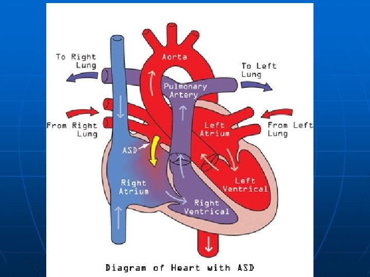
 Case 2 When 22 -year-old Ms. KS began experiencing left arm weakness, she went to a clinic only to be told that nothing was seriously wrong. But when her symptoms persisted, so did she. She was finally referred to the Medical Center, where neurologists confirmed that her symptoms were the result of a stroke.
 Case 2 n Further testing in the Coagulation Laboratory revealed activated protein C resistance, the result of a heterozygous mutation in factor V Leiden, as well as a homozygous mutation in the methylene tetrahydrofolate reductase gene, responsible for elevated homocystine
 Case 2 n Although she had only one clinical stroke, multiple other strokes were evident by imaging studies. Factors contributing to her hypercoagulable state included: 1. the methylene tetrahydrofolate reductase deficiency, which elevated her homocystine level, 2. the heterozygous state for factor V Leiden, causing activated protein C resistance, 3. oral contraceptives, 4. smoking, and 5. hypertension. She is doing well, without recurrence, on aspirin, and antihypertensive medicines, having stopped smoking and birth control pills.
 Case 3 n In another case, a 20 -year-old female college student was sent to the Medical Center after a brain scan showed what her physicians thought to be a brain tumor. Neurosurgeons quickly realized there was no tumor, and that the young woman had suffered a stroke. After thrombosis of intracranial venous sinuses was diagnosed she was treated with heparin, and then Warfarin. Repeat imaging revealed recanalization of the previously thrombosed sinuses.
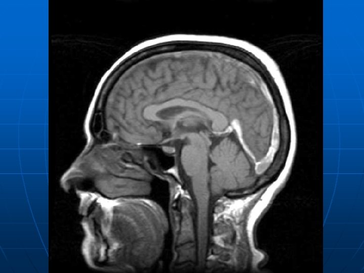
 Case 3 n Investigation in the Special Coagulation Laboratory revealed deficiency of antithrombin III, on two separate occasions, several weeks apart. This patient suffered a venous infarction secondary to a hypercoagulable state induced by inherited antithrombin III deficiency, probably exacerbated by use of oral contraceptives. With anticoagulation and careful monitoring, her prognosis is good.
 Case 4 n n n A 28 -year-old man has sudden weakness of the left arm and leg and slurred speech. Except for untreated hypertension, his medical history is unremarkable. He is a current smoker. On arrival at the emergency department 1 hour 15 minutes after the onset of symptoms, he reports no headache or vomiting. His blood pressure is 180/100 mm Hg, and his pulse is 76 beats per minute and is regular. Neurologic examination shows dysarthria, a left homonymous hemianopia, severe left-sided weakness, and a failure to register light touch on the left side of the body when both sides are touched simultaneously (left tactile extinction).
 Case 4 n He had sudden left-sided hemiparesis, strongly suggestive of a right hemisphere stroke.
 Summary n These are not isolated cases. Frequently young patients, even teenagers, who have suffered stroke are misdiagnosed. Though they may have presented with classic symptoms - dizziness, severe headache, weakness in a limb, or unilateral loss of sensation, for example - they often have been misdiagnosed by their provider, most likely because they do not fit the profile of the "typical" stroke patient.
 Summary n Misdiagnosis has included patients being told they had vertigo, migraine, alcohol intoxication or other conditions. They were discharged from clinic and later discovered to have suffered a stroke.
 Summary n The second important point is that the causes of stroke in young people are much more diverse, and less likely to be the ones that commonly cause stroke in older people, such as atherosclerosis of brainsupplying vessels and emboli from cardiac disorders related to coronary artery disease. Without the right resources, providers may be at a loss to pinpoint the cause in order to treat it and prevent further damage.
 Summary n Abuse of amphetamine-like drugs ("speed" and cocaine, for example) can lead to stroke in teenagers and young adults. These drugs are popular in rural and urban areas alike.
 Summary n However, one of the increasingly recognized causes of stroke in a young person is a blood disorder that predisposes to blood clotting. This allows clots to form too readily, often in the veins, leading to stroke. Once such a disorder has been identified, treatment can help prevent future damage. Diagnosis of a hereditary disorder can also lead to identification of other family members who may be at high risk.
 Summary n n The long term outcome of cerebral infarction is better in young adults than in older patients, morbidity and mortality are still significant. Despite a good functional and motor outcome young adult patients usually encounter difficulties in social and work caused by cognitive impairment.
 Summary n The bottom line: for young stroke patients, extensive testing and careful evaluation may be necessary to determine the underlying cause.