Spinal Injuries M. Jamous M. D. Spinal

- Размер: 2 Mегабайта
- Количество слайдов: 29
Описание презентации Spinal Injuries M. Jamous M. D. Spinal по слайдам
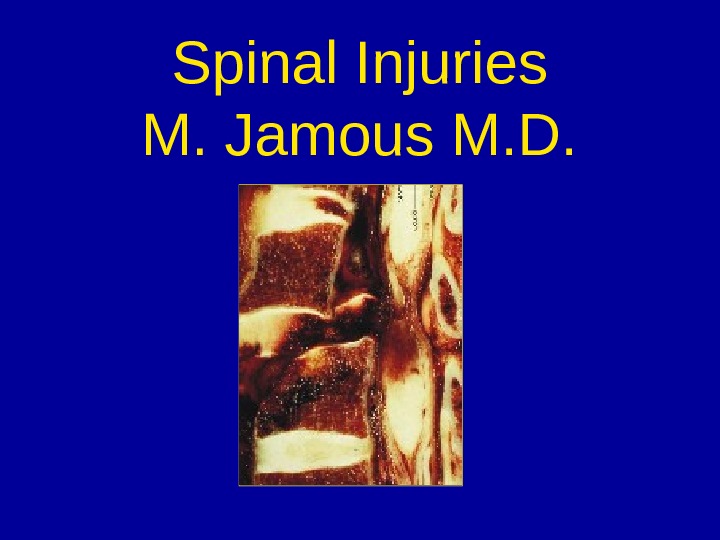
 Spinal Injuries M. Jamous M. D.
 Spinal Injuries • Incidence 30 -40/ 1, 000 person • The mortality rate 40 -50% • Most common in the cervical region (55%) • The peak incidence in the young age group (15 -25 year-old) • Motor vehicle accidents acounts for 50% followed by falls (25%), athletic accidents (15%), and penetraing injuries (10%)
 Acute evaluation and ER management • Strict spine precautions (immobilization) • Emergency resuscitation (ABC. . ) • Comprehensive approach • Always expect multiple trauma (neuroexam, chest, abdomin, muskuloskeletal…) • Differentiate hggic from neurogenic shock
 Neurological and radiological evaluation • In awake patients, both motor and sensory examinations in all extremeties • Unconcious patients: muscle tone, reflexes, rectal sphinctor tone, priapism
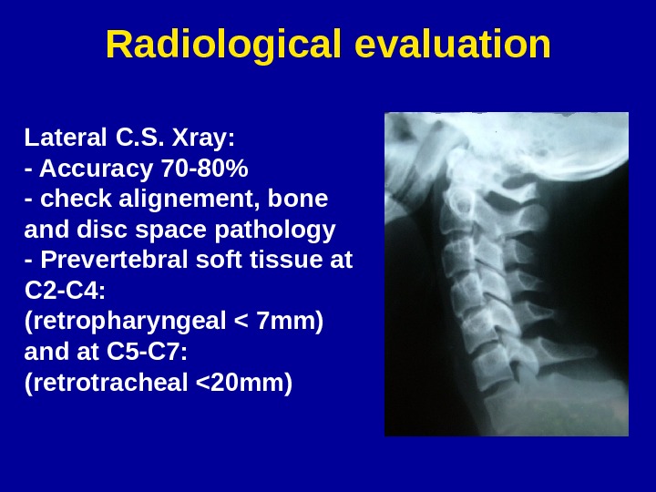
 Radiological evaluation Lateral C. S. Xray: — Accuracy 70 -80% — check alignement, bone and disc space pathology — Prevertebral soft tissue at C 2 -C 4: (retropharyngeal < 7 mm) and at C 5 -C 7: (retrotracheal <20 mm)
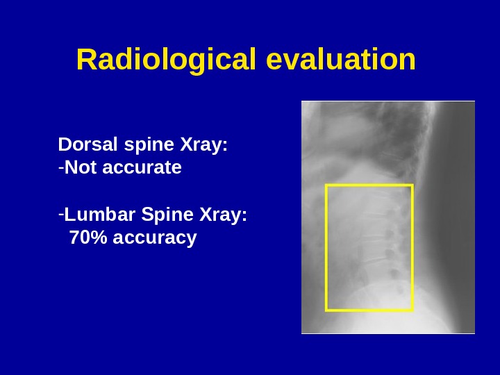
 Radiological evaluation Dorsal spine Xray: — Not accurate — Lumbar Spine Xray: 70% accuracy
 Radiological evaluation CT scan and MRI in case of clinical suspicion or abnormal Xray
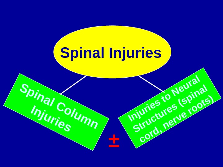
 Spinal Injuries S p in a l C o lu m n In ju rie s Injuries to N eural S tructures (spinal cord, nerve roots) ±
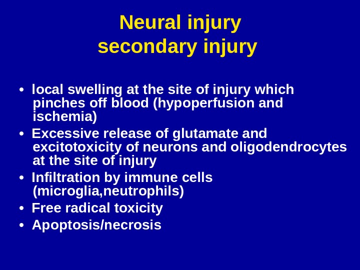
 Neural injury secondary injury • local swelling at the site of injury which pinches off blood (hypoperfusion and ischemia) • Excessive release of glutamate and excitotoxicity of neurons and oligodendrocytes at the site of injury • Infiltration by immune cells (microglia, neutrophils) • Free radical toxicity • Apoptosis/necrosis
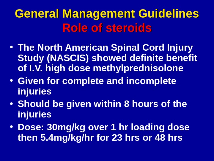
 General Management Guidelines Role of steroids • The North American Spinal Cord Injury Study (NASCIS) showed definite benefit of I. V. high dose methylprednisolone • Given for complete and incomplete injuries • Should be given within 8 hours of the injuries • Dose: 30 mg/kg over 1 hr loading dose then 5. 4 mg/kg/hr for 23 hrs or 48 hrs
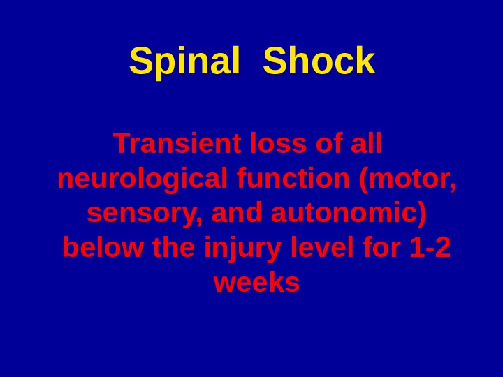
 Spinal Shock Transient loss of all neurological function (motor, sensory, and autonomic) below the injury level for 1 -2 weeks
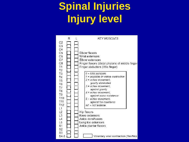
 Spinal Injuries Injury level
 Spinal Injuries ASIA IMPAIRMENT SCALE: A =Complete: No motor or sensory function is preserved B =Incomplete: Sensory but not motor function is preserved C =Incomplete: Non-useful motor function is pre-served below the neurological level D =Incomplete: Useful motor function is pre-served below the neurological level E =Normal: Motor and sensory func-tionare normal.
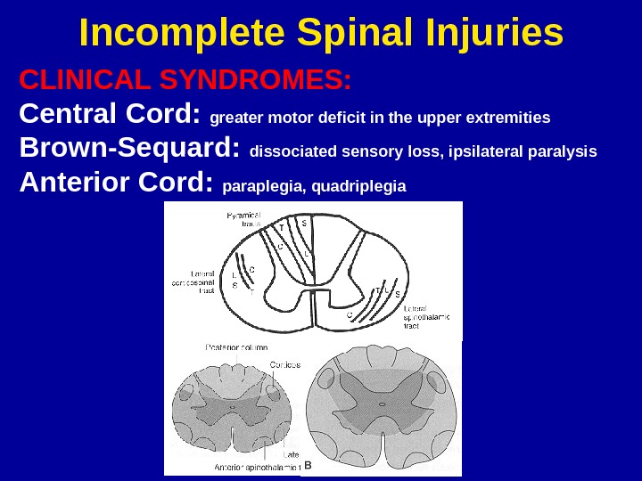
 Incomplete Spinal Injuries CLINICAL SYNDROMES: Central Cord: greater motor deficit in the upper extremities Brown-Sequard: dissociated sensory loss, ipsilateral paralysis Anterior Cord: paraplegia, quadriplegia
 Incomplete Spinal Injuries CLINICAL SYNDROMES: Conus Medullaris: saddle anesthesia, incontinence (painless, symmetrical) Cauda Equina: saddle anesthesia, incontinence (painful, asymmetrical)
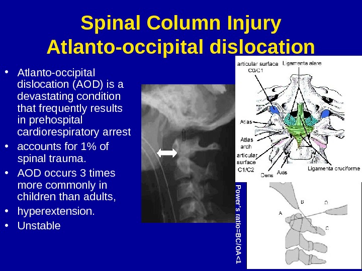
 Spinal Column Injury Atlanto-occipital dislocation • Atlanto-occipital dislocation (AOD) is a devastating condition that frequently results in prehospital cardiorespiratory arrest • accounts for 1% of spinal trauma. • AOD occurs 3 times more commonly in children than adults, • hyperextension. • Unstable. Pow er’s ratio=B C /O A <
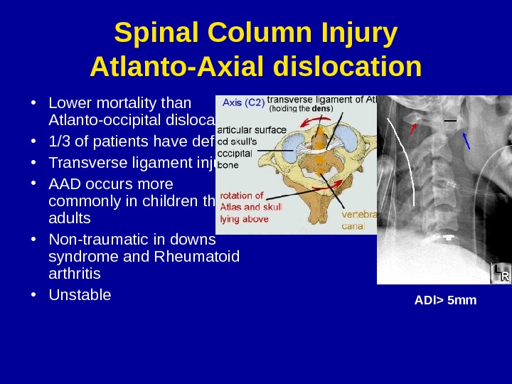
 Spinal Column Injury Atlanto-Axial dislocation • Lower mortality than Atlanto-occipital dislocation • 1/3 of patients have deficit • Transverse ligament injury • AAD occurs more commonly in children than adults • Non-traumatic in downs syndrome and Rheumatoid arthritis • Unstable ADI> 5 mm
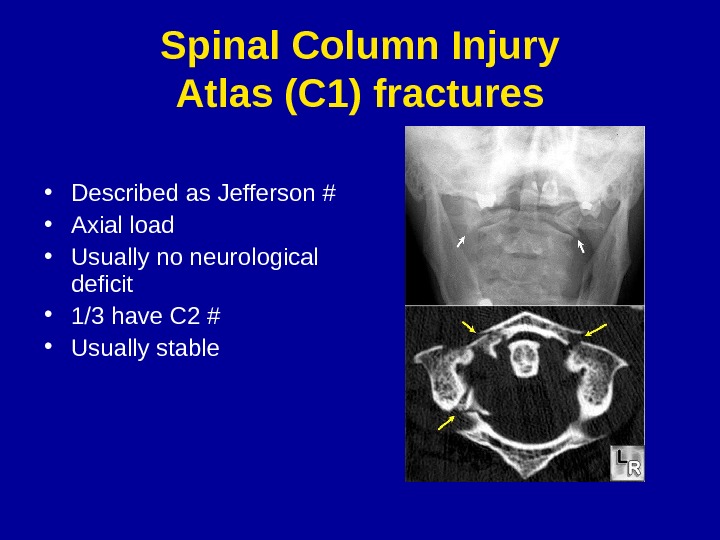
 Spinal Column Injury Atlas (C 1) fractures • Described as Jefferson # • Axial load • Usually no neurological deficit • 1/3 have C 2 # • Usually stable
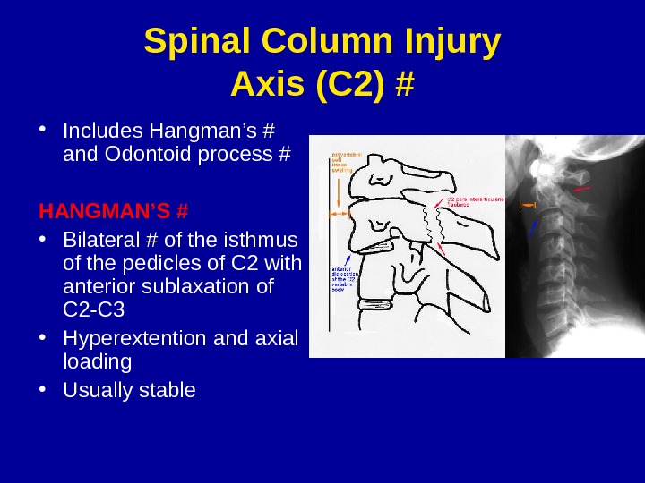
 Spinal Column Injury Axis (C 2) # • Includes Hangman’s # and Odontoid process # HANGMAN’S # • Bilateral # of the isthmus of the pedicles of C 2 with anterior sublaxation of C 2 -C 3 • Hyperextention and axial loading • Usually stable
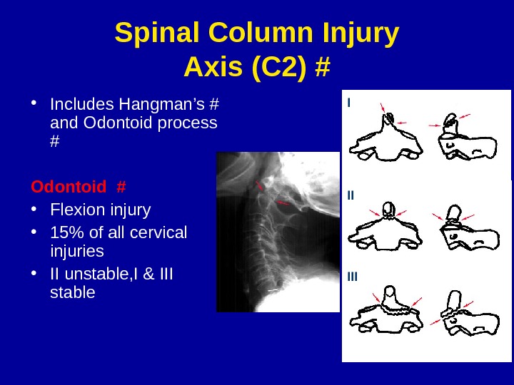
 Spinal Column Injury Axis (C 2) # • Includes Hangman’s # and Odontoid process # Odontoid # • Flexion injury • 15% of all cervical injuries • II unstable, I & III stable I II III
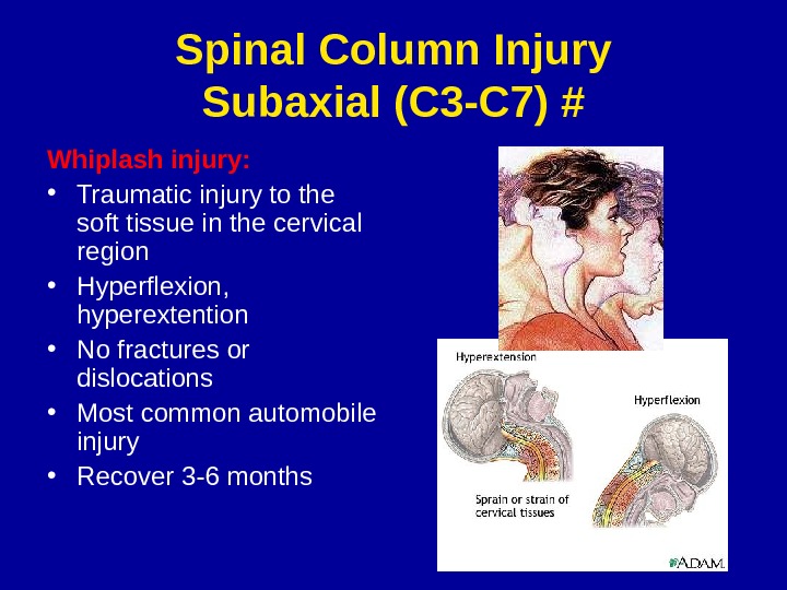
 Spinal Column Injury Subaxial (C 3 -C 7) # Whiplash injury: • Traumatic injury to the soft tissue in the cervical region • Hyperflexion, hyperextention • No fractures or dislocations • Most common automobile injury • Recover 3 -6 months
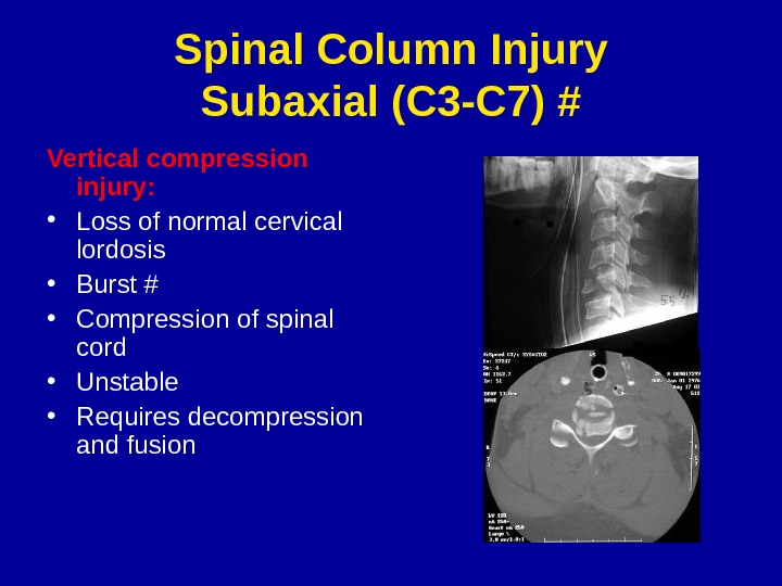
 Spinal Column Injury Subaxial (C 3 -C 7) # Vertical compression injury: • Loss of normal cervical lordosis • Burst # • Compression of spinal cord • Unstable • Requires decompression and fusion
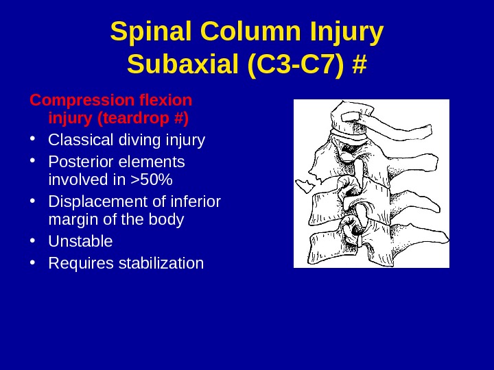
 Spinal Column Injury Subaxial (C 3 -C 7) # Compression flexion injury (teardrop #) • Classical diving injury • Posterior elements involved in >50% • Displacement of inferior margin of the body • Unstable • Requires stabilization
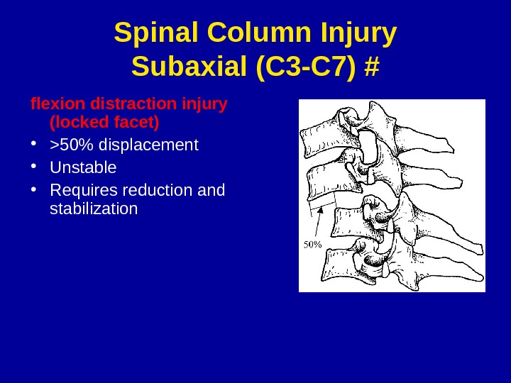
 Spinal Column Injury Subaxial (C 3 -C 7) # flexion distraction injury (locked facet) • >50% displacement • Unstable • Requires reduction and stabilization
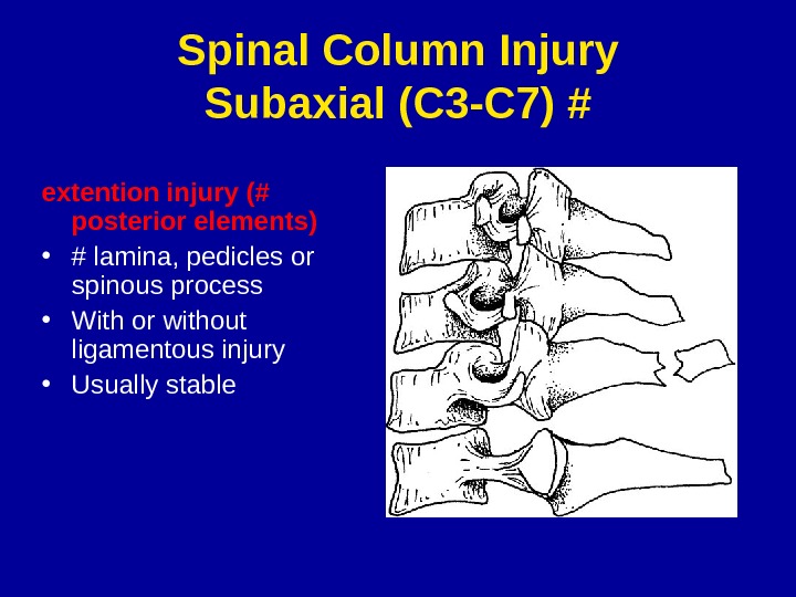
 Spinal Column Injury Subaxial (C 3 -C 7) # extention injury (# posterior elements) • # lamina, pedicles or spinous process • With or without ligamentous injury • Usually stable
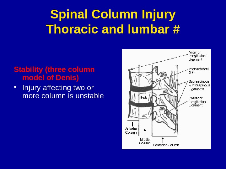
 Spinal Column Injury Thoracic and lumbar # Stability (three column model of Denis) • Injury affecting two or more column is unstable
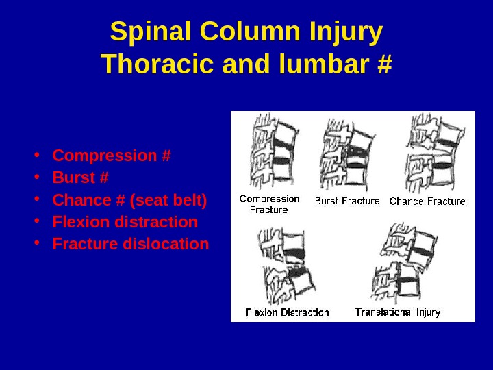
 Spinal Column Injury Thoracic and lumbar # • Compression # • Burst # • Chance # (seat belt) • Flexion distraction • Fracture dislocation
 General Management Guidelines • Strict spine precautions (immobilization) • Emergency resuscitation (ABC. . ) • Comprehensive approach • Neurological and Radiological assesment. • Always expect multiple trauma (neuroexam, chest, abdomin, muskuloskeletal…) • Differentiate hggic from neurogenic shock
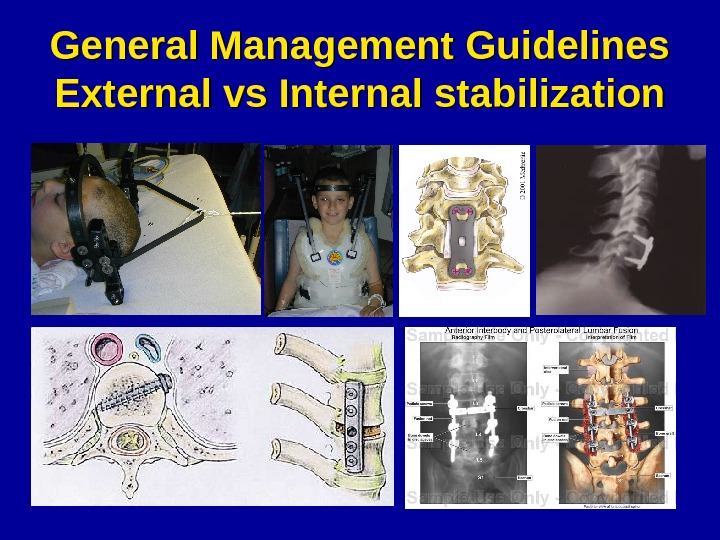
 General Management Guidelines External vs Internal stabilization

