

опухоли #2 .ppt
- Количество слайдов: 91
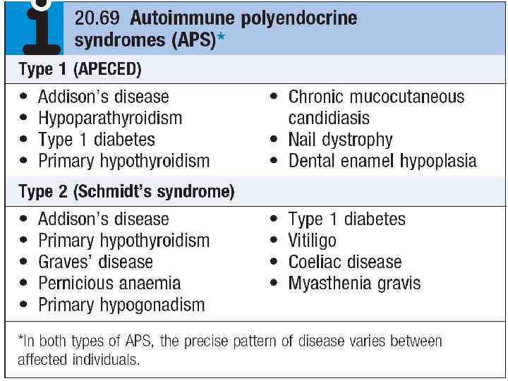
 синдром Сиппла Glucagonoma - 20% of cases, multiple endocrine neoplasia type I
 ПЛАН ЗАНЯТИЯ • КАНЦЕРОГЕНЫ • ПРЕОПУХОЛЕВЫЕ ЗАБОЛЕВАНИЯ И СОСТОЯНИЯ • ВОЗДЕЙСТВИЕ ОПУХОЛИ НА ОРГАНИЗМ • ОНКОМАРКЕРЫ
 КАНЦЕРОГЕНЫ
 Канцерогены v химические v физические v биологические протоонкогены
") Лев Александрович Зильбер (1894— 1966)
Лев Александрович Зильбер (1894— 1966)
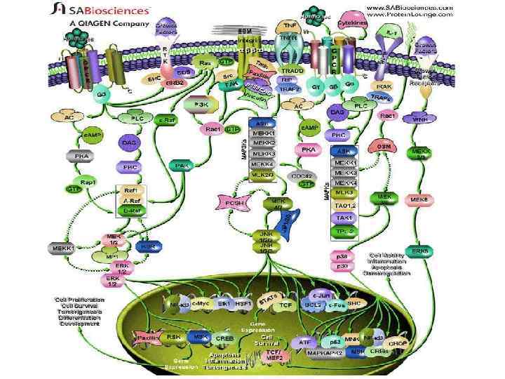
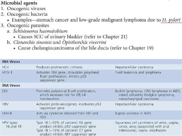
 revealed that its transforming function resided") Early studies of the Rous sarcoma virus (RSV) revealed that its transforming function resided in a single gene called src (25, 125, 210). In work for which they received the Nobel prize, Bishop and Varmus (210) showed that the viral src gene, v-src, was derived from a normal cellular protooncogene c-src. Clues to the machinery of intercellular signaling via growth factors and mitogens came when it was discovered that in normal cells protooncogenes function in signaling roles that regulate cell growth and proliferation (189, 256, 272). Cellular oncogenes were found to encode proteins that represent all major components of the growth factor response pathway: from the growth factor sis (42, 62, 133, 134) and growth factor receptor erb. B (64, 218), to the small G protein ras (59, 189, 256) or nonreceptor tyrosine kinase src (25, 125, 210), and finally nuclear proteins, such as myc (3, 71, 105, 146), fos (55), or jun (26, 88, 202). It was known that application of growth factors like platelet-derived growth factor (PDGF) leads to rapid activation of gene expression despite the presence of protein synthesis inhibitors such as cycloheximide. These rapid response genes were termed immediate-early genes (IEGs) (46). Nuclear run-off transcription assays after growth factor treatment revealed that the protooncogenes c-fos and c-myc were among the IEGs (96, 190). Several facts implicated c-fos in regulation of gene expression. It was known to encode a nuclear protein (55) associated with chromatin and capable of binding DNA cellulose in vitro (226, 233). It turned out that c-fos and c-jun are specific members of inducible gene families whose products associate combinatorially to form dimeric complexes that function as transcriptional activators (54). These transcription factors, in turn, help induce expression of second wave genes, termed delayed early genes.
Early studies of the Rous sarcoma virus (RSV) revealed that its transforming function resided in a single gene called src (25, 125, 210). In work for which they received the Nobel prize, Bishop and Varmus (210) showed that the viral src gene, v-src, was derived from a normal cellular protooncogene c-src. Clues to the machinery of intercellular signaling via growth factors and mitogens came when it was discovered that in normal cells protooncogenes function in signaling roles that regulate cell growth and proliferation (189, 256, 272). Cellular oncogenes were found to encode proteins that represent all major components of the growth factor response pathway: from the growth factor sis (42, 62, 133, 134) and growth factor receptor erb. B (64, 218), to the small G protein ras (59, 189, 256) or nonreceptor tyrosine kinase src (25, 125, 210), and finally nuclear proteins, such as myc (3, 71, 105, 146), fos (55), or jun (26, 88, 202). It was known that application of growth factors like platelet-derived growth factor (PDGF) leads to rapid activation of gene expression despite the presence of protein synthesis inhibitors such as cycloheximide. These rapid response genes were termed immediate-early genes (IEGs) (46). Nuclear run-off transcription assays after growth factor treatment revealed that the protooncogenes c-fos and c-myc were among the IEGs (96, 190). Several facts implicated c-fos in regulation of gene expression. It was known to encode a nuclear protein (55) associated with chromatin and capable of binding DNA cellulose in vitro (226, 233). It turned out that c-fos and c-jun are specific members of inducible gene families whose products associate combinatorially to form dimeric complexes that function as transcriptional activators (54). These transcription factors, in turn, help induce expression of second wave genes, termed delayed early genes.
 Харальд цур Хаузен
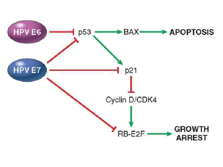
 HPV
HPV
 Уоррен Маршал
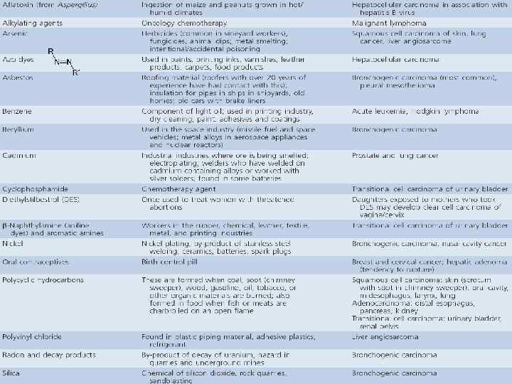
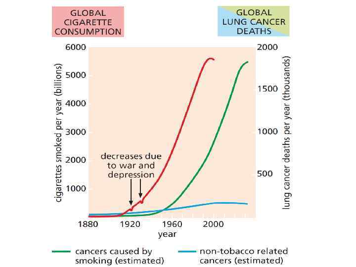
 Химический канцерогенез

") Герман Джозеф Мёллер(Hermann Joseph Muller; 1890 - 1967)
Герман Джозеф Мёллер(Hermann Joseph Muller; 1890 - 1967)
") Специальный механизм репарации (Excision-repair)
Специальный механизм репарации (Excision-repair)
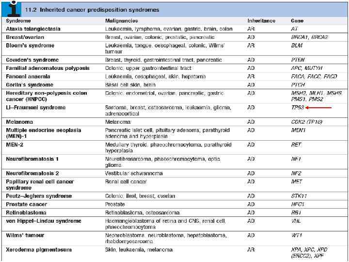
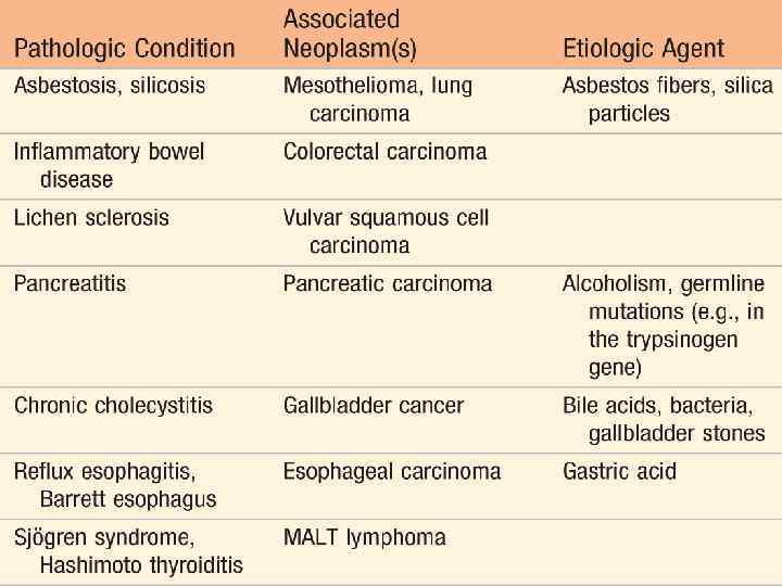
 ПРЕДОПУХОЛЕВЫЕ ЗАБОЛЕВАНИЯ И СОСТОЯНИЯ
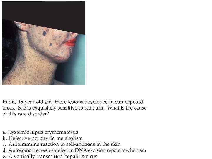
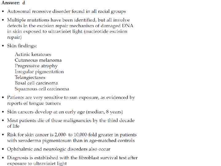
 Xeroderma pigmentosum. This is an autosomal recessive disease with defects in DNA repair. Note the numerous hyperpigmented lesions, and nodular and scaly growths on the face. Many of these lesions are precancerous or ultraviolet light–related cancers

 chronic infection and inflammation are among the most important epigenetic and environmental factors that can influence the establishment and progression of certain tumors. !

 NB Общие черты хронического воспаления и рака v миграция клеток v пролиферация клеток v ангиогенез v фиброгенез
 Воспаление способствует развитию опухоли
") Опухоль способствует воспалению (перитуморозному)
Опухоль способствует воспалению (перитуморозному)
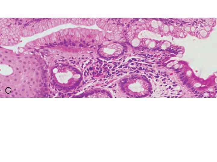
 пищевод Барретта Histologic appearance of the gastroesophageal junction in Barrett esophagus. Note the transition between esophageal squamous mucosa (left) and Barrett metaplasia, with abundant metaplastic goblet cells (right) нормальная слизистая пищевода нормальная слизистая кишечника
 Gross appearance of reflux esophagitis. Marked hyperemia with focal hemorrhage is present in the area of reflux. B, Barrett esophagus showing intestinal metaplasia (goblet cells; arrows). The normal squamous epithelium of the esophagus is on the left side of the biopsy
 keratosis. Note the pearly gray-white hyperkeratotic lesion (arrow) on the hand. The") Actinic (solar) keratosis. Note the pearly gray-white hyperkeratotic lesion (arrow) on the hand. The other lesions (circles) are good examples of solar lentigo. Both of these lesions are common in the elderly population and are located in sun-exposed areas. Squamous cancer occurs in 2% to 5% of cases
Actinic (solar) keratosis. Note the pearly gray-white hyperkeratotic lesion (arrow) on the hand. The other lesions (circles) are good examples of solar lentigo. Both of these lesions are common in the elderly population and are located in sun-exposed areas. Squamous cancer occurs in 2% to 5% of cases
 D. Junctional nevus. Note the oval, uniformally pigmented macular lesion. E, Compound nevus. Note the pigmented lesion with the slightly papillomatous appearing surface. F, Intradermal nevus. Note the raised, pigmented lesion with the papillomatous appearing surface. G, Dysplastic nevus syndrome. Note the numerous pigmented lesions over the back and neck. The inset shows a dysplastic nevus that is >6 mm in diameter and shows variable pigmentation. H, Superficial spreading malignant melanoma. The lesion on the patient’s forearm is black, is multinodular, and has an irregular border with areas of pale gray discoloration. I, Lentigo maligna melanoma. Note that the facial lesion shows asymmetry, border irregularity, color variation, and a diameter >6
 ABCD
ABCD
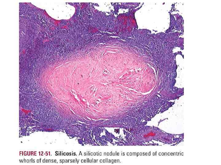
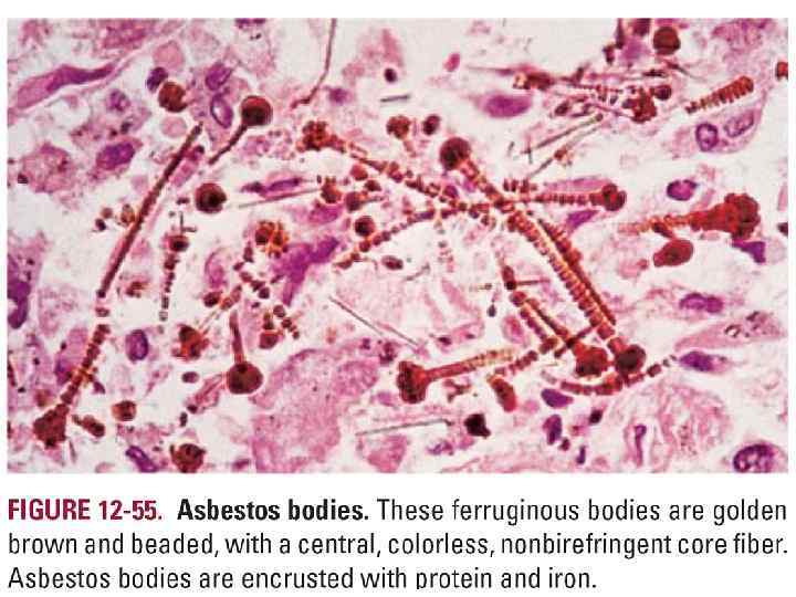
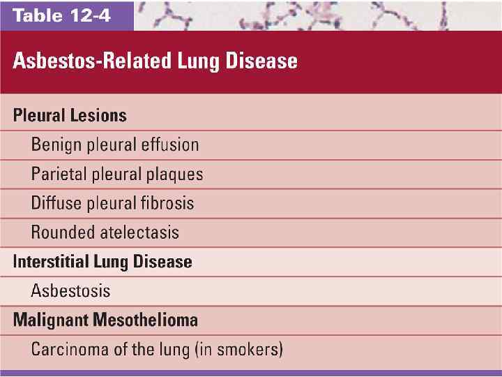
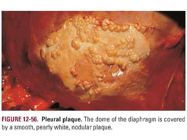
 +Caplan nodules
 А. Complete hydatidiform mole. The enlarged and edematous villi are interconnected by thin cord-like structures. No fetus is present. B, Ultrasound of a complete hydatidiform mole showing the classic “snowstorm” appearance
 ВОЗДЕЙСТВИЕ ОПУХОЛЕЙ НА ОРГАНИЗМ
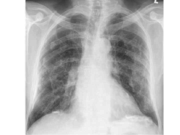
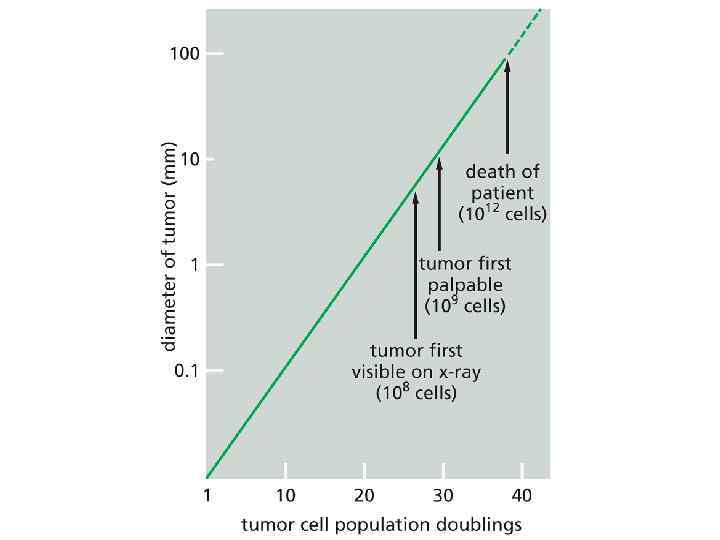
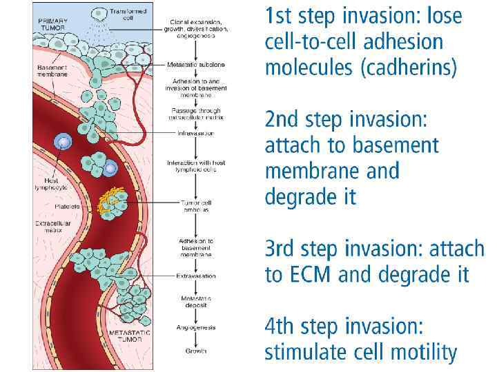
 q Инвазивность q Метастазирование") ВОЗДЕЙСТВИЕ ОПУХОЛИ НА ОРГАНИЗМ q Нарастание клеточной массы (+ ткань) q Инвазивность q Метастазирование q Кахексия q Анемия q Лихорадка q Паранеопластические синдромы q Синдром распада опухоли
ВОЗДЕЙСТВИЕ ОПУХОЛИ НА ОРГАНИЗМ q Нарастание клеточной массы (+ ткань) q Инвазивность q Метастазирование q Кахексия q Анемия q Лихорадка q Паранеопластические синдромы q Синдром распада опухоли

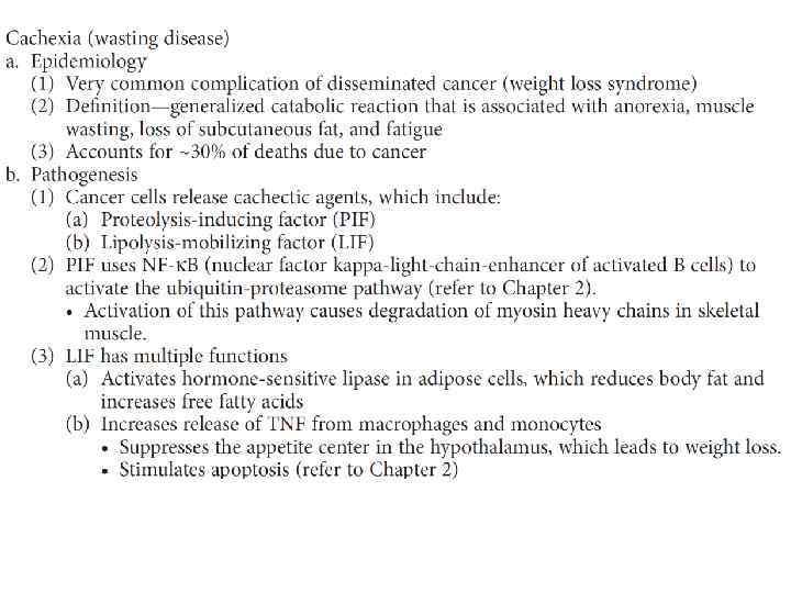
 К а х е к с и я
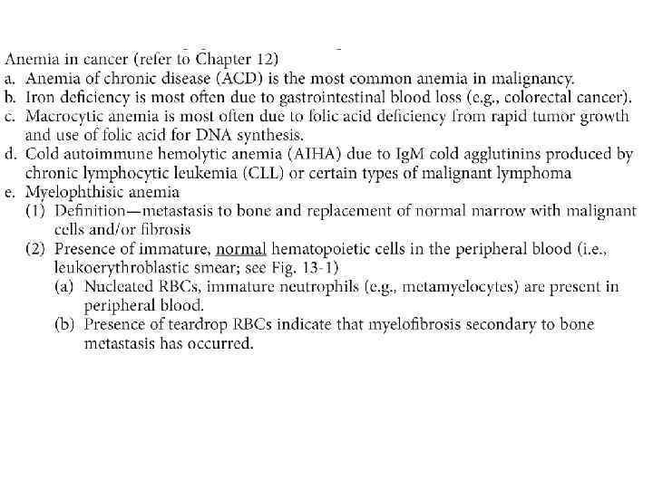
 a. Most often due to infection rather than pyrogens secreted from cancer cells b. Gram-negative sepsis from Escherichia coli or Pseudomonas aeruginosa is a common cause of death in cancer
 Sometimes the metastatic") Where these tumor emboli eventually settle depends on several factors. (a) Sometimes the metastatic site is the first capillary bed it encounters. (b) Sometimes it travels through the Batson paravertebral plexus and ends up in the vertebral column (discussed later). (c) Sometimes the primary cancer releases chemokines that go specifically to sites that have chemokine receptors similar to those in the primary tumor (d) Sometimes target organs release chemoattractants that signal tumor cells to deposit at that site
Where these tumor emboli eventually settle depends on several factors. (a) Sometimes the metastatic site is the first capillary bed it encounters. (b) Sometimes it travels through the Batson paravertebral plexus and ends up in the vertebral column (discussed later). (c) Sometimes the primary cancer releases chemokines that go specifically to sites that have chemokine receptors similar to those in the primary tumor (d) Sometimes target organs release chemoattractants that signal tumor cells to deposit at that site
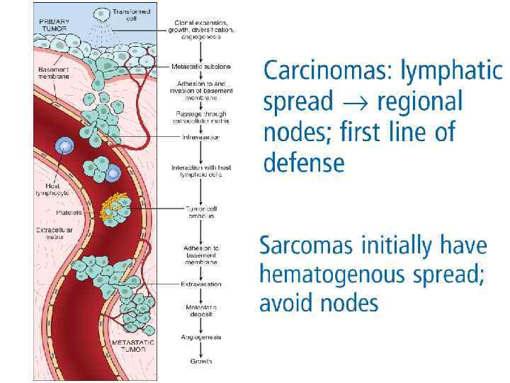
 b.") a. Lymph nodes (e. g. , metastatic breast and lung cancer most common) b. Lungs (e. g. , metastatic breast cancer most common) c. Liver (e. g. , metastatic lung cancer most common) d. Bone (e. g. , metastatic breast cancer most common) e. Brain (e. g. , metastatic lung cancer most common)
a. Lymph nodes (e. g. , metastatic breast and lung cancer most common) b. Lungs (e. g. , metastatic breast cancer most common) c. Liver (e. g. , metastatic lung cancer most common) d. Bone (e. g. , metastatic breast cancer most common) e. Brain (e. g. , metastatic lung cancer most common)
 Вероятность метастазирования различных видов опухолей
 connections with the vena cava and the vertebral bodies Breast cancer is the most common cancer metastatic to bone; second most common is prostate cancer
 Malignant cells in metastatic sites secrete cytokines that specifically activate osteoblasts,") Osteoblastic metastases (1) Malignant cells in metastatic sites secrete cytokines that specifically activate osteoblasts, which initiate reactive bone formation (a) Prostate cancer is the most common cancer producing osteoblastic metastases; second most common is breast cancer. (b) Serum alkaline phosphatase (ALP) is elevated, because osteoblasts use this enzyme in bone formation. (2) Bone formation in metastatic sites produces radiodensities that are identified in radiographs
Osteoblastic metastases (1) Malignant cells in metastatic sites secrete cytokines that specifically activate osteoblasts, which initiate reactive bone formation (a) Prostate cancer is the most common cancer producing osteoblastic metastases; second most common is breast cancer. (b) Serum alkaline phosphatase (ALP) is elevated, because osteoblasts use this enzyme in bone formation. (2) Bone formation in metastatic sites produces radiodensities that are identified in radiographs
 winking owl sign
 патологические переломы") гипер-Ca (остеолитические М, + других механизма) патологические переломы
гипер-Ca (остеолитические М, + других механизма) патологические переломы
 • • • Вирхова Крукенберга Шнитцлера Сестры Джозеф Блумера Айриша
 остеолитический М
 Синдром Мейгса
 Синдром распада опухоли
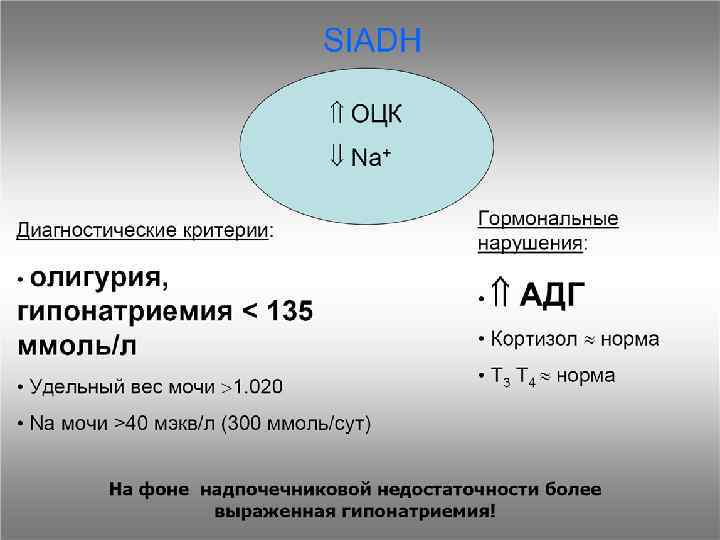
 ? Как объяснить развивающиеся на фоне бронхолегочной карциномы: 1. гипонатриемию 2. гипокалиемический алкалоз 3. симптомы миастении ?
 ТТГ
ТТГ
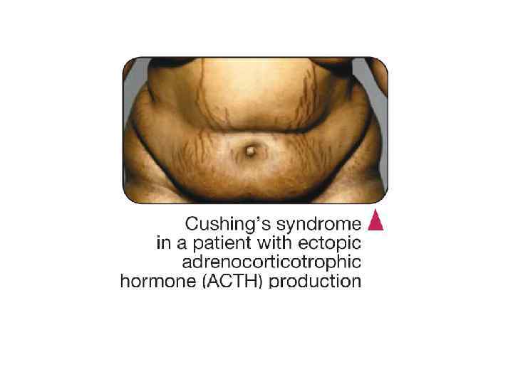
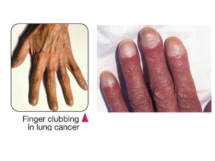
 Синдром Ламберта-Итона Синдром Труссо") • • • Синдром Кушинга Синдром Шварца-Бартера (SIADH/ Пархона) Синдром Ламберта-Итона Синдром Труссо Синдром Штоффера Синдром Лесера-Трела Синдром Мари-Бамбергера Синдром Свита Синдром Базекса …
• • • Синдром Кушинга Синдром Шварца-Бартера (SIADH/ Пархона) Синдром Ламберта-Итона Синдром Труссо Синдром Штоффера Синдром Лесера-Трела Синдром Мари-Бамбергера Синдром Свита Синдром Базекса …
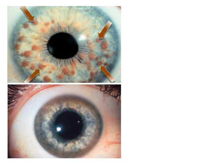
 Acanthosis nigricans. Note the pigmented verrucoid lesion in the axilla. Like the Leser-Trelat sign, these lesions may be associated with an underlying gastric adenocarcinoma or other disorders
 IGF-1 Obesity Type 2 diabetes mellitus Cushing’s disease Polycystic ovaries Thyroid disease Adenocarcinoma of the gastrointestinal tract Acromegaly Medications (e. g. , prednisone, nicotinic acid)
 > 40 - Adenocarcinoma of the gastrointestinal tract ?
 Seborrheic keratosis • Note the numerous raised, pigmented lesions with a verrucoid surface. • These lesions appeared suddenly (Leser-Trelat sign) in this patient indicating a possible underlying gastric adenocarcinoma. • In most cases, they are a common lesion in the elderly population where they frequently occur on the face and axilla. • ВИЧ-индикаторное заб.
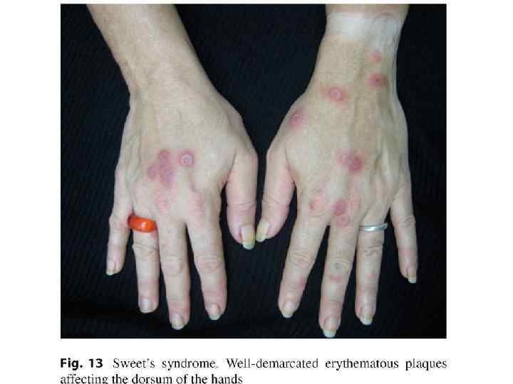
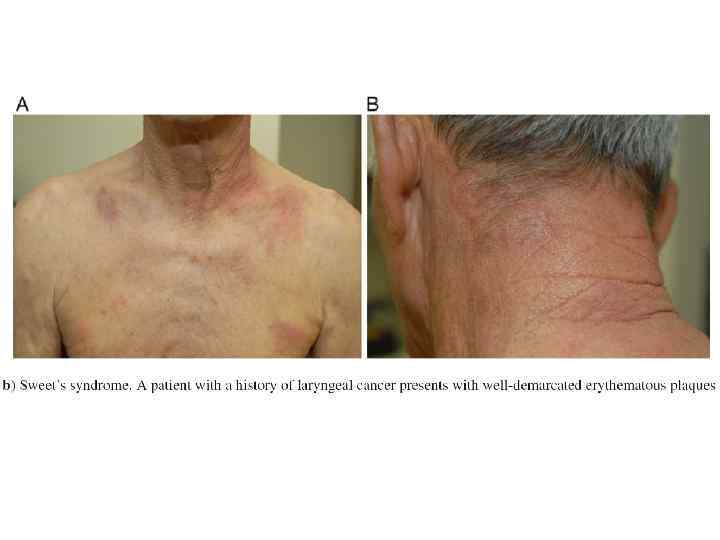
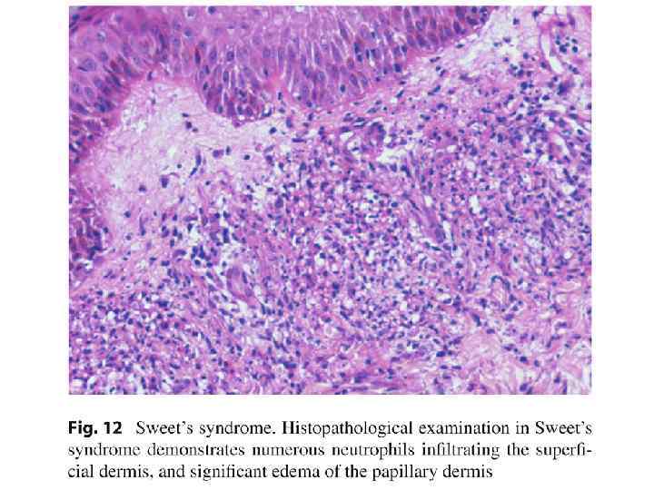
 Sweet’s Syndrome §Fever §Blood neutrophilic leukocytosis §Raised painful plaques on the limbs, face and neck §Histologic findings of dense infiltration of the dermis by mature neutrophils 20% of cases, the syndrome is associated with hematologic illness
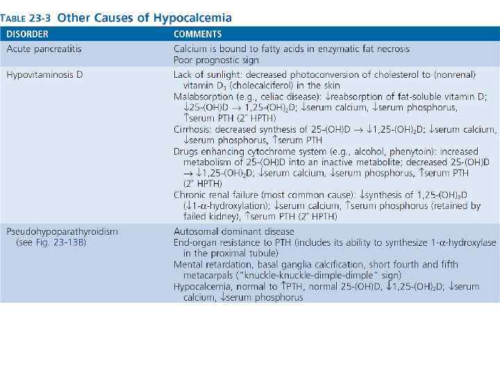
 account for ~80%") Primary HPTH and the hypercalcemia of malignancy (refer to Chapter 9) account for ~80% of all cases of hypercalcemia Primary differentiating feature between these two diagnoses is serum PTH. (1) Serum PTH is increased in primary HPTH. (2) Serum PTH is decreased in hypercalcemia of malignancy, whether it is due to lytic lesions (most common) or PTH-related peptide
Primary HPTH and the hypercalcemia of malignancy (refer to Chapter 9) account for ~80% of all cases of hypercalcemia Primary differentiating feature between these two diagnoses is serum PTH. (1) Serum PTH is increased in primary HPTH. (2) Serum PTH is decreased in hypercalcemia of malignancy, whether it is due to lytic lesions (most common) or PTH-related peptide
 Acrokeratosis Paraneoplastica Bazex
 ОНКОМАРКЕРЫ
 Pathologists use special stains and techniques that help define different types of cancer. a. Cytokeratin stain positive—epithelial tissue origin b. Vimentin stain positive—connective tissue origin c. CD 45 positive—malignant lymphoma
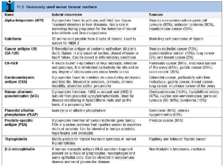
 HORMONES Human chorionic gonadotropin Trophoblastic tumors, nonseminomatous testicular tumors Calcitonin Medullary carcinoma of thyroid Catecholamine and metabolites Pheochromocytoma and related tumors Ectopic hormones See “Paraneoplastic Syndromes” ONCOFETAL ANTIGENS α-Fetoprotein Liver cell cancer, nonseminomatous germ cell tumors of testis Carcinoembryonic antigen Carcinomas of the colon, pancreas, lung, stomach, and heart ISOENZYMES Prostatic acid phosphatase Prostate cancer Neuron-specific enolase Small-cell cancer of lung, neuroblastoma SPECIFIC PROTEINS Immunoglobulins Multiple myeloma and other gammopathies Prostate-specific antigen and prostate-specific membrane antigen Prostate cancer MUCINS AND OTHER GLYCOPROTEINS CA-125 Ovarian cancer CA-19 -9 Colon cancer, pancreatic cancer CA-15 -3 Breast cancer NEW MOLECULAR MARKERS p 53, APC, RAS mutants in stool and serum Colon cancer p 53 and RAS mutants in stool and serum Pancreatic cancer p 53 and RAS mutants in sputum and serum Lung cancer p 53 mutants in urine Bladder cancer