

Recovered File 1.pptx
- Количество слайдов: 58
 Senses and nerves Oleg, Klasha, Matti, Ignatii SPO 10 S/SPO 12 S 2013
 Part number 1 Senses
 The 5 senses: • • • Sight Hearing Taste Smell Touch
 Sight
Sight
 to focus and detect") Sight or vision • is the capability of the eye(s) to focus and detect images of visible light on photoreceptors in the retina of each eye that generates electrical nerve impulses for varying colors, hues, and brightness.
Sight or vision • is the capability of the eye(s) to focus and detect images of visible light on photoreceptors in the retina of each eye that generates electrical nerve impulses for varying colors, hues, and brightness.
 Structure and location: • Eye has a complex structure consisting of a transparent lens that focuses light on the retina. • The retina is covered with two basic types of light -sensitive cells-rods and cones. • These cells are located around the fovea and are responsible for peripheral vision and night vision.
 Two types of photoreceptors: • Cell rods Rods are very sensitive to light, but do not distinguish colors. • Cones distinguish colors, but are less sensitive to dim light.
 Brain and eyes: • The eye is connected to the brain through the optic nerve and the point of this connection is called "blind spot”-insensitive to light. • The brain combines the input of our two eyes into a single three-dimensional image. • The image on the retina is upside-down because of the focusing action of the lens, the brain compensates and provides the right-side-up perception.
 Why we can see in the dark and in bright light? • In the dark, a substance produced by the rod cells increases the sensitivity of the eye so that it is possible to detect very dim light. • In strong light, the iris contracts reducing the size of the aperture that admits light into the eye and a protective obscure substance reduces the exposure of the light-sensitive cells.
 The spectrum of light: • The spectrum of light to which the eye is sensitive varies from the red to the violet. • Lower electromagnetic frequencies in the infrared are sensed as heat, but cannot be seen. • Higher frequencies in the ultraviolet and beyond cannot be seen either, but can be sensed as tingling of the skin or eyes depending on the frequency.
 Eye disease: Blindness - the inability to see. • Blindness may result from damage to the eyeball, especially to the retina, damage to the optic nerve that connects each eye to the brain. • Temporary or permanent blindness can be caused by poisons or medications.
 Blindsight • People who are blind from degradation or damage to the visual cortex, but still have functional eyes, are actually capable of some level of vision and reaction to visual stimuli but not a conscious perception.
 Daltonism • Color blindness is a common abnormality in human vision that makes it impossible to differentiate colors accurately. • One type of color blindness results in the inability to distinguish red from green.
 Hearing -is the sense of sound perception.
 Ear structure and function: • The outer ear protrudes away from the head and is shaped like a cup to direct sounds toward the tympanic membrane. • The tympanic membrane transmits vibrations to the inner ear through a series of small bones in the middle ear called the malleus, incus and stapes.
 • The inner ear, or cochlea, is a spiral-shaped chamber covered internally by nerve fibers that react to the vibrations. • The cochlea transmit impulses to the brain via the auditory nerve. • The brain combines the input of our two ears to determine the direction and distance of sounds.
 Brain and ears: • The brain combines the input of our two ears to determine the direction and distance of sounds. • The inner ear has a vestibular system formed by three semicircular canals that are approximately at right angles to each other and which are responsible for the sense of balance and spatial orientation.
 • The inner ear has chambers filled with a viscous fluid and small particles (otoliths) containing calcium carbonate. • The movement of these particles over small hair cells in the inner ear sends signals to the brain that are interpreted as motion and acceleration.
 Why can we hear? • The human ear can perceive frequencies from 16 cycles per second, which is a very deep bass, to 28, 000 cycles per second, which is a very high pitch. • The human ear can detect pitch changes as small as 3 hundredths of one percent of the original frequency in some frequency ranges. • Some people have "perfect pitch", which is the ability to map a tone precisely on the musical scale without reference to an external standard.
 Ear disease: Deafness or hearing impairment-Inability to hear. • Sound can also be detected as vibrations conducted through the body by tactition. • Lower frequencies than can be heard are detected this way.
 Taste
Taste
 Taste or gustation: • is one of the traditional five senses. It refers to the capability to detect the taste of substances such as food, certain minerals, and poisons.
 Structure and location: • The receptors for taste, called taste buds, are situated chiefly in the tongue, but they are also located in the roof of the mouth and near the pharynx. • They are able to detect four basic tastes: salty, sweet, bitter, and sour. The tongue also can detect a sensation called "umami" from taste receptors sensitive to amino acids.
 Taste buds: • The taste buds close to the tip of the tongue are sensitive to sweet tastes, whereas those in the back of the tongue are sensitive to bitter tastes. • The taste buds on top and on the side of the tongue are sensitive to salty and sour tastes. • At the base of each taste bud there is a nerve that sends the sensations to the brain.
 • The sense of taste functions in coordination with the sense of smell. • The number of taste buds varies substantially from individual to individual, but greater numbers increase sensitivity. • Women, in general, have a greater number of taste buds than men. • As in the case of color blindness, some people are insensitive to some tastes.
 Smell
Smell
 Structure and function: • The nose is the organ responsible for the sense of smell. • The cavity of the nose is lined with mucous membranes that have smell receptors connected to the olfactory nerve. • The smells themselves consist of vapors of various substances.
 • The smell receptors interact with the molecules of these vapors and transmit the sensations to the brain. • The nose also has a structure called the vomeronasal organ. • Vomeronasal organ has not been determined, but which is suspected of being sensitive to pheromones that influence the reproductive cycle.
 Smell receptors: • The smell receptors are sensitive to seven types of sensations that can be characterized as camphor, musk, flower, mint, ether, acrid, or putrid. • The sense of smell is sometimes temporarily lost when a person has a cold. • Dogs have a sense of smell that is many times more sensitive than man's.
 Touch
Touch
 Touch or taction: • is a perception resulting from activation of neural receptors, generally in the skin including hair follicles, but also in the tongue, throat, and mucosa.
 Structure and function: • The sense of touch is distributed throughout the body. • Nerve endings in the skin and other parts of the body transmit sensations to the brain. • The touch sense of itching caused by insect bites or allergies involves special itch-specific neurons in the skin and spinal cord.
 • Some parts of the body have a larger number of nerve endings and, therefore, are more sensitive. • Four kinds of touch sensations can be identified: cold, heat, contact, and pain. • Hairs on the skin magnify the sensitivity and act as an early warning system for the body.
 Diseases: Tactile anesthesia • The loss or impairment of the ability to feel anything touched. Paresthesia • is a sensation of tingling, pricking, or numbness of the skin that may result from nerve damage and may be permanent or temporary.
 Part number 2 Nerves
 3 groups based on the direction that signals are conducted: • Afferent nerves • Efferent nerves • Mixed nerves 2 groups based on where they connect to the central nervous system: • Spinal nerves • Cranial nerves
 • In the nervous system there is a "closed loop" system of sensation, decision, and reactions. This process is carried out through the activity of afferent neurons, interneurons, and efferent neurons. • The Affective and Effective Nerves are the part of the central nerves system (CNS) • The Spinal Nerves are part of the peripheral nervous system (PNS).
") Group number 1 Afferent nerves (INPUT)
Group number 1 Afferent nerves (INPUT)
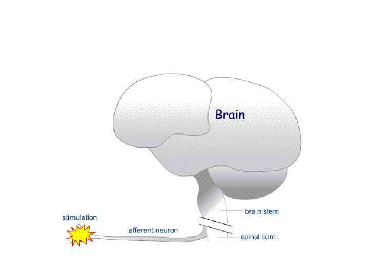
 • Conduct signals from sensory neurons to the central nervous system, for example from the mechanoreceptors in skin. • Communicate with specialized interneurons. • A touch or painful stimulus, for example, creates a sensation in the brain only after information about the stimulus travels there via afferent nerve pathways.
 Structure: • Afferent neurons are pseudounipolar neurons, that have a single long dendrite and a short axon, and a smooth and rounded cell body. • The dendrite is structurally and functionally similar to an axon, and is myelinated; it is these axon-like dendrites that make up the afferent nerves. • Just outside the spinal cord, thousands of afferent neuronal cell bodies are aggregated in a swelling in the dorsal root known as the dorsal root ganglion.
 Transportation: • Afferent neurons Somas are located in the peripheral nervous system and the axons of these cells travel from ganglion to ganglion and lead back to the spinal cord. • Have a single axon leaving the cell body and is sent towards the sensory organ. • Somatosensory receptors include senses such as pain, touch, temperature, and itch. • All of these sensations travel along the same general pathway towards the brain, from the dorsal root ganglion they travel to the spinal cord.
") Efferent nerves (OUTPUT)
Efferent nerves (OUTPUT)
 • Conduct signals from the central nervous system along motor neurons to their target muscles and glands. • The motor nerves are efferent nerves involved in muscular control.
 Structure: • The cell body of the efferent neuron is connected to a single, long axon and several shorter dendrites projecting out of the cell body itself. • This axon then forms a neuromuscular junction with the effectors. • The cell body of the motor neuron is satelliteshaped.
 • The motor neuron is present in the grey matter of the spinal cord and medulla oblongata, and forms an electrochemical pathway to the effector organ or muscle. • Besides motor nerves, there are efferent sensory nerves that often serve to adjust the sensitivity of the signal relayed by the afferent sensory nerve.
 Transportation:
 Mixed nerve • contain both afferent and efferent axons, and thus conduct both incoming sensory information and outgoing muscle commands in the same bundle.
 Group number 2 Spinal nerves
 • Innervate much of the body, and connect through the spinal column to the spinal cord. • They are given letter-number designations according to the vertebra through which they connect to the spinal column. • http: //www. britannica. com/EBchecked/topi c/560071/spinal-nerve
 Structure: Humans have 31 left-right pairs of spinal nerves, each roughly corresponding to a segment of the vertebral column: • 8 cervical spinal nerve pairs (C 1 -C 8), • 12 thoracic pairs (T 1 -T 12), • 5 lumbar pairs (L 1 -L 5), • 5 sacral pairs (S 1 -S 5), • 1 coccygeal pair.
 • Each spinal nerve is formed by the combination of nerve fibers from the dorsal and ventral roots of the spinal cord. • The dorsal roots carry afferent sensory axons, while the ventral roots carry efferent motor axons. • The spinal nerve emerges from the spinal column through an opening (intervertebral foramen) between adjacent vertebrae. • This is true for all spinal nerves except for the first spinal nerve pair, which emerges between the occipital bone and the atlas (the first vertebra).
 Outside the vertebral column, the nerve divides into branches: • The dorsal branch • The ventral branch • The meningeal branches Some ventral branches merge with adjacent ventral rami to form a nerve plexus, a network of interconnecting nerves.
 Cranial nerves
 • Innervate parts of the head, and connect directly to the brain (especially to the brainstem). They are typically assigned Roman numerals from 1 to 12, although cranial nerve zero is sometimes included. • In addition, cranial nerves have descriptive names.
 Structure: • In humans, there are traditionally twelve pairs of cranial nerves. Only the first and the second pair emerge from the cerebrum; the remaining ten pairs emerge from the brainstem.
 List of cranial nerves: • http: //www. meddean. luc. edu/lumen/Med. Ed/ grossanatomy/h_n/cn/cn 1/table 1. htm • http: //www. youtube. com/watch? v=tl. Mx 8 Be. P T 2 w
 Thank you for attention