

Zhumanov_E_242_GMF_Appendix.pptx
- Количество слайдов: 15
 SEMEY STATE MEDICAL UNIVERSITY THE CHAIR OF ANATOMY AND HISTOLOGY SIW Variety of a position of the Appendix Made by Zhumanov Eldar 242 group, GMF Checked by Milyushina Y. A. Semey 2014
 Plan 1. Introduction 2. Main part a. About the Appendix b. Positions of vermiform appendix by direction c. Positions of vermiform appendix by Wakeley 3. Conclusion 4. References
 large intestine • The large intestine is the last part of the digestive system in vertebrates. Water is absorbed here and the remaining waste material is stored as feces before being removed by defecation • The small intestine (or small bowel) is the part of the gastrointestinal tract following the stomach and followed by the large intestine, and is where much of the digestion and absorption of food takes place. The small intestine is composed of a duodenum, jejunum, and ileum. It receives bile juice and pancreatic juice through the hepatopancreatic duct, controlled by the sphincter of Oddi
 Sections The cecum and the vermiform appendix The ascending colon The right colic flexure (hepatic) The transverse colon The transverse mesocolon The left colic flexure (splenic) The descending colon The sigmoid colon – the v-shaped region of the large intestine . The duodenum The jejunum The ileum
 small intestine
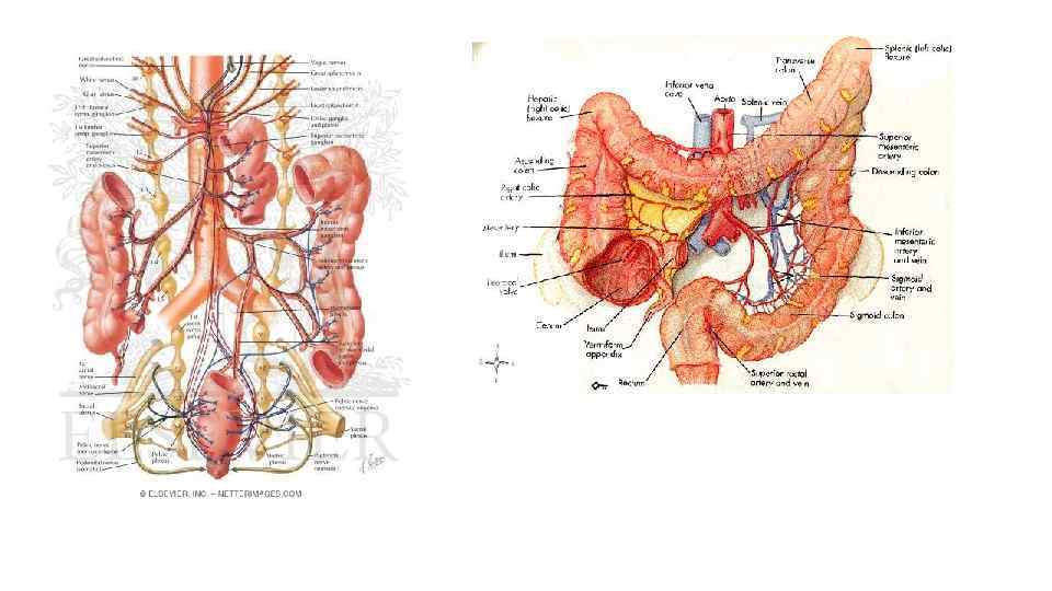
 Blood supply Arterial supply to the colon comes from branches of the superior mesenteric artery (SMA) and inferior mesenteric artery (IMA). Flow between these two systems communicates via a "marginal artery" that runs parallel to the colon for its entire length. Historically, it has been believed that the arc of Riolan, or the meandering mesenteric artery (of Moskowitz), is a variable vessel connecting the proximal SMA to the proximal IMA that can be extremely important if either vessel is occluded The duodenum is supplied proximally by a branch of the common hepatic artery called the gastroduodenal artery that stems from the celiac trunk and gives the superior anterior and posterior pancreaticoduodenal arteries. It also receives blood distally from the anterior and posterior inferior pancreaticoduodenal arteries which are branches of the superior mesenteric artery. The terminal branches of the duodenal arteries form an important anastomoses between the celiac trunk and the superior mesenteric artery.
 . However, recent studies conducted with improved imaging technology have questioned the actual existence of this vessel, with some experts calling for the abolition of the terms from future medical literature. citation needed Venous drainage usually mirrors colonic arterial supply, with the inferior mesenteric vein draining into the splenic vein, and the superior mesenteric vein joining the splenic vein to form the hepatic portal vein that then enters the liver. The main arterial supply of the jejunum and the ileum is from a single artery known as the superior mesenteric and between fifteen to eighteen of its branches which form anastomoses loops known as arterial arcades with terminal vasa recta or straight branches. It
 Innervation Similarly to the small intestine, there are pacemaker cells that generate an action potential. It was shown in acute and chronic degeneration Cells are able to function as a syncytium due to studies that the nerve elements of the cat small gap junctions, allowing the action potential to intestine belong to two according to whether they are extrinsic or intrinsic in origin. Part of spread. Contractions are generated in the extrinsic nerve fibres originate from the forward (peristaltic) and backward vagus nerve and can be found on both intestinal (antiperistaltic) directions. Antiperistaltic plexuses, the other fibres are processes of the contractions move ingesta into the caecum in some species. The formation of action potentials coeliac ganglion and occur in the first place in is under a much stronger neural influence than the vascular tunica adventitia. in the stomach and small intestine. The large intestine receives sympathetic and parasympathetic innervation.
 The sympathetic have coeliac, cranial mesenteric and caudal mesenteric ganglia. As the sympathetic fibres leave the ganglia, they surround their respective artery. Parasympathetic innervation increases the frequency of action potentials and thus stimulates peristalsis. Sympathetic innervation has the opposite effect. Neurones interact with the myenteric plexus to affect contractility, and with the submucosal plexus to affect secretions. Motility of the large intestine increases during meals, possibly as a result of gastrin and cholecystokinin secretion. The intrinsic nerve cells may be classified into three groups on grounds their ultrastructure the number of synapses and processes on their surface. The synaptic connections between the different nerve cells of the internal plexuses were confirmed on chronic isolated of intestinal loops. The local reflexes provided by these connections are held responsible for peristalsis, the functional modifications of the mucosal pattern and the movements of the villi.
 Function The large intestine takes about 16 hours to finish the digestion of the food. It removes water and any remaining absorbable nutrients from the food before sending the indigestible matter to the rectum. The colon absorbs vitamins that are created by the colonic bacteria - such as vitamin K (especially important as the daily ingestion of vitamin K is not normally enough to maintain adequate blood coagulation), vitamin B 12, thiamine and riboflavin. It also compacts feces, and stores fecal matter in the rectum until it can be discharged via the anus in defecation. The large intestine also secretes K+ and Cl-. Chloride secretion increases in cystic fibrosis. Recycling of various nutrients takes place in colon. Examples include fermentation of carbohydrates, short chain fatty acids, and urea cycling Digestion The small intestine is where most chemical digestion takes place Most of the digestive enzymes that act in the small intestine are secreted by the pancreas and enter the small intestine via the pancreatic duct. Enzymes enter the small intestine in response to the hormone cholecystokinin, which is produced in the small intestine in response to the presence of nutrients. The hormone secretin also causes bicarbonate to be released into the small intestine from the pancreas in order to neutralize the potentially harmful acid coming from the stomach.
 The large intestine differs in physical form from the small intestine in being much wider and in showing the longitudinal layer of the muscularis have been reduced to 3 strap-like structures known as the taeniae coli. The wall of the large intestine is lined with simple columnar epithelium. Instead of having the evaginations of the small intestine (villi), the large intestine has invaginations (the intestinal glands). While both the small intestine and the large intestine have goblet cells, they are abundant in the large intestine Absorption Digested food is now able to pass into the blood vessels in the wall of the intestine through either diffusion or active transport. The small intestine is the site where most of the nutrients from ingested food are absorbed. The inner wall, or mucosa, of the small intestine is lined with simple columnar epithelial tissue. Structurally, the mucosa is covered in wrinkles or folds called plicae circulares, which are considered permanent features in the wall of the organ. They are distinct from rugae which are considered non-permanent or temporary allowing for distention and contraction. From the plicae circulares project microscopic finger-like pieces of tissue called villi (Latin for "shaggy hair"). The individual epithelial cells also have finger-like projections known as microvilli. The functions of the plicae circulares, the villi, and the microvilli are to increase the amount of surface area available for the absorption of nutrients, and to limit the loss of said nutrients to intestinal fauna.
 The intestinal microflora is a complex ecosystem containing over 400 bacterial species. Anaerobes outnumber facultative The large intestine houses over 700 species of bacteria that anaerobes. The flora is sparse in the stomach and upper perform a variety of functions. intestine, but luxuriant in the lower bowel. Bacteria occur both in the lumen and attached to the mucosa, but do not normally The large intestine absorbs some of the products formed by the penetrate the bowel wall. bacteria inhabiting this region. Undigested polysaccharides (fiber) are metabolized to short-chain fatty acids by bacteria in the large intestine and absorbed by passive diffusion. The intestinal microflora may prevent infection by interfering bicarbonate that the large intestine secretes helps to neutralize with pathogens. The flora includes low populations of the increased acidity resulting from the formation of these fatty potentially pathogenic organisms such as Clostridium difficile. acids Antibiotics that upset the balance of the normal flora can favor both infection by exogenous pathogens and overgrowth by These bacteria also produce large amounts of vitamins, endogenous pathogens. If the bowel wall is breached, enteric especially vitamin K and biotin (a B vitamin), for absorption bacteria can escape into the peritoneum and cause peritonitis into the blood. Although this source of vitamins, in general, and abscesses. provides only a small part of the daily requirement, it makes a significant contribution when dietary vitamin intake is low. An individual who depends on absorption of vitamins formed by bacteria in the large intestine may become vitamin-deficient if treated with antibiotics that inhibit other species of bacteria as well as the disease-causing bacteria

