Section 4 Regulation of the Respiration I.


section_4_regulation_of_the_respiration.ppt
- Размер: 1.1 Mегабайта
- Количество слайдов: 33
Описание презентации Section 4 Regulation of the Respiration I. по слайдам
 Section 4 Regulation of the Respiration
 I. Respiratory Center and Formation of the Respiratory Rhythm 1 Respiratory Center


 Respiratory Centers
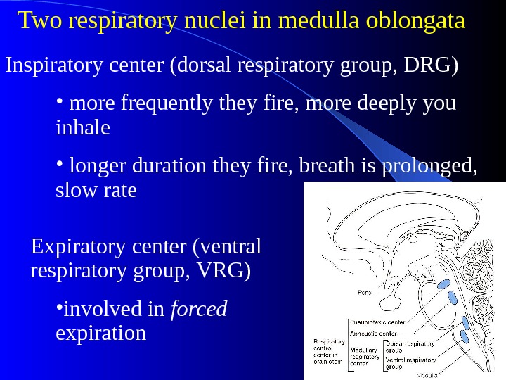
 Two respiratory nuclei in medulla oblongata Expiratory center (ventral respiratory group, VRG) • involved in forced expiration. Inspiratory center (dorsal respiratory group, DRG) • more frequently they fire, more deeply you inhale • longer duration they fire, breath is prolonged, slow rate
 Respiratory Centers in Pons Apneustic center (lower pons) • Sends continual inhibitory impulses to inspiratory center of the medulla oblongata, • As impulse frequency rises, breathe faster and shallower • Stimulation causes apneusis • Integrates inspiratory cutoff information. Pneumotaxic center (upper pons)
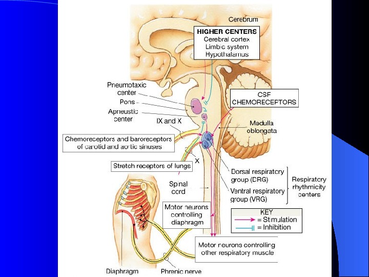
 Respiratory Structures in Brainstem
 2. Rhythmic Ventilation (Inspiratory Off Switch) • Starting inspiration – Medullary respiratory center neurons are continuously active (spontaneous) – Center receives stimulation from receptors and brain concerned with voluntary respiratory movements and emotion – Combined input from all sources causes action potentials to stimulate respiratory muscles
 • Increasing inspiration – More and more neurons are activated • Stopping inspiration – Neurons receive input from pontine group and stretch receptors in lungs. – Inhibitory neurons activated and relaxation of respiratory muscles results in expiration. – Inspiratory off swithch.
 3. Higher Respiratory Centers Modulate the activity of the more primitive controlling centers in the medulla and pons. Allow the rate and depth of respiration to be controlled voluntarily. During speaking, laughing, crying, eating, defecating, coughing, and sneezing. …. Adaptations to changes in environmental temperature —Panting
 II Pulmonary Reflex 1. Chemoreceptor Reflex
 Two Sets of Chemoreceptors Exist • Central Chemoreceptors – Responsive to increased arterial PCO 2 – Act by way of CSF [H + ] . • Peripheral Chemoreceptors – Responsive to decreased arterial PO 2 – Responsive to increased arterial PCO 2 – Responsive to increased H + ion concentration.
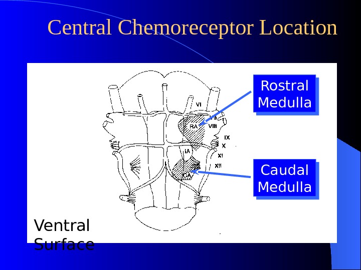
 Central Chemoreceptor Location Rostral Medulla Caudal Medulla Ventral Surface
 Central Chemoreceptor Stimulation Arterial CSF CO 2 COHO HCOH 22 3 C e n t r a l C h e m o r e c e p t o r H +slow ? ? ?
 Peripheral Chemoreceptor Pathways
 Peripheral Chemoreceptors • Carotid bodies – Sensitive to: P a O 2 , P a CO 2 , and p. H – Afferents in glossopharyngeal nerve. • Aortic bodies – Sensitive to: P a O 2 , P a CO 2 , but not p. H – Afferents in vagus

 Carotid Body Function • High flow per unit weight: (2 L/min/100 g) • High carotid body VO 2 consumption: (8 ml O 2 /min/100 g) • Tiny a-v O 2 difference: Receptor cells see arterial PO 2. • Responsiveness begins at P a O 2 (not the oxygen content) below about 60 mm. Hg.
 Carotid Body Response Critical PO 2 Hypercapnea Acidosis Hypocapnea Alkalosis
 Carbon Dioxide, Oxygen and p. H Influence Ventilation (through peripheral receptor) • Peripheral chemoreceptorssensitive to P O 2 , P CO 2 and p. H • Receptors are activated by increase in P CO 2 or decrease in P O 2 and p. H • Send APs through sensory neurons to the brain • Sensory info is integrated within the medulla • Respiratory centers respond by sending efferent signals through somatic motor neurons to the skeletal muscles • Ventilation is increased (decreased)
 Effects of Hydrogen Ions (through central chemoreceptors) • p. H of CSF (most powerful respiratory stimulus) • Respiratory acidosis (p. H 43 mm. Hg – CO 2 easily crosses blood-brain barrier, in CSF the CO 2 reacts with water and releases H + , central chemoreceptors strongly stimulate inspiratory center – corrected by hyperventilation, pushes reaction to the left by “blowing off ” CO 2 (expired) + H 2 O H 2 CO 3 HCO 3 — + H +
 Carbon Dioxide • Indirect effects – through p. H as seen previously • Direct effects CO 2 may directly stimulate peripheral chemoreceptors and trigger ventilation more quickly than central chemoreceptors • If the PCO 2 is too high, the respiratory center will be inhibited.
 Oxygen • Direct inhibitory effect of hypoxemia on the respiratory center • Chronic hypoxemia, PO 2 < 60 mm. Hg, can significantly stimulate ventilation – emphysema, pneumonia – high altitudes after several days
 Overall Response to. Pco 2, Po 2 and p. H

 2. Neuroreceptor reflex
 Hering-Breuer Reflex or Pulmonary Stretch Reflex • Including pulmonary inflation reflex and pulmonary deflation reflex • Receptor: Slowly adapting stretch receptors (SARs) in bronchial airways. • Afferent: vagus nerve • Pulmonary inflation reflex: – Terminate inspiration. – By speeding inspiratory termination they increase respiratory frequency. – Sustained stimulation of SARs: causes activation of expiratory neurons

 Significance of Hering-Breuer • Normal adults. Receptors are not activated at end normal tidal volumes. – Become Important during exercise when tidal volume is increased. – Become Important in Chronic obstructive lung diseases when lungs are more distended. • Infants. Probably help terminate normal inspiration.
 Brainstem Transection Normal Pattern Gasping Patterns. Apneustic Breathing Respiratory Arrest. Increase d Inspirato ry Depth
 Factors Influencing Respiration


