
SDU. LIZHENHUARegional anatomy of neck 山山山山山山山


- Размер: 6.3 Mегабайта
- Количество слайдов: 64
Описание презентации SDU. LIZHENHUARegional anatomy of neck 山山山山山山山 по слайдам
SDU. LIZHENHUARegional anatomy of neck 山山山山山山山 山山山
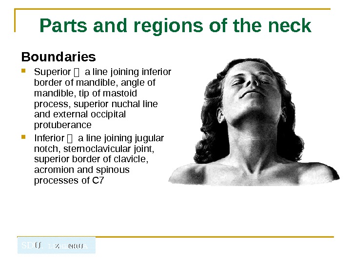
SDU. LIZHENHUAParts and regions of the neck Boundaries Superior - a line joining inferior border of mandible, angle of mandible, tip of mastoid process, superior nuchal line and external occipital protuberance Inferior - a line joining jugular notch, sternoclavicular joint, superior border of clavicle, acromion and spinous processes of
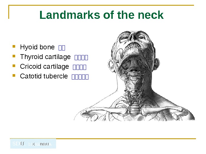
SDU. LIZHENHUA Landmarks of the neck Hyoid bone -- Thyroid cartilage ---- Cricoid cartilage ---- Catotid tubercle -----
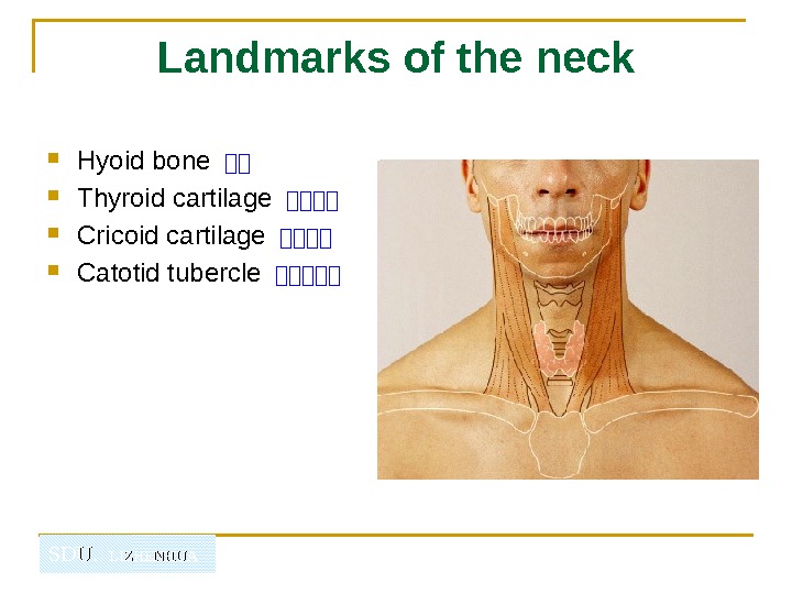
SDU. LIZHENHUA Landmarks of the neck Hyoid bone -- Thyroid cartilage ---- Cricoid cartilage ---- Catotid tubercle -----
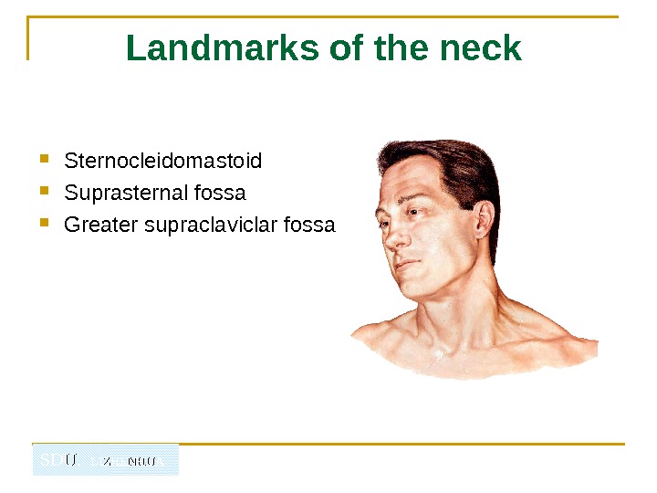
SDU. LIZHENHUA Landmarks of the neck Sternocleidomastoid Suprasternal fossa Greater supraclaviclar fossa
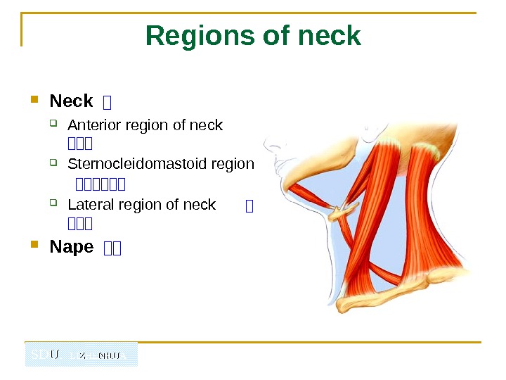
SDU. LIZHENHUA Regions of neck Neck 山 Anterior region of neck 山山山 Sternocleidomastoid region 山山山山山山 Lateral region of neck 山 山山山 Nape 山山
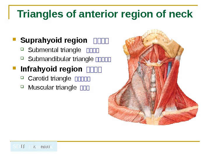
SDU. LIZHENHUATriangles of anterior region of neck Suprahyoid region 山山山山 Submental triangle 山山山山 Submandibular triangle 山山山山山 Infrahyoid region 山山山山 Carotid triangle 山山山山山 Muscular triangle 山山山
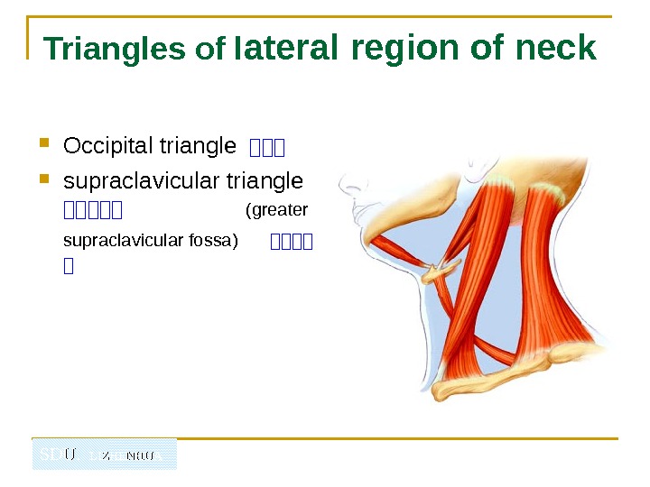
SDU. LIZHENHUATriangles of l ateral region of neck Occipital triangle 山山山 supraclavicular triangle 山山山山山 (greater supraclavicular fossa) 山山山山 山
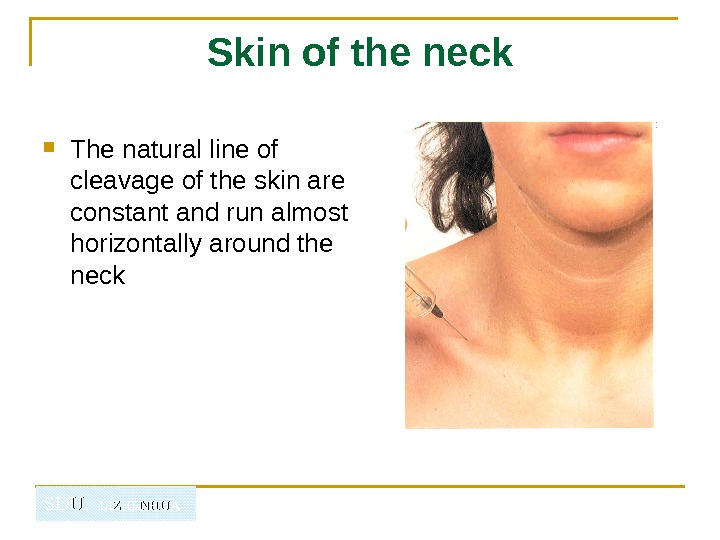
SDU. LIZHENHUA Skin of the neck The natural line of cleavage of the skin are constant and run almost horizontally around the neck
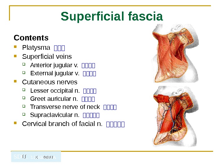
SDU. LIZHENHUA Superficial fascia Contents Platysma --- Superficial veins Anterior jugular v. ---- External jugular v. ---- Cutaneous nerves Lesser occipital n. ---- Greet auricular n. ---- Transverse nerve of neck ---- Supraclavicular n. ----- Cervical branch of facial n. -----
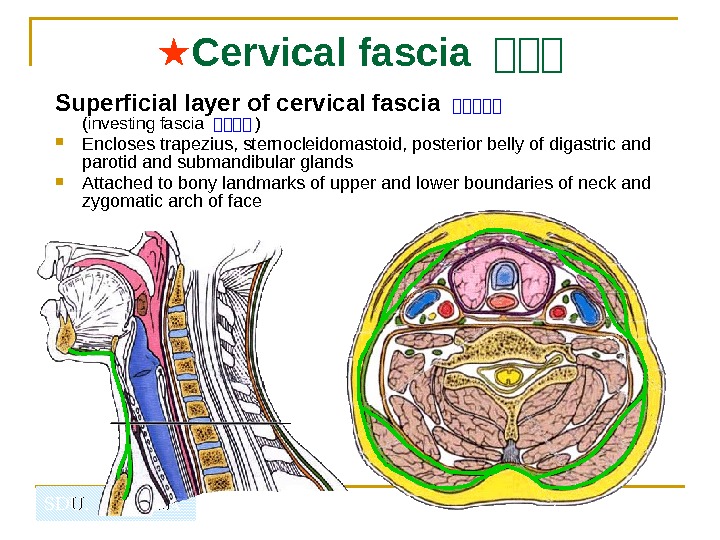
SDU. LIZHENHUA ★ Cervical fascia 山山山 Superficial layer of cervical fascia 山山山山山 (investing fascia 山山山山 ) Encloses trapezius, sternocleidomastoid, posterior belly of digastric and parotid and submandibular glands Attached to bony landmarks of upper and lower boundaries of neck and zygomatic arch of face
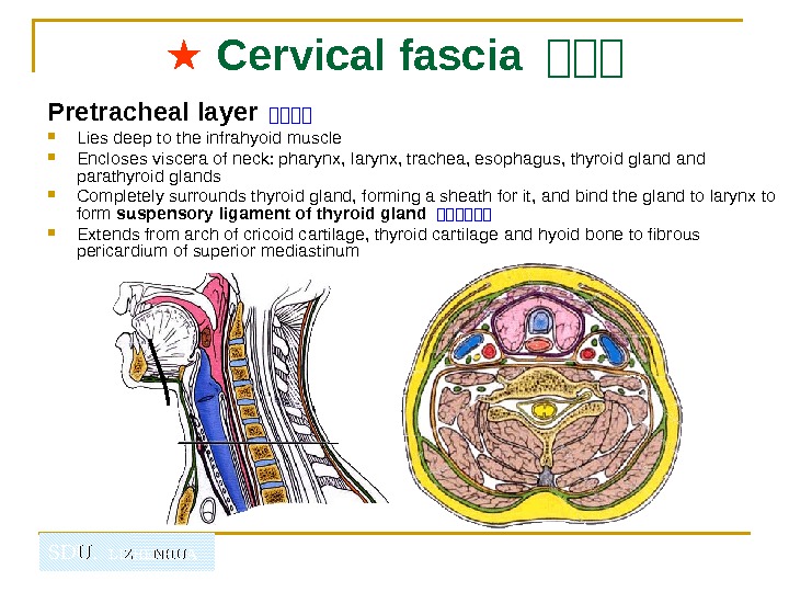
SDU. LIZHENHUA ★ Cervical fascia 山山山 Pretracheal layer 山山山山 Lies deep to the infrahyoid muscle Encloses viscera of neck: pharynx, larynx, trachea, esophagus, thyroid gland parathyroid glands Completely surrounds thyroid gland, forming a sheath for it, and bind the gland to larynx to form suspensory ligament of thyroid gland 山山山山山山 Extends from arch of cricoid cartilage, thyroid cartilage and hyoid bone to fibrous pericardium of superior mediastinum
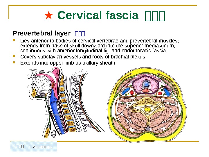
SDU. LIZHENHUA ★ Cervical fascia 山山山 Prevertebral layer 山山山 Lies anterior to bodies of cervical vertebrae and prevertebral muscles; extends from base of skull downward into the superior mediastinum, continuous with anterior longitudinal lig. and endothoracic fascia Covers subclavian vessels and roots of brachial plexus Extends into upper limb as axillary sheath
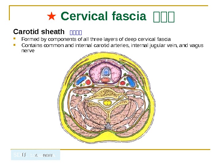
SDU. LIZHENHUA ★ Cervical fascia 山山山 Carotid sheath 山山山山 Formed by components of all three layers of deep cervical fascia Contains common and internal carotid arteries, internal jugular vein, and vagus nerve
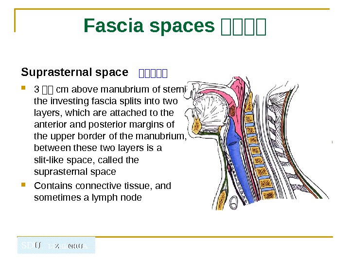
SDU. LIZHENHUA Fascia spaces 山山山山 Suprasternal space 山山山山山 3 -- cm above manubrium of sterni the investing fascia splits into two layers, which are attached to the anterior and posterior margins of the upper border of the manubrium, between these two layers is a slit-like space, called the suprasternal space Contains connective tissue, and sometimes a lymph node
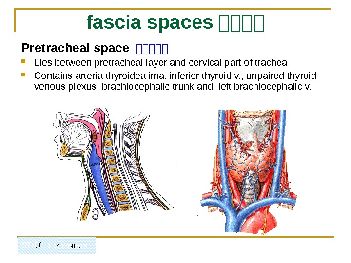
SDU. LIZHENHUA fascia spaces 山山山山 Pretracheal space 山山山山山 Lies between pretracheal layer and cervical part of trachea Contains arteria thyroidea ima, inferior thyroid v. , unpaired thyroid venous plexus, brachiocephalic trunk and left brachiocephalic v.
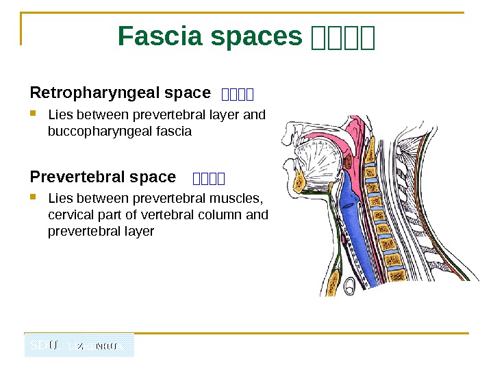
SDU. LIZHENHUA Fascia spaces 山山山山 Retropharyngeal space 山山山山 Lies between prevertebral layer and buccopharyngeal fascia Prevertebral space 山山山山 Lies between prevertebral muscles, cervical part of vertebral column and prevertebral layer
SDU. LIZHENHUAAnterior region of neck 山山山
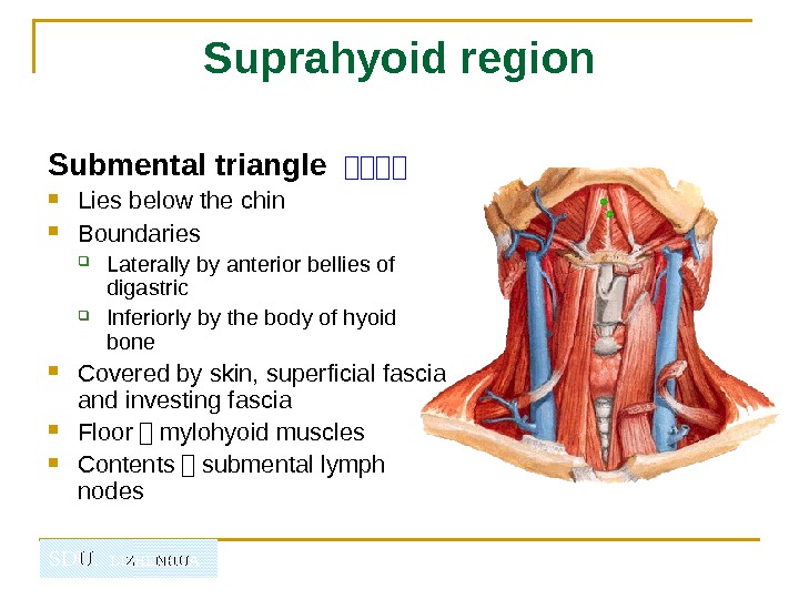
SDU. LIZHENHUA Suprahyoid region Submental triangle 山山山山 Lies below the chin Boundaries Laterally by anterior bellies of digastric Inferiorly by the body of hyoid bone Covered by skin, superficial fascia and investing fascia Floor - mylohyoid muscles Contents - submental lymph nodes
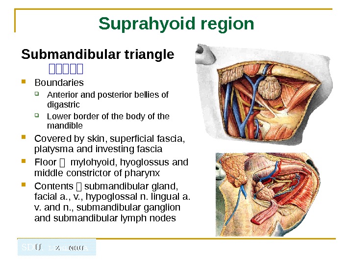
SDU. LIZHENHUA Suprahyoid region Submandibular triangle 山山山山山 Boundaries Anterior and posterior bellies of digastric Lower border of the body of the mandible Covered by skin, superficial fascia, platysma and investing fascia Floor - mylohyoid, hyoglossus and middle constrictor of pharynx Contents - submandibular gland, facial a. , v. , hypoglossal n. lingual a. v. and n. , submandibular ganglion and submandibular lymph nodes
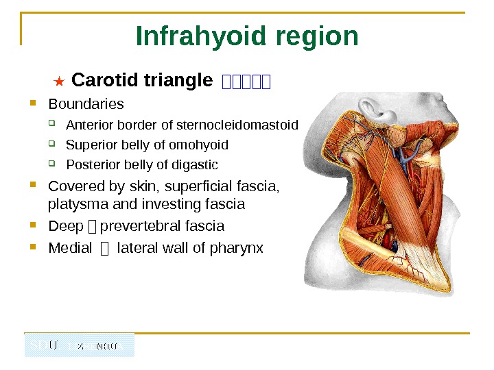
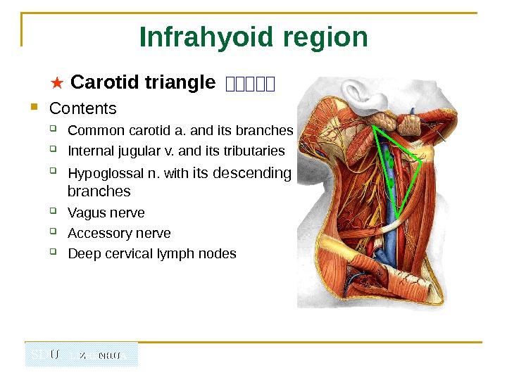
SDU. LIZHENHUA Infrahyoid region ★ Carotid triangle 山山山山山 Boundaries Anterior border of sternocleidomastoid Superior belly of omohyoid Posterior belly of digastic Covered by skin, superficial fascia, platysma and investing fascia Deep - prevertebral fascia Medial - lateral wall of pharynx
SDU. LIZHENHUA Infrahyoid region ★ Carotid triangle 山山山山山 Contents Common carotid a. and its branches Internal jugular v. and its tributaries Hypoglossal n. with its descending branches Vagus nerve Accessory nerve Deep cervical lymph nodes
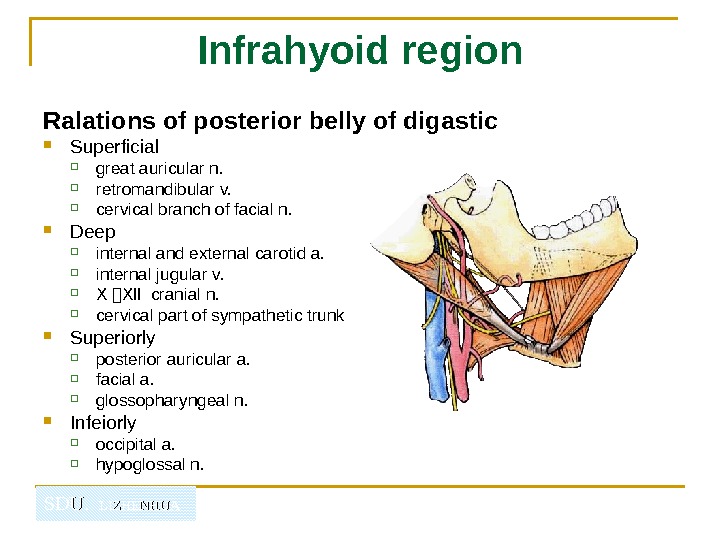
SDU. LIZHENHUA Infrahyoid region Ralations of posterior belly of digastic Superficial great auricular n. retromandibular v. cervical branch of facial n. Deep internal and external carotid a. internal jugular v. Ⅹ -Ⅻ cranial n. cervical part of sympathetic trunk Superiorly posterior auricular a. facial a. glossopharyngeal n. Infeiorly occipital a. hypoglossal n.
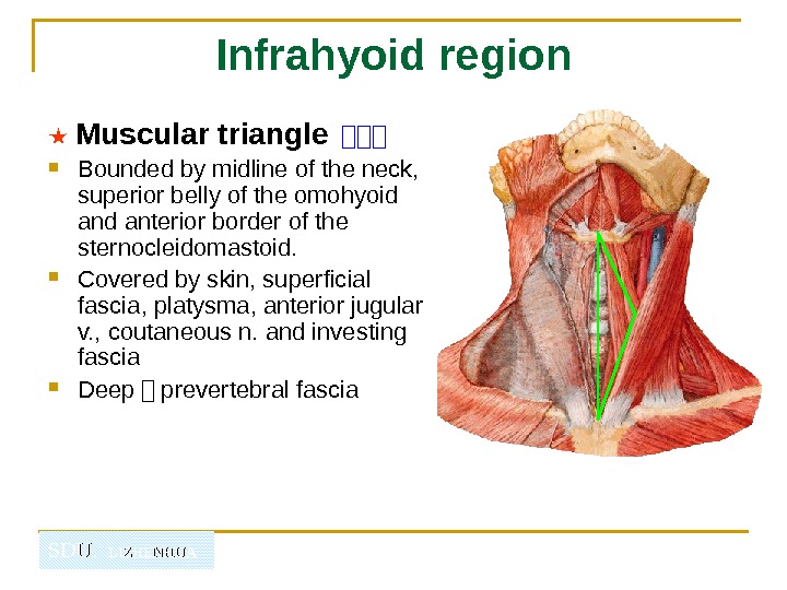
SDU. LIZHENHUA Infrahyoid region ★ Muscular triangle 山山山 Bounded by midline of the neck, superior belly of the omohyoid anterior border of the sternocleidomastoid. Covered by skin, superficial fascia, platysma, anterior jugular v. , coutaneous n. and investing fascia Deep - prevertebral fascia
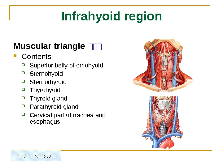
SDU. LIZHENHUA Infrahyoid region Muscular triangle 山山山 Contents Superior belly of omohyoid Sternohyoid Sternothyroid Thyrohyoid Thyroid gland Parathyroid gland Cervical part of trachea and esophagus
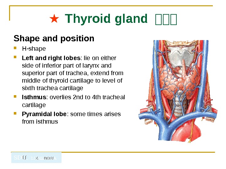
SDU. LIZHENHUA ★ Thyroid gland 山山山 Shape and position H-shape Left and right lobes : lie on either side of inferior part of larynx and superior part of trachea, extend from middle of thyroid cartilage to level of sixth trachea cartilage Isthmus : overlies 2 nd to 4 th tracheal cartilage Pyramidal lobe : some times arises from isthmus
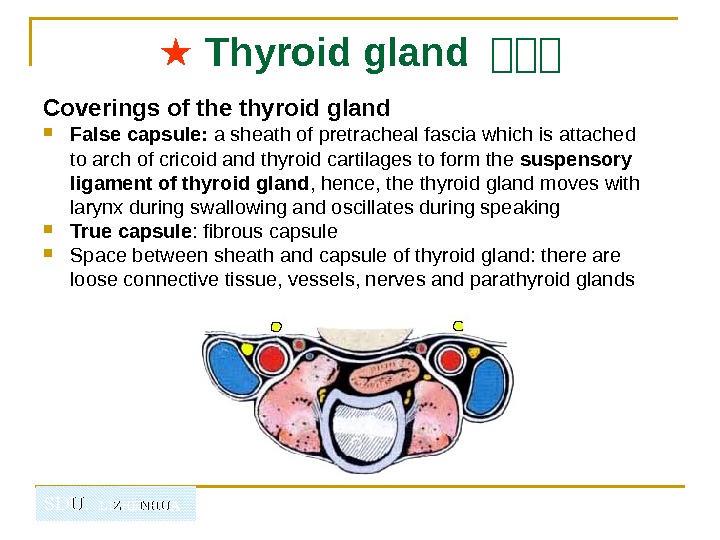
SDU. LIZHENHUA ★ Thyroid gland 山山山 Coverings of the thyroid gland False capsule: a sheath of pretracheal fascia which is attached to arch of cricoid and thyroid cartilages to form the suspensory ligament of thyroid gland , hence, the thyroid gland moves with larynx during swallowing and oscillates during speaking True capsule : fibrous capsule Space between sheath and capsule of thyroid gland: there are loose connective tissue, vessels, nerves and parathyroid glands
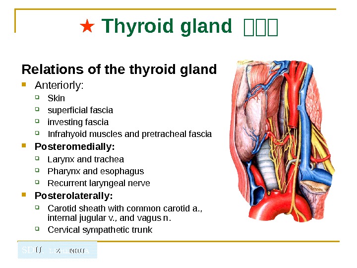
SDU. LIZHENHUA ★ Thyroid gland 山山山 Relations of the thyroid gland Anteriorly: Skin superficial fascia investing fascia Infrahyoid muscles and pretracheal fascia Posteromedially: Larynx and trachea Pharynx and esophagus Recurrent laryngeal nerve Posterolaterally: Carotid sheath with common carotid a. , internal jugular v. , and vagus n. Cervical sympathetic trunk
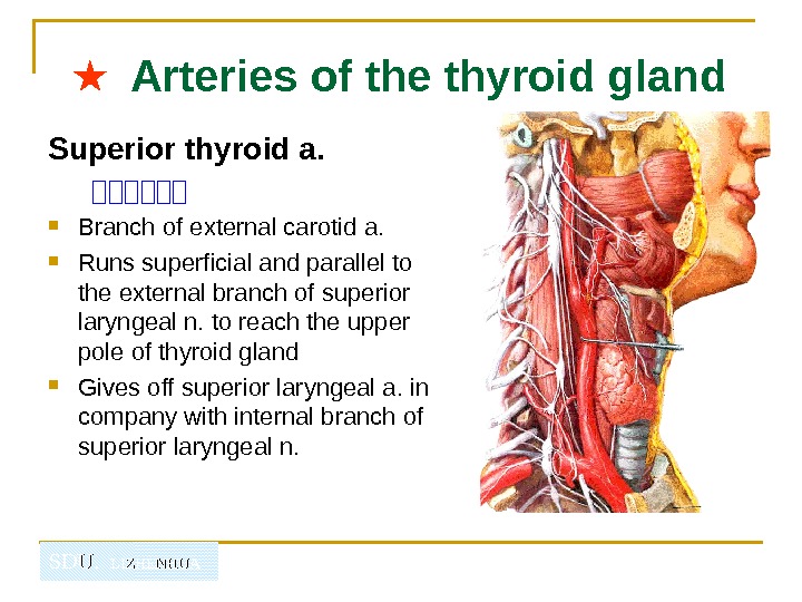
SDU. LIZHENHUA★ Arteries of the thyroid gland Superior thyroid a. 山山山山山山 Branch of external carotid a. Runs superficial and parallel to the external branch of superior laryngeal n. to reach the upper pole of thyroid gland Gives off superior laryngeal a. in company with internal branch of superior laryngeal n.
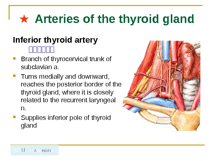
SDU. LIZHENHUA★ Arteries of the thyroid gland Inferior thyroid artery 山山山山山山 Branch of thyrocervical trunk of subclavian a. Turns medially and downward, reaches the posterior border of the thyroid gland, where it is closely related to the recurrent laryngeal n. Supplies inferior pole of thyroid gland
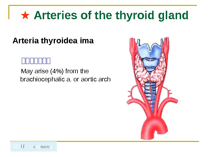
SDU. LIZHENHUA★ Arteries of the thyroid gland Arteria thyroidea ima 山山山山山山山 May arise (4%) from the brachiocephalic a. or aortic arch
SDU. LIZHENHUA ★ Nerves of the larynx Superior laryngeal n. 山山山山 Internal branch 山山 - which pierces thyrohyoid membrane to innervates mucous membrane of larynx above fissure of glottis External branch 山山 - is fine n. , which descends in company with the superior thyroid a. and supplies cricothyroid
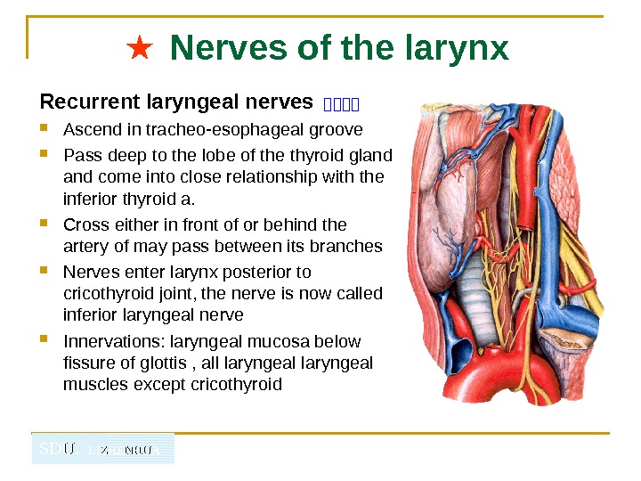
SDU. LIZHENHUA ★ Nerves of the larynx Recurrent laryngeal nerves 山山山山 Ascend in tracheo-esophageal groove Pass deep to the lobe of the thyroid gland come into close relationship with the inferior thyroid a. Cross either in front of or behind the artery of may pass between its branches Nerves enter larynx posterior to cricothyroid joint, the nerve is now called inferior laryngeal nerve Innervations: laryngeal mucosa below fissure of glottis , all laryngeal muscles except cricothyroid
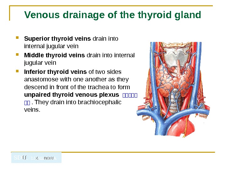
SDU. LIZHENHUAVenous drainage of the thyroid gland Superior thyroid veins drain into internal jugular vein Middle thyroid veins drain into internal jugular vein Inferior thyroid veins of two sides anastomose with one another as they descend in front of the trachea to form unpaired thyroid venous plexus 山山山山山 山山. They drain into brachiocephalic veins.
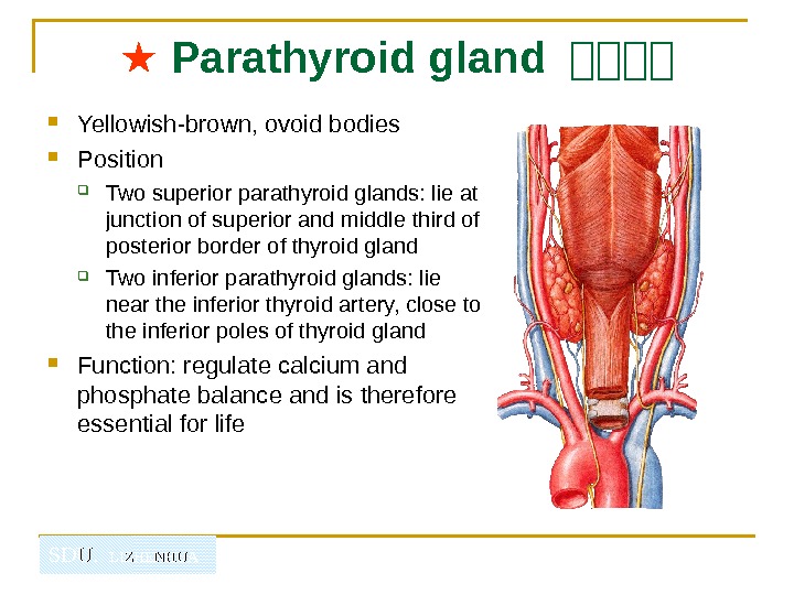
SDU. LIZHENHUA ★ Parathyroid gland 山山山山 Yellowish-brown, ovoid bodies Position Two superior parathyroid glands: lie at junction of superior and middle third of posterior border of thyroid gland Two inferior parathyroid glands: lie near the inferior thyroid artery, close to the inferior poles of thyroid gland Function: regulate calcium and phosphate balance and is therefore essential for life
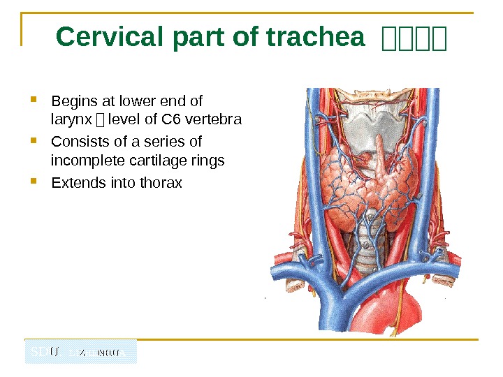
SDU. LIZHENHUACervical part of trachea 山山山山 Begins at lower end of larynx - level of C 6 vertebra Consists of a series of incomplete cartilage rings Extends into thorax
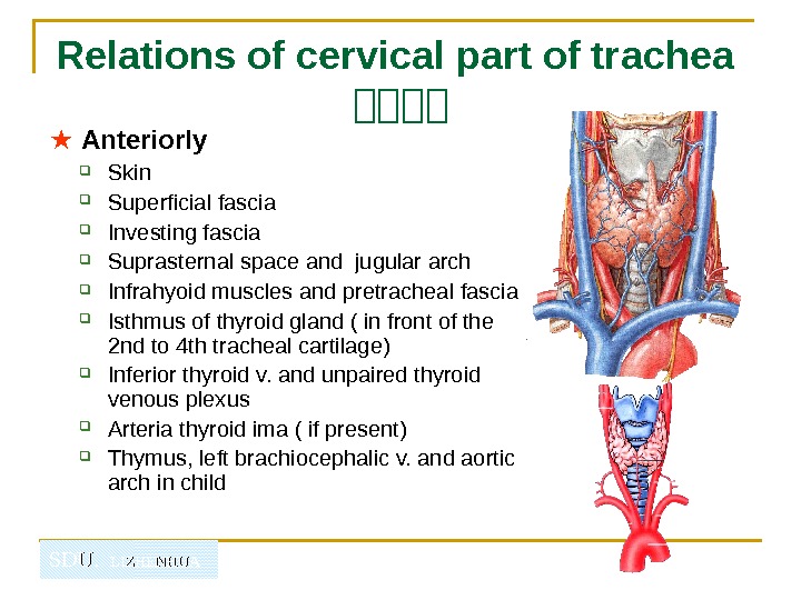
SDU. LIZHENHUARelations of cervical part of trachea 山山山山 ★ Anteriorly Skin Superficial fascia Investing fascia Suprasternal space and jugular arch Infrahyoid muscles and pretracheal fascia Isthmus of thyroid gland ( in front of the 2 nd to 4 th tracheal cartilage) Inferior thyroid v. and unpaired thyroid venous plexus Arteria thyroid ima ( if present) Thymus, left brachiocephalic v. and aortic arch in child
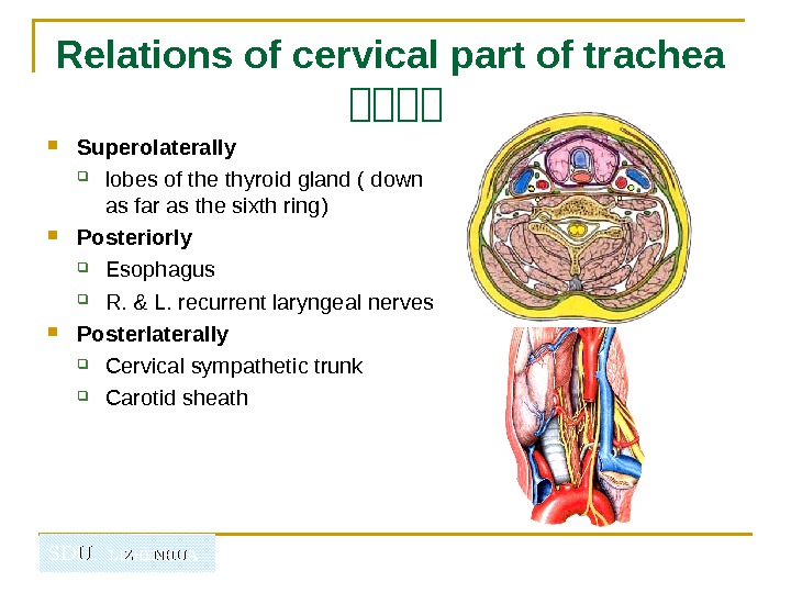
SDU. LIZHENHUARelations of cervical part of trachea 山山山山 Superolaterally lobes of the thyroid gland ( down as far as the sixth ring) Posteriorly Esophagus R. & L. recurrent laryngeal nerves Posterlaterally Cervical sympathetic trunk Carotid sheath
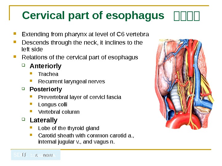
SDU. LIZHENHUACervical part of esophagus 山山山山 Extending from pharynx at level of C 6 vertebra Descends through the neck, it inclines to the left side Relations of the cervical part of esophagus Anteriorly Trachea Recurrent laryngeal nerves Posteriorly Prevertebral layer of cervicl fascia Longus colli Vertebral column Laterally Lobe of the thyroid gland Carotid sheath with common carotid a. , internal jugular v. , and vagus n.
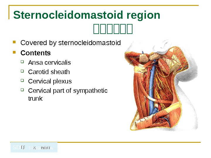
SDU. LIZHENHUASternocleidomastoid region 山山山山山山 Covered by sternocleidomastoid Contents Ansa cervicalis Carotid sheath Cervical plexus Cervical part of sympathetic trunk
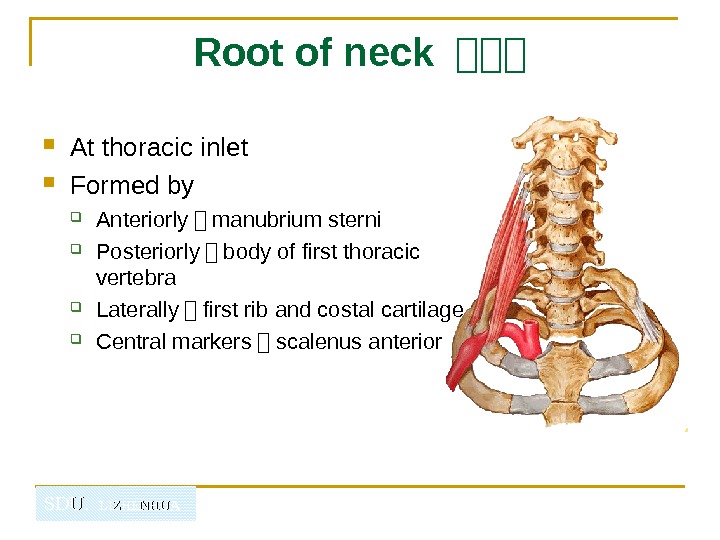
SDU. LIZHENHUA Root of neck 山山山 At thoracic inlet Formed by Anteriorly - manubrium sterni Posteriorly - body of first thoracic vertebra Laterally - first rib and costal cartilage Central markers - scalenus anterior
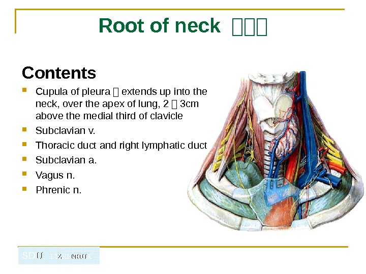
SDU. LIZHENHUA Root of neck 山山山 Contents Cupula of pleura - extends up into the neck, over the apex of lung, 2 - 3 cm above the medial third of clavicle Subclavian v. Thoracic duct and right lymphatic duct Subclavian a. Vagus n. Phrenic n.
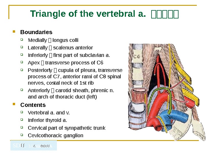
SDU. LIZHENHUA Triangle of the vertebral a. 山山山山山 Boundaries Medially - longus colli Laterally - scalenus anterior Inferiorly - first part of subclavian a. Apex - transverse process of C 6 Posteriorly - cupula of pleura, transverse process of C 7, anterior rami of C 8 spinal nerves, costal neck of 1 st rib Anteriorly - carotid sheath, phrenic n. and arch of thoracic duct (left) Contents Vertebral a. and v. Inferior thyroid a. Cervical part of sympathetic trunk Cevicothoracic ganglion
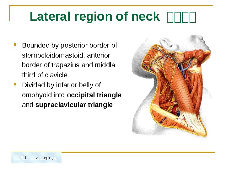
SDU. LIZHENHUALateral region of neck 山山山山 Bounded by posterior border of sternocleidomastoid, anterior border of trapezius and middle third of clavicle Divided by inferior belly of omohyoid into occipital triangle and supraclavicular triangle
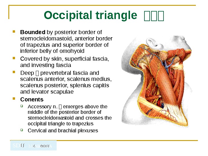
SDU. LIZHENHUA Occipital triangle 山山山 Bounded by posterior border of sternocleidomastoid, anterior border of trapezius and superior border of inferior belly of omohyoid Covered by skin, superficial fascia, and investing fascia Deep - prevertebral fascia and scalenus anterior, scalenus medius, scalenus posterior, splenius capitis and levator scapulae Conents Accessory n. - emerges above the middle of the posterior border of sternocleidomastoid and crosses the occipital triangle to trapezius Cervical and brachial plexuses
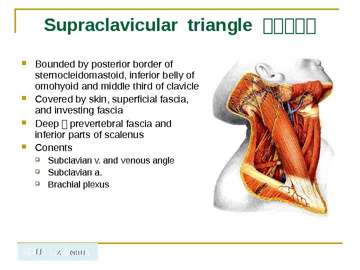
SDU. LIZHENHUASupraclavicular triangle 山山山山山 Bounded by posterior border of sternocleidomastoid, inferior belly of omohyoid and middle third of clavicle Covered by skin, superficial fascia, and investing fascia Deep - prevertebral fascia and inferior parts of scalenus Conents Subclavian v. and venous angle Subclavian a. Brachial plexus
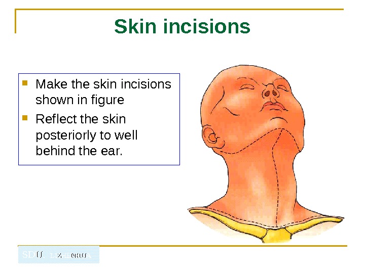
SDU. LIZHENHUA Skin incisions Make the skin incisions shown in figure Reflect the skin posteriorly to well behind the ear.
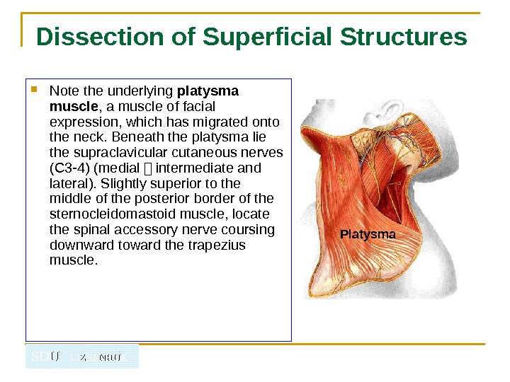
SDU. LIZHENHUADissection of Superficial Structures Note the underlying platysma muscle , a muscle of facial expression, which has migrated onto the neck. Beneath the platysma lie the supraclavicular cutaneous nerves (C 3 -4) (medial - intermediate and lateral). Slightly superior to the middle of the posterior border of the sternocleidomastoid muscle, locate the spinal accessory nerve coursing downward toward the trapezius muscle. Platysma
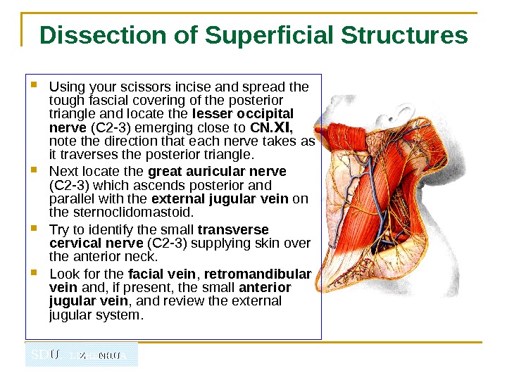
SDU. LIZHENHUADissection of Superficial Structures Using your scissors incise and spread the tough fascial covering of the posterior triangle and locate the lesser occipital nerve (C 2 -3) emerging close to CN. , Ⅺ note the direction that each nerve takes as it traverses the posterior triangle. Next locate the great auricular nerve (C 2 -3) which ascends posterior and parallel with the external jugular vein on the sternoclidomastoid. Try to identify the small transverse cervical nerve (C 2 -3) supplying skin over the anterior neck. Look for the facial vein , retromandibular vein and, if present, the small anterior jugular vein , and review the external jugular system.
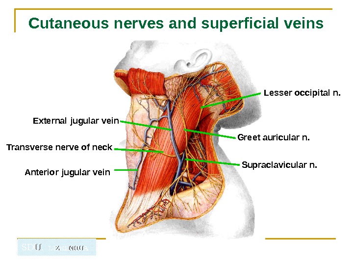
SDU. LIZHENHUACutaneous nerves and superficial veins External jugular vein Anterior jugular vein Lesser occipital n. Greet auricular n. Transverse nerve of neck Supraclavicular n.
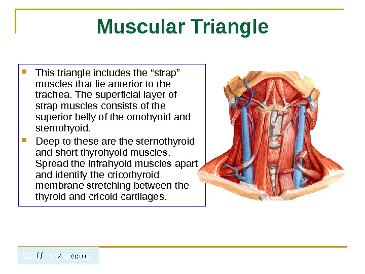
SDU. LIZHENHUA Muscular Triangle This triangle includes the “strap” muscles that lie anterior to the trachea. The superficial layer of strap muscles consists of the superior belly of the omohyoid and sternohyoid. Deep to these are the sternothyroid and short thyrohyoid muscles. Spread the infrahyoid muscles apart and identify the cricothyroid membrane stretching between the thyroid and cricoid cartilages.
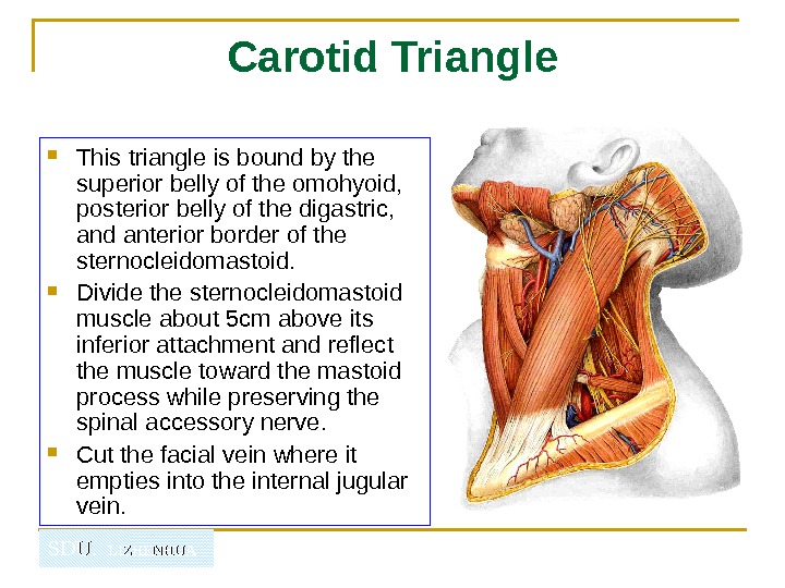
SDU. LIZHENHUA Carotid Triangle This triangle is bound by the superior belly of the omohyoid, posterior belly of the digastric, and anterior border of the sternocleidomastoid. Divide the sternocleidomastoid muscle about 5 cm above its inferior attachment and reflect the muscle toward the mastoid process while preserving the spinal accessory nerve. Cut the facial vein where it empties into the internal jugular vein.
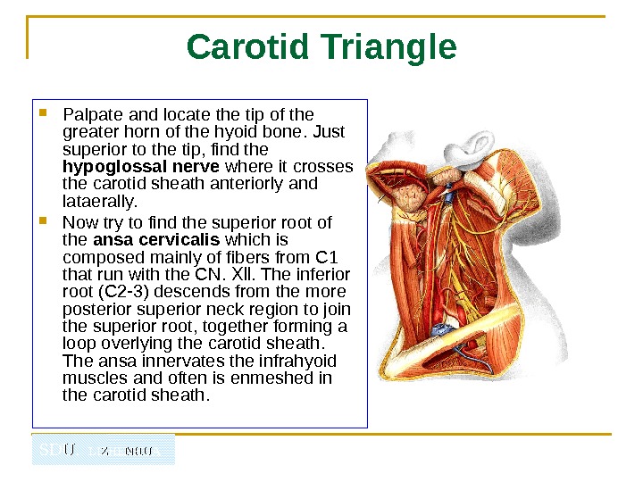
SDU. LIZHENHUA Carotid Triangle Palpate and locate the tip of the greater horn of the hyoid bone. Just superior to the tip, find the hypoglossal nerve where it crosses the carotid sheath anteriorly and lataerally. Now try to find the superior root of the ansa cervicalis which is composed mainly of fibers from C 1 that run with the CN. . The inferior Ⅻ root (C 2 -3) descends from the more posterior superior neck region to join the superior root, together forming a loop overlying the carotid sheath. The ansa innervates the infrahyoid muscles and often is enmeshed in the carotid sheath.
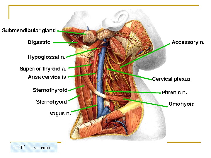
SDU. LIZHENHUASubmendibular gland Omohyoid. Sternothyroid. Superior thyroid a. Cervical plexus Phrenic n. Ansa cervicalis Vagus n. Hypoglossal n. Accessory n. Digastric
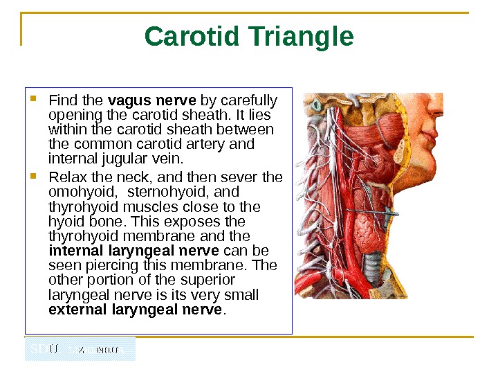
SDU. LIZHENHUA Carotid Triangle Find the vagus nerve by carefully opening the carotid sheath. It lies within the carotid sheath between the common carotid artery and internal jugular vein. Relax the neck, and then sever the omohyoid, sternohyoid, and thyrohyoid muscles close to the hyoid bone. This exposes the thyrohyoid membrane and the internal laryngeal nerve can be seen piercing this membrane. The other portion of the superior laryngeal nerve is its very small external laryngeal nerve.
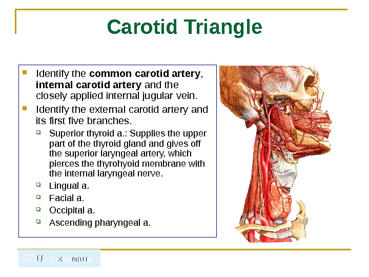
SDU. LIZHENHUA Carotid Triangle Identify the common carotid artery , internal carotid artery and the closely applied internal jugular vein. Identify the external carotid artery and its first five branches. Superior thyroid a. : Supplies the upper part of the thyroid gland gives off the superior laryngeal artery, which pierces the thyrohyoid membrane with the internal laryngeal nerve. Lingual a. Facial a. Occipital a. Ascending pharyngeal a.
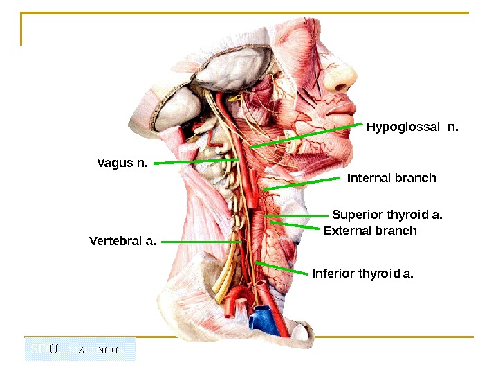
SDU. LIZHENHUA Superior thyroid a. External branch Internal branch Hypoglossal n. Vagus n. Vertebral a. Inferior thyroid a.
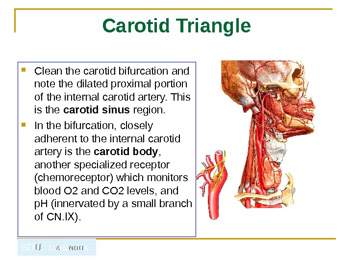
SDU. LIZHENHUA Carotid Triangle Clean the carotid bifurcation and note the dilated proximal portion of the internal carotid artery. This is the carotid sinus region. In the bifurcation, closely adherent to the internal carotid artery is the carotid body , another specialized receptor (chemoreceptor) which monitors blood O 2 and CO 2 levels, and p. H (innervated by a small branch of CN. ). Ⅸ
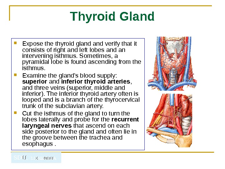
SDU. LIZHENHUA Thyroid Gland Expose thyroid gland verify that it consists of right and left lobes and an intervening isthmus. Sometimes, a pyramidal lobe is found ascending from the isthmus. Examine the gland’s blood supply: superior and inferior thyroid arteries , and three veins (superior, middle and inferior). The inferior thyroid artery often is looped and is a branch of the thyrocervical trunk of the subclavian artery. Cut the isthmus of the gland to turn the lobes laterally and probe for the recurrent laryngeal nerves that ascend on each side posterior to the gland often lie in the groove between the trachea and esophagus.
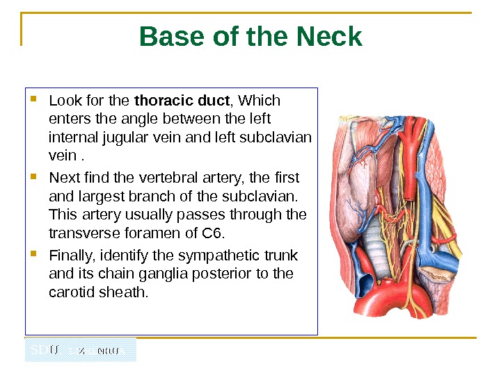
SDU. LIZHENHUA Base of the Neck Look for the thoracic duct , Which enters the angle between the left internal jugular vein and left subclavian vein. Next find the vertebral artery, the first and largest branch of the subclavian. This artery usually passes through the transverse foramen of C 6. Finally, identify the sympathetic trunk and its chain ganglia posterior to the carotid sheath.
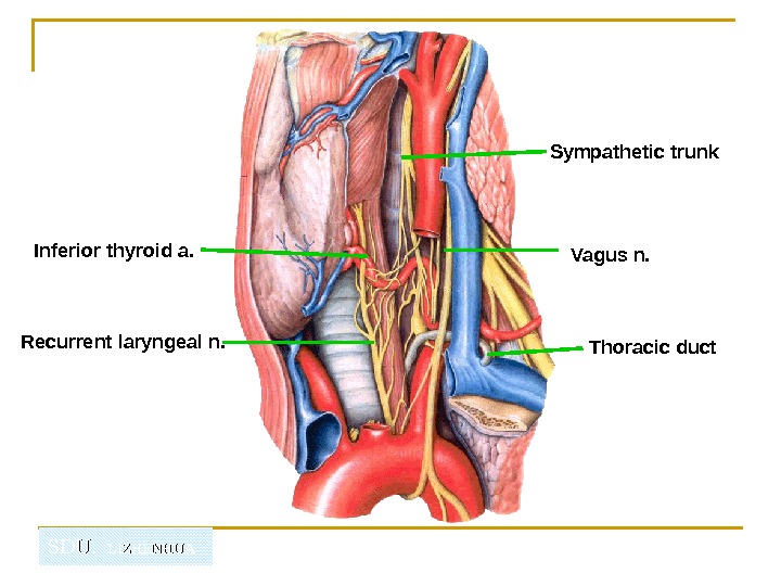
SDU. LIZHENHUAInferior thyroid a. Recurrent laryngeal n. Thoracic duct Vagus n. Sympathetic trunk
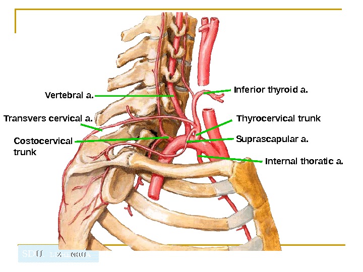
SDU. LIZHENHUAVertebral a. Thyrocervical trunk. Inferior thyroid a. Internal thoratic a. Transvers cervical a. Costocervical trunk Suprascapular a.
SDU. LIZHENHUA★ You must identify follow structures 山 Muscles Sternocleidomastoid Sternohyoid Sternothyroid Thyrohyoid Omohyoid Scalenus anterior Scalenus medius Scalenus posterior Arteries Common carotid a. Internal external carotid artery External carotid artery Superior thyroid a. Lingual a. Facial a. Occipital a. Subclavian a. Vertebral a. Internal thoracic a. Thyrocervical trunk Inferior thyroid a.
SDU. LIZHENHUA★ You must identify follow structures 山 Veins External jugular vein Internal jugular v. Subclavian v. Lymph duct Thoracic duct R ight lymphatic duct Nerves Lesser occipital n. Great auricular n. Transverse nerve of neck Supraclavicular n. Phrenic n. Ansa cervicalis Accesory n. Vagus n. Internal branch of superior laryngeal n External branch of superior laryngeal n. Recurrent laryngeal nerves H ypoglossal n. C ervical part of sympathetic trunk Organs Submandibular gland Thyroid gland