Описание презентации RPE Rip Red-free Photograph Image courtesy of по слайдам
 RPE Rip
 Red-free Photograph Image courtesy of J Slakter
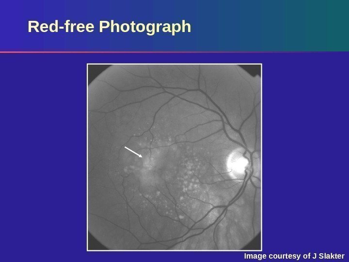
 Red-free Photograph Red-free photograph: – Shallow subretinal fluid – Drusen Note the area just temporal to the centre of the macula that appears to be hypopigmented (arrow)
 Early-phase Fluorescein Angiogram Image courtesy of J Slakter
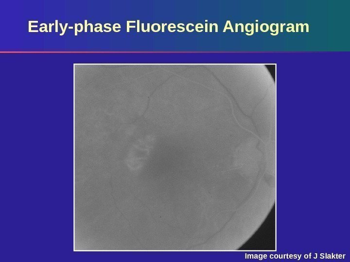
 Early-phase Fluorescein Angiogram Early (arterial) phase: – The most important frame Note the oval area of mild hyperfluorescence occurring when the retinal arteries are just filling The choroid is also just beginning to fill – Horizontally oriented vessels represent larger choroidal vessels seen through a rip of the RP
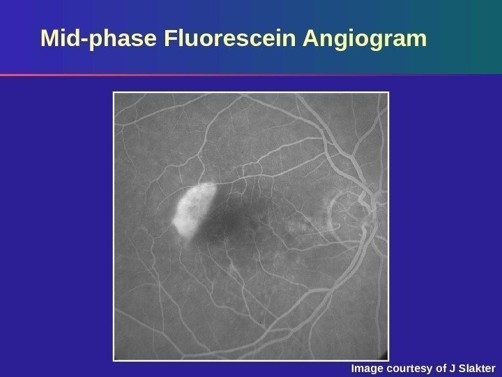 Mid-phase Fluorescein Angiogram Image courtesy of J Slakter
 Mid-phase Fluorescein Angiogram Mid-phase: – Well-circumscribed hyperfluorescent area with curved outer border and almost straight line margin adjacent to an area of relative hypofluorescence – Findings are typical of RPE rips and should act as clues to the diagnosis
 Mid-phase Fluorescein Angiogram By only examining this image and the one that follows, this can be mistaken for classic CNV – The very early image helps to make the correct diagnosis This can also be misdiagnosed as geographic atrophy, which has: – A border that is geographic not linear – A lesion with no overlying retinal detachment
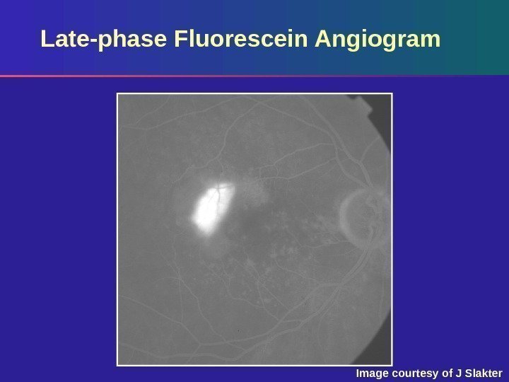 Late-phase Fluorescein Angiogram Image courtesy of J Slakter
 Late-phase Fluorescein Angiogram Late phase: – Increased hyperfluorescence with mild leakage at margins Again, this can easily be confused with the pattern seen with classic CNV if the entire study is not examined, including the red-free image


