rheumatic_endocarditis.pptx
- Количество слайдов: 13


Rheumatic Endocarditis Student`s Name: Rahat Gulnaz; Ashimbai Anar Group: 16 -1 Faculty: OM
The definition of “endocarditis” 2)Classification • 3)The causes • 4)Rheumatic endocarditis –")
Plan: • 1)The definition of “endocarditis” 2)Classification • 3)The causes • 4)Rheumatic endocarditis – the main form of endocarditis • 5)The symptoms of the rheumatic endocarditis • 6)The examination of the rheumatic endocarditis

• The definition of "endocarditis" includes damage internal lining of the heart of an inflammatory nature. Mainly affected the valve unit, rarely changes are localized on the endocardium endings.

Classification of endocarditis • The basic meaning of the clinic are three forms of endocarditis: • 1 Rheumatoid. • 2 Tightening (subacute) septic. • 3 Acute septic

• It is well known that myocardial damage rheumatism observed in all cases. The defeat of the valve apparatus "valvulity, ” after the first attack of rheumatic fever is observed in 90% of cases in children and approximately 40% of patients older than 30 years. In other words endocarditis is the main form of rheumatism.

• Etiology and pathogenesis of rheumatic endocarditis are the same and rheumatism. As is now common etiologic relationship of rheumatic fever with streptococcal infection.

Rheumatism - a systemic inflammatory disease affecting mainly the connective tissue of the cardiovascular system. Cause of rheumatic fever is a betahemolytic streptococcus group A. In most cases, rheumatism develops after a sore throat. Rheumatic fever affects all layers of the heart: the endocardium, myocardium and pericardium.
")
• The clinical picture of rheumatic endomiocarditis determined by the following factors: 1) the activity of rheumatic process; 2) the state of the valve apparatus and the heart muscle; 3) focal infection.

• The symptoms -a general malaise, early fatigue on exertion, cardiac discomfort and body temperature to a subfebrile level for a prolonged period of time. The pulse becomes irregular and accelerated on physical exertion.


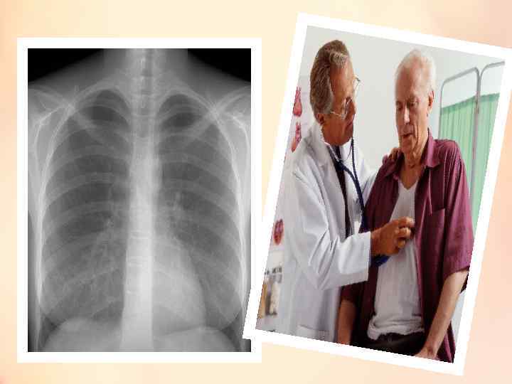
• The blood analysis revealed moderate leucocytosis and an elevated ESR. The electrocardiogram showed the changes in the most important readings. On percussion the doctor determined the heart to be slightly enlarged. These findings of the physical examination were confirmed by the X-ray examination.

rheumatic_endocarditis.pptx