Rh Isoimmunization Immunologic disorder that occurs in pregnant



388-rh_isoimmunisation_3.ppt
- Количество слайдов: 31
 Rh Isoimmunization
 Immunologic disorder that occurs in pregnant Rh negative lady carrying Rh positive fetus
 Pathophysiology The rhesus system which comprises number of antigens C, D, E, c, e. A person who lacks D antigen is called Rh negative. 15% of Caucasians, 5% African Americans and 2 % of Asians are Rh negative. Rh isoimmunisation is due to D antigen in more than 90% of cases. Occasionally hemolytic disease of the newborn is a result of maternal immunization to Irregular RBC antigens other than Rh group like anti- Kell and anti- Duffy
 Pathophysiology Initial response is forming IgM antibodies for short period followed by production of IgG which crosses placenta IgG antibodies adhere to the antigen site on the surface of erythrocytes causing hemolysis. The excessive removal of circulatory RBCs leads to severe anemia and hypoxia. Erythropoiesis results in hepatosplenomegaly. Tissue hypoxia and hypoproteinemia results in cardiac and circulatory failure, with generalized odema and hydrops
 Pathophysiology Maternal immune system becomes sensitized when there is fetal blood leak into the maternal circulation. Although leaks are common only 8% are sensitized within 6 months after first del of ABO compatible preg. 16% are sensitized after second full term pregnancy of Rh positive ABO compatible pregnancy. The risk of sensitization after ABO incompatible pregnancy is only 2% Risk after spontaneous miscarriage is 3.5% Risk after induce abortion is 5.5% Risk after ectopic pregnancy is about 1%
 Natural History 50% of affected infants have no or mild anemia, requiring either phototherapy or no treatment. 25% have some degree of hepatosplenomegaly and moderate anemia and progressive jundice culminating in kernicterus, neonatal death or severe handicap. 25% are hydropic and usually die in utero or in the neonatal period ( half of these the hydrops develops before 34 weeks gestation ).
 The aim of antenatal management To predict which pregnancy is at risk To predict whether or not the fetus is severely affected. To correct anemia and reverse hydrops by intrauterine transfusion. To deliver the baby at the appropriate time, weighing the risks of prematurity against these of intrauterine transfusion.
 Recognition of pregnancy at risk First ante-natal visit check blood group, antibody screening. If indirect coombs test is positive, the father’s Rh should be tested. Serial maternal Anti D titers should be done every 2- 4 weeks. If titer is less than 1/16 the fetus is not at risk. If titer is more than 1/16 then severity of condition should be evaluated.
 Prediction of the severity of fetal hemolysis History of previous affected pregnancies The levels of maternal hemolytic antibodies Amniocentesis Biophysical surveillance Fetal blood sampling
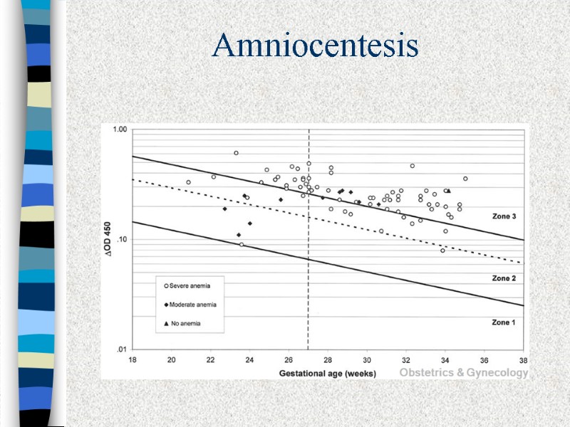
 Amniocentesis There is an excellent correlation between the amount of bilirubin in amniotic fluid and fetal hematocrit. The optical density deviation at 450 nm measures the amniotic fluid unconjugated bilirubin.
 Amniocentesis
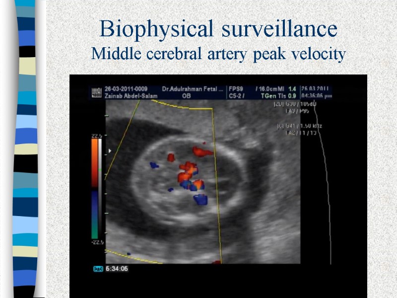
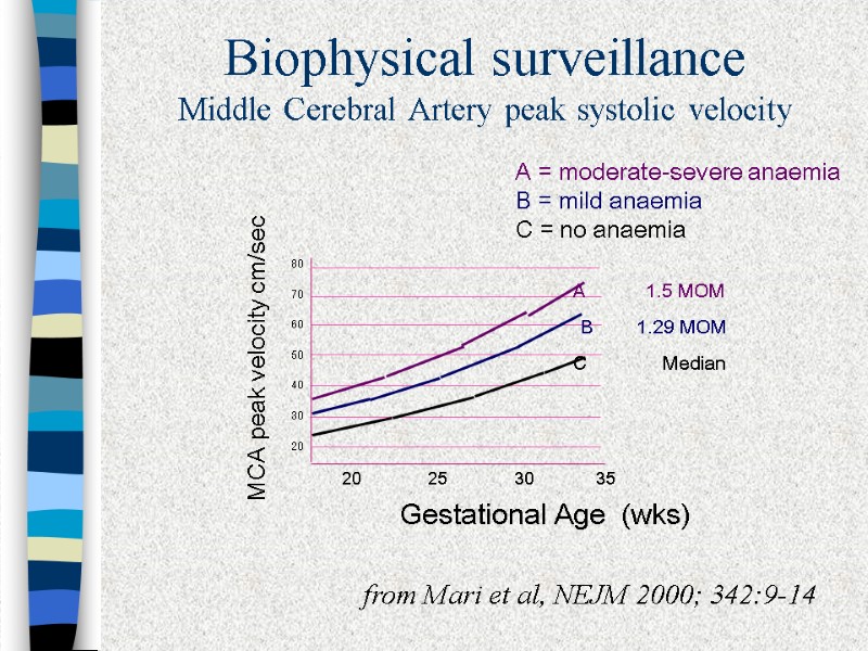
 Ultrasound detection of Rh Sensitization Serial U/S examination for fetal well being. Placental size and thickness and hepatic size. Fetal hydrops is easy to diagnose when finding one or more of the following: Ascites, pleural effusion, pericardial effusion, or skin edema. Doppler assessment of peak velocity of fetal middle cerebral artery proved to valuable in predicting fetal anemia
 Biophysical surveillance Middle cerebral artery peak velocity
 Biophysical surveillance Middle Cerebral Artery peak systolic velocity C Median 80 70 60 50 40 30 20 20 25 30 35 A 1.5 MOM B 1.29 MOM Gestational Age (wks) MCA peak velocity cm/sec from Mari et al, NEJM 2000; 342:9-14 A = moderate-severe anaemia B = mild anaemia C = no anaemia
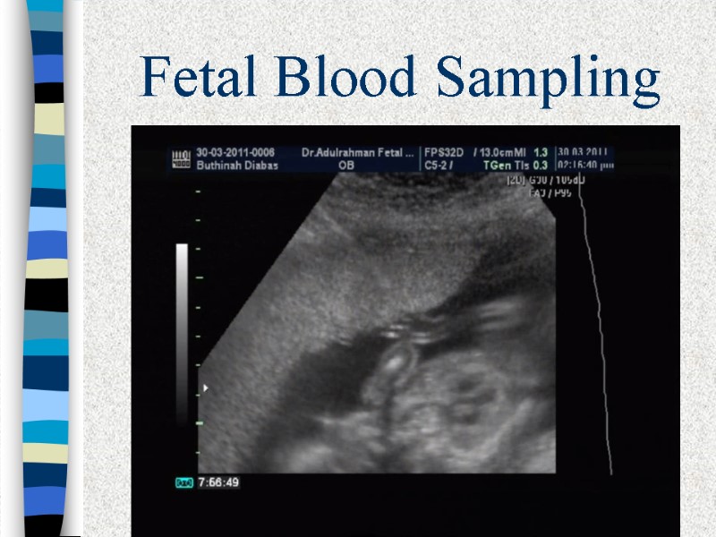
 Fetal Blood Sampling FBS Percutaneous Umbilical Blood Sampling BUBS Allows measurement of fetal Hb, Hct, pH, reticulocytes and nucleated RBCs. Risk fetal exanguination and fetal death 1%.
 Fetal Blood Sampling
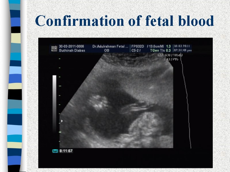
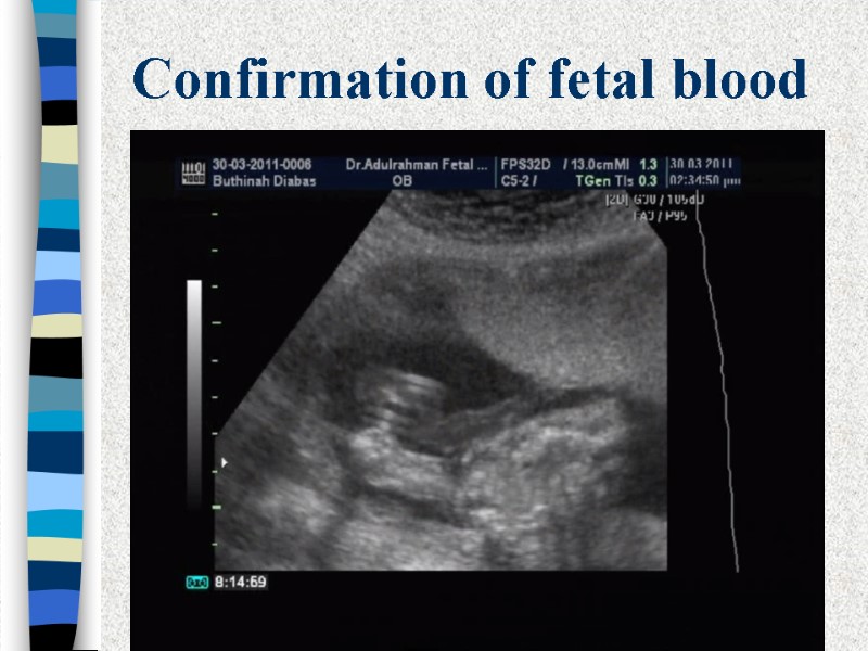
 Confirmation of fetal blood Visualizing the needle tip within the cord vessel Turbulance when injecting a small amount of saline into the vessel. Fetal blood has a higher mean corpuscular volume ( MCV usually 100 fL An alkali elusion method can be used Acid elution Betke-Kleihauer test
 Confirmation of fetal blood
 Confirmation of fetal blood
 Intrauterine Transfusion Radiologically guided intraperitoneal transfusion by Liley in 1963. Adoption of ultrasound guided IPT resulted in dramatic improvement in survival rates. Fetoscopic intravascular route was renewed by Rodeck in 1981. Percutaneous transfusion into the intrahepatic umbilical vein, Bang 1982. Cordocentesis, Daffos 1983. The use of cordocentesis for fetal top up transfusion by Berkowitz.
 Intravascular transfusion IVT has superseded intraperitoneal transfusion in most centres. It has superior results, regarding survival rates, quality of survivors, prolongation of gestation and the higher rate of vaginal delivery. Access to the fetal circulation is extremely essential to avoid the lethal mistake of transfusing the Rh negative fetus or the nonanemic Rh positive fetus. Decisions regarding transfusion and delivery are made on the basis of fetal blood group, Hb estimation and other hematologic parameters . IVT results are better in the compromised hydropic fetus.
 Intrauterine blood transfusion
 Donor Blood Washed, filtered or irradiated with 2500 rad Gamma ray packed red blood cells with Hct 75% Group O negative heterologous blood Maternal blood
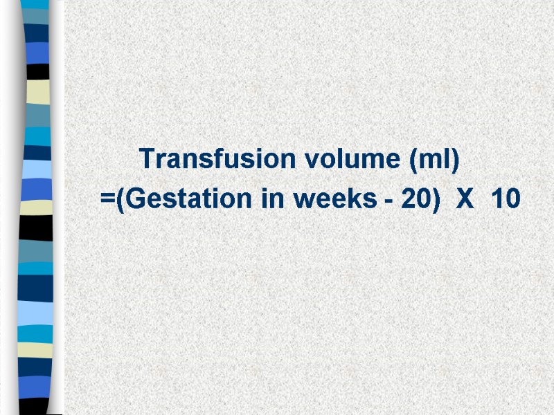
 Transfusion volume (ml) =(Gestation in weeks - 20) X 10
 Timing of transfusions Subsequent transfusions are timed on the basis of the fetal Hct achieved at the end of the previous transfusion and the rate of fall in fetal Hct. The latter has been reported to be on average equal to 1% of Hct/day
 Complications of cordocentesis and intravascular transfuaion Hemorrhage. Hematomas.. Bradycardias. Fetomaternal hemorrhage. Infection. Abruptio placetae. Preterm labor.
 Timing of Delivery Weighing the risk of fetal loss including that related to intrauterine transfusion against the risk of prematurity.
 Prevention The widespread use of anti D prophylaxis in the late 1960s has led to a great reduction in isoimmunisation. Following delivery and caesarean section Routine anti D administration at 28 weeks. Following spontaneous and induced abortion. Vaginal bleeding and threatened miscarriage. Performing amniocentesis, CVS, Abdomenal trauma and external cephalic version.
 Prevention The usual dose is 300 µg within 72 hours following delivery. This covers 30 ml of fetal blood leak into maternal circulation. If greater transplacental hemorrhage is suspected the dose should be tailored after Kleihauer- Betke test to determine the volume of hemorrhage.
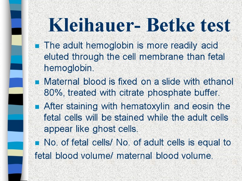
 Kleihauer- Betke test The adult hemoglobin is more readily acid eluted through the cell membrane than fetal hemoglobin. Maternal blood is fixed on a slide with ethanol 80%, treated with citrate phosphate buffer. After staining with hematoxylin and eosin the fetal cells will be stained while the adult cells appear like ghost cells. No. of fetal cells/ No. of adult cells is equal to fetal blood volume/ maternal blood volume.
 Thank you

