
RENAL DISEASE Acute glomerulonephritis Acute

- Размер: 870 Кб
- Количество слайдов: 57
Описание презентации RENAL DISEASE Acute glomerulonephritis Acute по слайдам
RENAL DISEAS
Acute glomerulonephritis Acute GN is characterized by the abrupt onset of hematuria and proteinuria, often accompanied by azotemia and renal salt and water retention after infection (the most often streptococcal).
Etiology Infectious – Streptococcal – Nonstreptococcal postinfectious glomerulonephritis Bacterial Viral Parasitic Noninfectious – Multisystemic diseases – Primary glomerular diseases
Pathogenesis Previously M-protein of the organism was felt to be responsible for PSGN. Recently, nephritis-associated streptococcal cationic protease and its zymogen precursor (NAPR) has been identified as a glyceraldehyde-3 -phosphate dehydrogenase that functions as a plasmin(ogen) receptor. Antibody levels to NAPR are elevated in streptococcal infections (of group A, C, and G) associated with glomerulonephritis, but are not elevated in streptococcal infections without glomerulonephritis, where as anti-streptolysin-O titers are elevated in both circumstances.
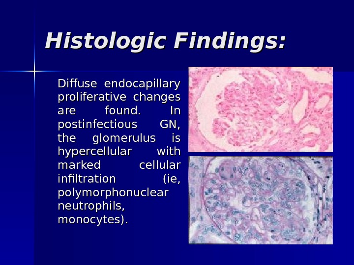
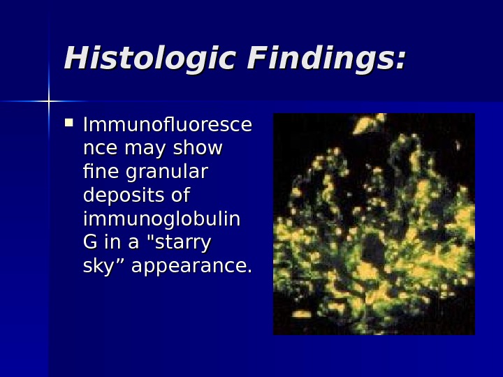
Pathology Diffuse endocapillary proliferative changes are found. In postinfectious GN, the glomerulus is hypercellular with marked cellular infiltration (ie, polymorphonuclear neutrophils, monocytes). Immunofluorescence may show fine granular deposits of immunoglobulin G in a «starry sky” appearance. Large subepithelial deposits may be observed on electron microscopy. Crescents may be observed.
Clinical Manifestations edemas, decreased volume and frequency of urination, systemic hypertension, uremic symptoms, costovertebral tenderness, gross hematuria, rash (ie, vasculitis, Henoch-Schönlein purpura), pp allor. .
Clinical syndromes urinary (haematuria, proteinuria), nephritic (edemas, hypertension, gross haematuria, proteinuria), nephrotic (edemas, proteinuria, hypoproteinemia, hypercholesterolemia), mixed.
Workup Lab Studies: Urinalysis Blood, urea, and nitrogen (BUN); serum creatinine; and serum electrolytes (especially serum potassium level) Complete blood cell count Erythrocyte sedimentation rate Twenty-four–hour urine test for total protein and creatinine clearance: Antistreptolysin-O titer (ASOT) Antibody to NAPR: Levels are elevated in streptococcal infections with GN but not in streptococcal infections without GN.
Imaging Studies: Abdominal ultrasound – Assesses renal size – Assesses echogenicity of renal cortex Excludes obstruction
Histologic Findings: Generally, a renal biopsy is not necessary for diagnosis of acute PSGN; however, in most cases, it is important because histology guides both prognosis and therapy.
Histologic Findings: Diffuse endocapillary proliferative changes are found. In postinfectious GN, the glomerulus is hypercellular with marked cellular infiltration (ie, polymorphonuclear neutrophils, monocytes).
Histologic Findings: Immunofluoresce nce may show fine granular deposits of immunoglobulin G in a «starry sky” appearance.
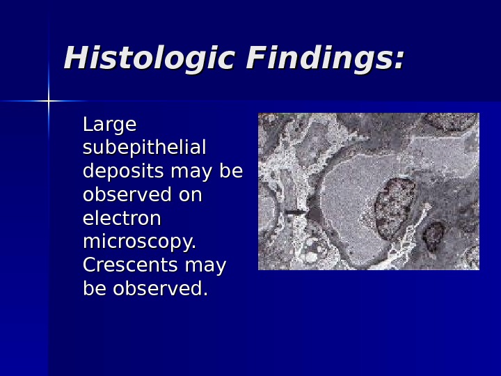
Histologic Findings: Large subepithelial deposits may be observed on electron microscopy. Crescents may be observed.
Differentials Crescentic Glomerulonephritis, Diffuse Proliferative Glomerulonephritis, Membranoproliferative Glomerulonephritis, Rapidly Progressive Glomerulonephritis, Goodpasture Syndrome, Hemolytic-Uremic Syndrome, Interstitial Nephritis, Lupus Nephritis.
Treatment Treat the underlying infections when acute GN is associated with chronic infections. Antimicrobial therapy – Antibiotics (eg, penicillin) are used to control local symptoms and to prevent spread of infection to close contacts. – Antimicrobial therapy does not appear to prevent the development of GN, except if given within the first 36 hours. Loop diuretic therapy – Loop diuretics may be required in patients who are edematous and hypertensive in order to remove excess fluid and to correct hypertension. – Relieves edema and controls volume, thereby helping to control volume-related elevation in BP. Vasodilator drugs (eg, nitroprusside, nifedipine, hydralazine, diazoxide) may be used if severe hypertension or encephalopathy is present Diet: Sodium and fluid restriction Protein restriction for azotemic patients Activity: Recommend bed rest until signs of glomerular inflammation and circulatory congestion subside.
Prognosis of acute PSGN is generally excellent in children. Within a week or so of onset, most patients with PSGN begin to experience spontaneous resolution of fluid retention and hypertension. Approximately 15% of patients at 3 years and 2% of patients at 7 -10 years may have persistent mild proteinuria. Long-term prognosis is not necessarily benign. Some patients may develop hypertension, proteinuria, and renal insufficiency as long as 10 -40 years after the initial illness.
Chronic gg lomerulonephritis The condition is characterized by irreversible and progressive glomerular and tubulointerstitial fibrosis, ultimately leading to a reduction in the glomerular filtration rate (GFR) and retention of uremic toxins. If disease progression is not halted with therapy, the net result is chronic kidney disease (CKD), end-stage renal disease (ESRD), and cardiovascular disease. The diagnosis of CKD can be made without knowledge of the specific cause.
Etiology Nearly all forms of acute glomerulonephritishave a tendency toprogress to chronic glomerulonephritis. The progression from acute glomerulonephritis to chronic glomerulonephritis is variable. Whereas complete recovery of renal function is the rule for patients with poststreptococcal glomerulonephritis, several other glomerulonephritides, such as immunoglobulin A (Ig. A) nephropathy, often have a relatively benign course and many do not progress to ESRD.
Pathogenesis Reduction in nephron mass from the initial injury reduces the GFR. This reduction leads to hypertrophy and hyperfiltration of the remaining nephrons and to the initiation of intraglomerular hypertension. These changes occur in order to increase the GFR of the remaining nephrons, thus minimizing the functional consequences of nephron loss. The changes, however, are ultimately detrimental because they lead to glomerulosclerosis and further nephron loss.
Histologic Findings In early stages, the glomeruli may still show some evidence of the primary disease. In advanced stages, the glomeruli are hyalinized and obsolescent. The tubules are disrupted and atrophic, and marked interstitial fibrosis and arterial and arteriolar sclerosis occur.
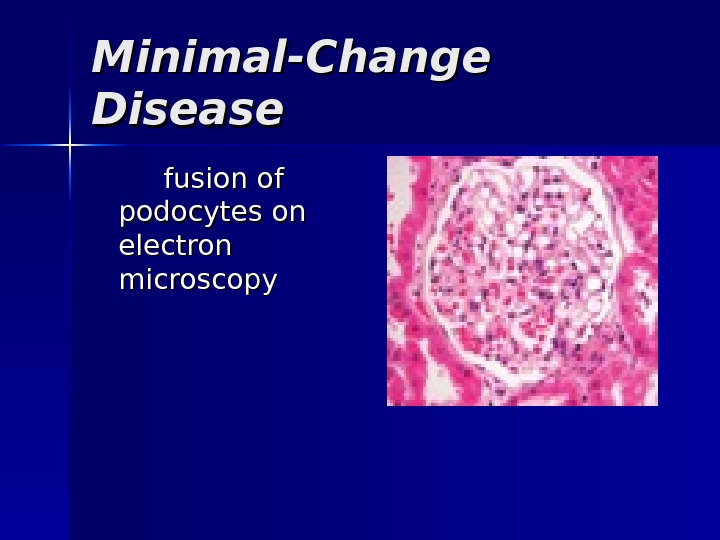
Minimal-Change Disease fusion of podocytes on electron microscopy
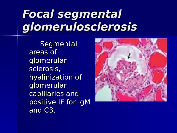
Focal segmental glomerulosclerosis Segmental areas of glomerular sclerosis, hyalinization of glomerular capillaries and positive IF for Ig. M and C 3.
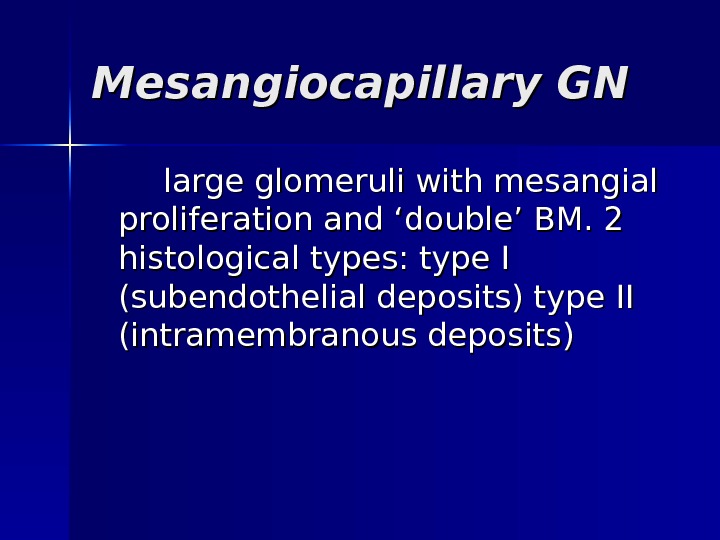
Mesangiocapillary GN large glomeruli with mesangial proliferation and ‘double’ BM. 2 histological types: type I (subendothelial deposits) type II (intramembranous deposits)
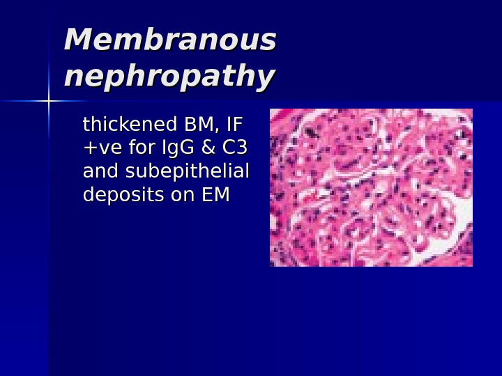
Membranous nephropathy thickened BM, IF +ve for Ig. G & C 3 and subepithelial deposits on EM
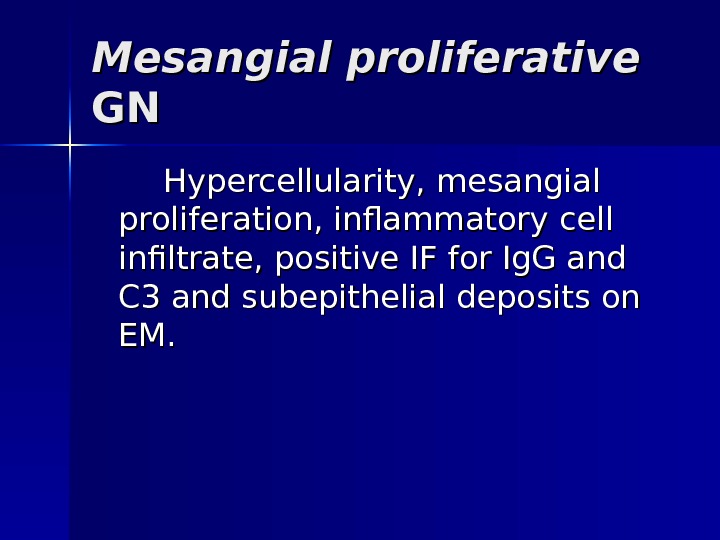
MM esangial proliferative GNGN Hypercellularity, mesangial proliferation, inflammatory cell infiltrate, positive IF for Ig. G and C 3 and subepithelial deposits on EM.
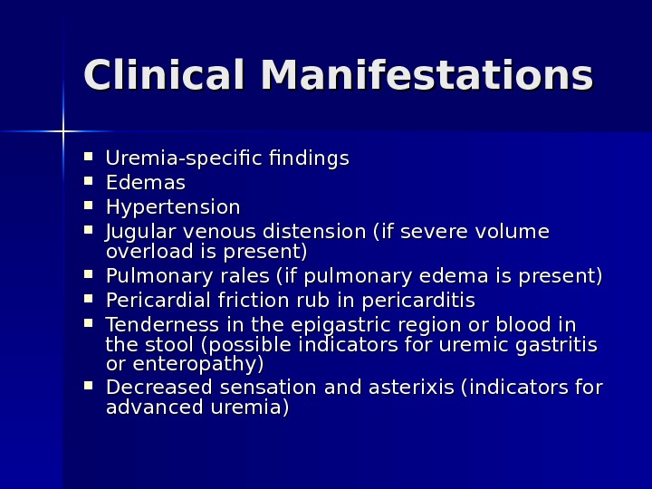
Clinical Manifestations Uremia-specific findings Edemas Hypertension Jugular venous distension (if severe volume overload is present) Pulmonary rales (if pulmonary edema is present) Pericardial friction rub in pericarditis Tenderness in the epigastric region or blood in the stool (possible indicators for uremic gastritis or enteropathy) Decreased sensation and asterixis (indicators for advanced uremia)
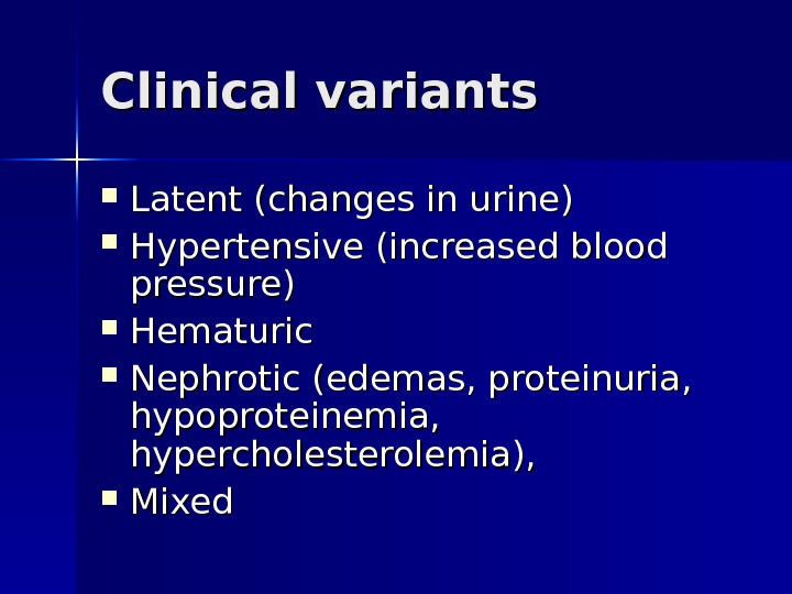
Clinical variants Latent (changes in urine) Hypertensive (increased blood pressure) Hematuric Nephrotic (edemas, proteinuria, hypoproteinemia, hypercholesterolemia), Mixed
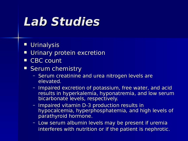
Lab Studies Urinalysis Urinary protein excretion CBC count Serum chemistry – Serum creatinine and urea nitrogen levels are elevated. – Impaired excretion of potassium, free water, and acid results in hyperkalemia, hyponatremia, and low serum bicarbonate levels, respectively. – Impaired vitamin D-3 production results in hypocalcemia, hyperphosphatemia, and high levels of parathyroid hormone. – Low serum albumin levels may be present if uremia interferes with nutrition or if the patient is nephrotic.
Imaging Studies Renal ultrasonogram – Obtain a renal ultrasonogram to determine renal size, to assess for the presence of both kidneys, and to exclude structural lesions that may be responsible for azotemia. – Small kidneys often indicate an irreversible process. Procedures Kidney biopsy
Differentials Azotemia , , Chronic Renal Failure , Acute Glomerulonephritis, Nonstreptococcal Associated With Infection, Poststreptococcal Glomerulonephritis, Rapidly Progr essive , , Uremia
Treatment The target pressure for patients with proteinuria greater than 1 g/d is less than 125/75 mm Hg; for patients with proteinuria less than 1 g/d, the target pressure is less than 130/80 mm Hg. – Angiotensin-converting enzyme inhibitors (ACEIs) – angiotensin II receptor blockers (ARBs) – combination therapy with ACEIs and ARBs. – Diuretics are often required because of decreased free-water clearance, and high doses may be required to control edema and hypertension when the GFR falls to less than 25 m. L/min. – Beta-blockers, calcium channel blockers, central alpha-2 agonists (eg, clonidine), alpha-1 antagonists, and direct vasodilators (eg, minoxidil, nitrates) may be used to achieve the target pressure.
Treatment Renal osteodystrophy can be managed early by replacing vitamin D and by administering phosphate binders. Seek and treat nonuremic causes of anemia, such as iron deficiency, before instituting therapy with erythropoietin. Discuss options for renal replacement therapy (eg, hemodialysis, peritoneal dialysis, renal transplantation). Treat hyperlipidemia (if present) Expose patients to educational programs for early rehabilitation from dialysis or transplantation.
Treatment Minimal change glomerulonephritis (MCGN) Corticosteroids induce remission in >90% of children and 80% of adults (slower response). Indications for immunosuppression: (cyclophosphamide, ciclosporin (=cylosporin)): early/ frequent relapses; steroid SEs/dependence. Prognosis: 1% progress to ESRF.
Treatment Focal segmental glomerulosclerosis Poor response to corticosteroids (10– 30%). Cyclophosphamide or ciclosporin (=cylosporin) may be used in steroid-resistant cases. Prognosis: 30– 50% progress to ESRF.
Treatment Mesangiocapillary GN Treatment: None is of proven benefit. Prognosis: 50% develop ESRF.
Treatment Membranous nephropathy If renal function deteriorates, consider corticosteroids and chlorambucil (Ponticelli regimen). Prognosis: Untreated, 15% complete remission, 9% ESRF at 2– 5 yrs and 41% at 15 yrs.
Treatment MM esangial pp roliferative GN Antibiotics, diuretics, and antihypertensives as necessary. Dialysis is rarely required. Prognosis: Good.
Rapidly Progressive Glomerulonephritis Rapidly progressive glomerulonephritis (RPGN) is a disease of the kidney that results in a rapid decrease in the glomerular filtration rate of at least 50% over a short period, from a few days to 3 months.
Etiology The cause of RPGN is unknown. A genetic predisposition may exist for the development of this disease. Patients with WG are more likely to have abnormal alpha 1 -antitrypsin phenotypes. Patients who have the Z phenotype are more likely to have aggressive disease. Multiple studies have demonstrated that ANCA-activated neutrophils attack vascular endothelial cells. Because 97% of patients have a flulike prodrome, a viral etiology is possible. However, to date, no evidence exists to support this postulate.
Pathogenesis In the mid 1970 s, a group of patients was described who fit the clinical criteria for RPGN but in whom no cause could be established. Many of these cases were associated with systemic signs of vascular inflammation (systemic vasculitis), but some were characterized only by renal disease. A distinct feature of these cases was the virtual absence of antibody deposition after immunofluorescence staining of the biopsy specimens, which led to the label pauci-immune RPGN. More than 80% of patients with pauci-immune RPGN were subsequently found to have circulating antineutrophil cytoplasmic antibodies (ANCA), and thus, this form of RPGN is now termed ANCA-associated vasculitis. The link between ANCA and the pathogenesis of ANCA-associated disease is unknown, but it is postulated that neutrophils and mononuclear phagocytes are directly activated by ANCA and these activated cells, in turn, attack vessel walls, producing injury similar to that produced by anti-GBM antibodies or immune complexes.
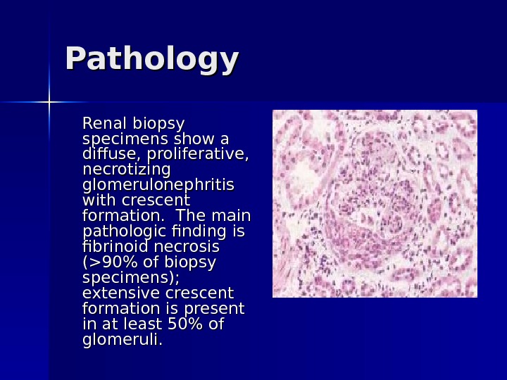
Pathology Renal biopsy specimens show a diffuse, proliferative, necrotizing glomerulonephritis with crescent formation. The main pathologic finding is fibrinoid necrosis (>90% of biopsy specimens); extensive crescent formation is present in at least 50% of glomeruli.
Classification RPGN is classified pathologically into 3 categories: (1) anti-GBM antibody disease (approximately 3% of cases), (2) immune complex disease (45% of cases), (3) pauci-immune disease (50% of cases).
Clinical Manifestations Symptoms and signs of renal failure , , loin pain, haematuria, systemic symptoms (fever, malaise, myalgia, weight loss).
Workup: Lab Studies The most important requirement in the diagnosis ofof antineutrophil cytoplasmic antibodies (ANCA) ANCA-associated disease is a high index of suspicion. Rapid diagnosis is essential for organ preservation. Laboratory studies include the following: – CBC count – Routine chemistry: The most common abnormality is an increased serum creatinine level. – Urinalysis with microscopy: – Antinuclear antibody (ANA) titer: – ANCA Urine and serum protein electrophoresis: Perform this in any middle-aged or elderly person presenting with RPGN to exclude the presence of light-chain disease or overt multiple myeloma as a cause of the clinical findings.
Differentials Amyloidosis, Antiphospholipid Syndrome, Churg-Strauss Syndrome, Cryoglobulinemia, Diffuse Proliferative Glomerulonephritis, Membranoproliferative Glomerulonephritis, Goodpasture Syndrome, Malignant Hypertension, Light-Chain Deposition Disease, Microscopic Polyangiitis, Multiple Myeloma, Lupus Nephritis, Polyarteritis Nodosa, Wegener Granulomatosis
Treatment High-dose corticosteroids; cyclophosphamide ± plasma exchange/ renal transplantation. Prognosis: Poor if initial serum creatinine >600µmol/L.
Chronic Pyelonephritis Chronic pyelonephritis is renal injury induced by recurrent or persistent renal infection.
Etiology E. coli is the commonest (>70% in the community and 41% in hospital). Others include Staphylococcus saprophyticus, Enterococcus faecalis, Proteus mirabilis, Klebsiella species, Enterobacter species, Acinetobacter species, Pseudomonas aeruginosa, and Serratia marascens.
Pathogenesis It occurs almost exclusively in patients with major anatomic anomalies, including urinary tract obstruction, struvite calculi, renal dysplasia, or, most commonly, vesicoureteral reflux (VUR) in young children. Sometimes, this diagnosis is established based on radiologic evidence obtained during an evaluation for recurrent urinary tract infection (UTI) in young children. VUR is a congenital defect that results in incompetence of the ureterovesical valve due to a short intramural segment. The condition is present in 30 -40% of young children with symptomatic UTIs and in almost all children with renal scars. VUR may also be acquired by patients with a flaccid bladder due to spinal cord injury. VUR is classified into 5 grades (I-V), according to the increasing degree of reflux.
Clinical Manifestations Fever Lethargy Nausea and vomiting Flank pain or dysuria Hypertension
Workup Lab Studies: Urinalysis – Urinalysis results may reveal pyuria. – Obtain a urine culture, which often isolates gram-negative bacteria such as Escherichia coli or or Proteus species. – A negative result from urine culture does not exclude a diagnosis of chronic pyelonephritis. – Proteinuria may be present and is a negative prognostic factor for this disease. Serum creatinine and blood urine nitrogen levels are elevated (azotemia).
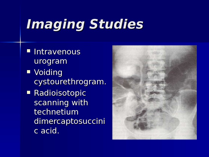
Imaging Studies Intravenous urogram Voiding cystourethrogram. Radioisotopic scanning with technetium dimercaptosuccini c acid.
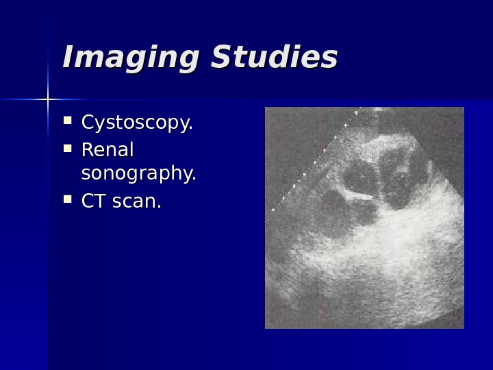
Imaging Studies Cystoscopy. Renal sonography. CT scan.
Differentials Azotemia, Chronic Renal Failure, Hypertension, Nephrolithiasis, Perinephric Abscess, Acute Pyelonephritis, Pyonephrosis, Tuberculosis, Uremia, Xanthogranulomatous Pyelonephritis, Analgesic abuse nephropathy, Renal tuberculosis, Renal malakoplakia
Treatment Medical therapy with antibiotics such as amoxicillin, trimethoprim/sulfamethoxazole (Bactrim), trimethoprim alone, or nitrofurantoin is usually sufficient.
Surgical Care The following are indications for surgical therapy: – Failure to comply with medical regimen – Breakthrough infections occurring in patients who are compliant – Women of childbearing age who prefer surgical therapy Surgery entails the reimplantation of the ureters with the creation of an adequate submucosal tunnel and detrusor support. Diet: Progressive renal injury can be reduced by restricting dietary protein intake.
Prognosis Although most children with chronic pyelonephritis due to VUR may experience spontaneous resolution of reflux, approximately 2% can still progress to renal failure and 5 -6% can have long-term complications, including hypertension. The Birmingham Reflux Study clearly shows that medical and surgical management are equally effective in preventing subsequent renal damage. Almost all children should receive a trial of medical management.