

9842f38b2f16571f5bdcd0503940dee0.ppt
- Количество слайдов: 29
 Refractive Anomalies
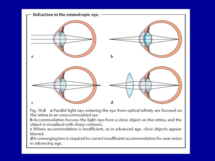
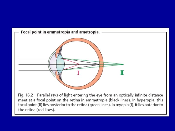
 EMETROPIA • Light rays coming from objects within our visual field are focused by the natural eye lens system (Cornea + Lens) so that an image of the object is brought onto the retina. • The optical system is so arranged that a match exists between the diopteric power and the AP length of the eye. So, with the ciliary muscle relaxed parallel light rays coming from distant objects are focused onto the retina (Emetropia).
 AMETROPIA • If a mismatch between the eye optical power and the eye AP length exists, the image will not fall onto the retina (Ametropia)
 REFRACTIVE ERRORS 1 - MYOPIA 2 - HYPERMETROPIA 3 - ASTIGMATISM
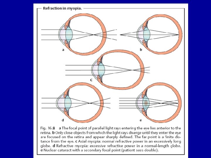
 , parallel") MYOPIA • A mismatch …so that (with the ciliary body relaxed ) , parallel rays from distant objects will be focused in front of the retina. 1 - Axial (e. g. congenital glaucoma). 2 - Curvature (e. g. keratoconus, spherophakia ). 3 - Index (e. g. nuclear sclerosis).
MYOPIA • A mismatch …so that (with the ciliary body relaxed ) , parallel rays from distant objects will be focused in front of the retina. 1 - Axial (e. g. congenital glaucoma). 2 - Curvature (e. g. keratoconus, spherophakia ). 3 - Index (e. g. nuclear sclerosis).
 • Ocular Associations of Myopia: 1 - Exo Deviations 2 - Pseudo Eso deviation 3 - Open angle glaucoma 4 - Cataract 5 - Myopic fundus degeneration (maculopathy, retinal tears, RD) 6 - Impaired convergence 7 - Impaired accommodation 8 - Late onset of presbyopia
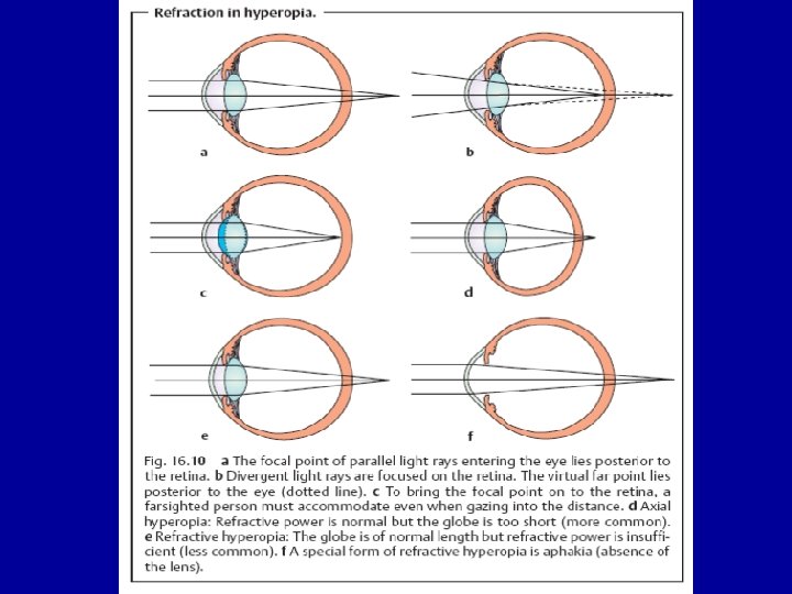
, parallel rays coming from") Hypermetropia A mismatch … so that (with the accommodation relaxed), parallel rays coming from distant objects will be focused behind the retina. 1 - Axial (e. g. microphthalmos) 2 - Curvature (e. g. cornea plana) 3 - Index (e. g. cortical cataract)
Hypermetropia A mismatch … so that (with the accommodation relaxed), parallel rays coming from distant objects will be focused behind the retina. 1 - Axial (e. g. microphthalmos) 2 - Curvature (e. g. cornea plana) 3 - Index (e. g. cortical cataract)
 • Ocular Association of Hypermetropia 1 - Eso Deviations 2 - Pseudo Exo deviation 3 - Angle closure glaucoma 4 - Early onset presbyopia 5 - Retinoschisis
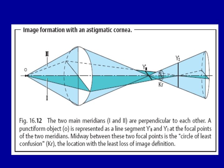
") ASTIGMATISM • A non point image to a point object (No single focal point) • Usually the problem is corneal (being spherocylinderical) • Different meridians of the cornea have different curvature (refraction) so that parallel light rays passing through different meridians will form images on different planes in relation to the retina (Different types)
ASTIGMATISM • A non point image to a point object (No single focal point) • Usually the problem is corneal (being spherocylinderical) • Different meridians of the cornea have different curvature (refraction) so that parallel light rays passing through different meridians will form images on different planes in relation to the retina (Different types)
 Clinically • Patient presents with insidious onset painless loss of vision that usually varies with distance. • Course and progression • Assessment of the refractive state is done using either manual retinoscopy or automated refraction • Anisometropia (difference in the refractive state between the two eyes) is frequently seen
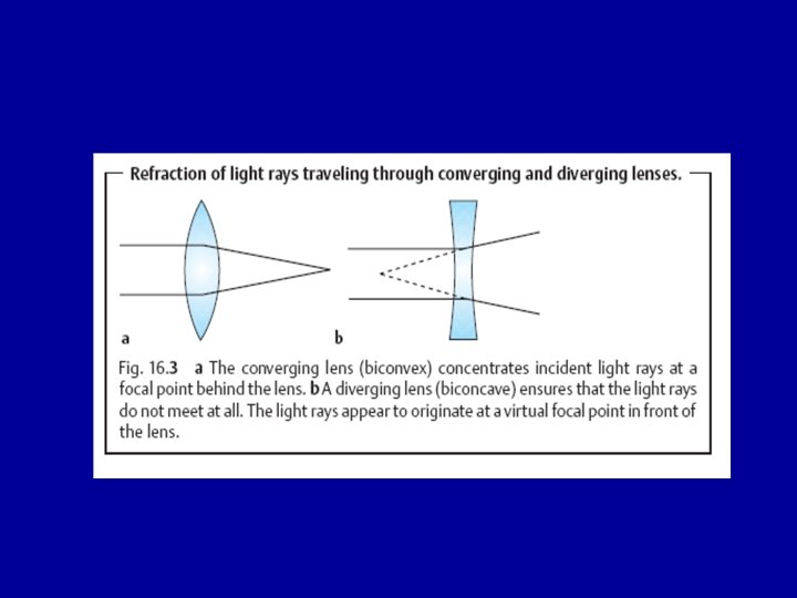
 Correction 1 - GLASSES Types; , Spherical, Cylinderical, Spherocyliderical, Monofocal, Multifocal 2 - CONTACT LENSES Types R , S, Therapeutic, Diagnostic 3 - REFRACTIVE SURGERY Types
 RELAXING INCISIONS") Surgical Correction Option LASIK INTRALASE PRK LASEK EPILASIK CORNEAL STROMAL RINGS (INTACS) RELAXING INCISIONS RADIAL KERATOTOMY
Surgical Correction Option LASIK INTRALASE PRK LASEK EPILASIK CORNEAL STROMAL RINGS (INTACS) RELAXING INCISIONS RADIAL KERATOTOMY
 How LASIK is Performed Step 1. • A suction ring is centered over the cornea of the eye
 Step 2: The microkeratome creates a partial flap in the cornea of uniform thickness
 Step 3: The corneal flap is folded back on the hinge exposing the middle portion of the cornea.
 Step 4: The excimer laser is then used to remove tissue and reshape the center of the cornea.
 Step 5: In the final step, the hinged flap is folded back into its original position.
 CHOROIDAL MELANOMA • Most common intra ocular malignant tumour in adult • Presentation : By chance, reduced Visual acuity, Visual field defect • Diagnosis : Indirect Ophthalmoscopy , Slit lamp biomicroscopy using +90 diopter lens, B-Scan Ultrasound • Subretinal dome shaped elevation, Pigmentation is variable. There may be associated retinal detachment.
 Treatment • Different modalities • Tailored to the patient (size, site, extension, state of fellow eye, patient factors) • 1 -Brachythrapy 2 - External radiotherapy 3 - Transpupillary thermathrapy (ttt) 4 - Transscleral local resection 5 - Enucleation 6 - Exentration 7 - Palliative
 Rhabdomyosarcoma • The most common primary malignant orbital tumour in children • Highly malignant, in its early stages may be mistaken as orbital cellulitis • 7 years • Present as rapidly progressive proptosis, other signs include: 1. palpable mass 2. ptosis 3. swelling & injection of overlying skin (but not hot)
 Rhabdomyosarcoma
 Investigations: 1. Biopsy for diagnosis 2. Systemic assessment for metastasis by CXR, LFT, BMA, LP, skeletal survey. . Treatment: Local radiotherapy + chemotherapy (Good response) IF no response Exentration