2ec53e57336f0755df51b68fedc40190.ppt
- Количество слайдов: 139
 Radiology Differential Diagnosis Richard D. Lackman, MD Director Orthopaedic Oncology Center Cooper University Hospital
 Bone Forming Tumors • • • Osteoid Osteoma Osteoblastoma Osteochondroma Osteosarcoma Blastic Mets Paget’s Cartilage Forming Tumors • • • Enchondroma Osteochondroma Chondromyxoid fibroma Chondroblastoma Chondrosarcoma 3 rd List • • • Infection Mets, myeloma Fibrous dysplasia Nonossifying fibroma LC Histiocytosis GCT ABC Simple Cyst Stress fx Round Cell Tumor Metabolic condition
 Matrix Formation • None ( Lytic ) • Calcified Matrix – Cartilage – Bone – Other
 Margin – width of interface with adjacent medullary bone. Geographic 1 mm. Motheaten 2 -4 mm. Permeative 1 -4 cm.
 Periosteal Reactions Benign Aggressive with Codman Triangles Aggressive Onion Skin Aggressive Hair on End or Sunburst
 Periosteal Reaction • Does Not Occur If: – The tumor is not out in the periosteum • Giant Cell Tumor – The periosteum does not recognize the tumor cells as foreign • Lymphoma
 Osteoid Osteoma • Small lytic nidus with surrounding sclerosis, constant pain relieved by NSAIDS • Prostaglandin secretion • MRI-edema, Best seen on CT scan
 20 y/o with constant pain
 32 y/o with Constant Leg Pain


 Osteoblastoma • Lytic lesion • Painful • Posterior elements of the spine


 Osteochondroma • Grows out from the medullary canal • Cortex of the bone becomes cortex of the lesion • Never sits on an intact cortex • Malignant if cartilage cap is thicker than 2. 5 cm in aduts


 Osteosaroma • Age 10 to 30, second peak in Paget’s • Usually metaphyseal • Permeative margin with cortical destruction and soft tissue extension • Osteoblastic, chondroblastic, fibroblastic, telanectatic (secondary ABC)
 Osteosarcoma

 Periosteal Osteosarcoma

 Osteosarcoma
 Parosteal osteosaroma • Posterior Distal femur • Slightly older age group • Crawls along the surface of the bone • Difficulty invading the medullary canal

 35 y/o with Knee Pain


 Parosteal Osteosarcoma

 Blastic Mets • 90% of prostate mets are blastic • LS Spine via Batson’s plexus • Breast may be mixed
 Pagets Disease • Uncoupled resorption-formation • Spectrum of associated radiographic findings • Pain: stress fracture vs. malignant transformation

 Enchondroma • Stippled calcification • Does nothing bad to the bone • Not painful • Always hot on bone scan

 Enchondroma
 Enchondroma
 Enchondroma
 Enchondroma

 Enchondroma
 Chondrosarcoma • Intralesional lysis • Endosteal scalloping • Cortical thinning/expansion • pain
 Chondrosarcoma in Enchondroma

 Chondrosarcoma

 Chondrosarcoma
 Chondromyxoid fibroma • Large benign looking lytic lesion • Proximal tibia • Young adults (20 -40) • Painful
 13 y/o with Knee Pain
 Periosteal chondroma • Benign cartilage tumor on the surface • Periosteal scalloping • May be painful
 Periosteal Chondroma
 Chondroblastoma • Lytic lesion in the epiphysis of a child • Painful • Significant surrounding edema • Mimics infection

 Chondroblastoma
 Chondroblastoma
 16 y/o with Shoulder Pain
 Infection • Mimics everything • Poorly marginated • Significant edema

 Osteomyelitis
 Lytic Mets • Most common aggressive lesion in older adults • Soft issue mass suggests sarcoma but occurs with kidney, lung • Usually multiple
 Plasmacytoma/myeloma • Punched out lytic lesions in bone • May mimic osteoporosis • Very lytic within the lesion

 Fibrous dysplasia • In the diff dx of every benign lesion • Long lesion in a long bone • Ground glass deformity • May be polyostotic • May be small and mimic NOF


 30 y/o with Hip Pain

 NOF
NOF


 18 y/o with Leg Pain


 LC Histiocytosis • Intra-medullary lytic lesion in a child • Poorly marginated • Periosteal reaction • Very inflammatory • Painful
 12 y/o with Hip Pain

 Giant Cell Tumor of Bone • Juxta-articular • Lytic Lesion • Moth Eaten Margin • Cortical Thinning or Erosion • No Periosteal Reaction


 Giant Cell Tumor


 Unicameral bone cyst • Full width lytic lesion • Cortical thinning • No periosteal reaction • Slight expansion • Proximal Humerus


 Simple Bone Cyst (? ABC)

 UBC
UBC
 Calcaneal UBC
 Aneurysmal bone cyst • Eccentric lytic lesion • Very aneurysmal • Fluid/fluid levels • May be primary or secondary to vascular tumors

 20 y/o with Elbow Pain


 Lymphoma • Marrow replacement • Permeative, not destructive • Minimal bone changes • Late soft tissue extension • Minimal periosteal reaction


 Ewing’s Sarcoma • Children and young adults • Diaphyseal • Onion skin periosteal reaction • Large soft tissue mass • 11/22, CD 99, Ews-fli
 Ewing’s Sarcoma
 20 y/o with Leg Pain
 18 y/o with Elbow Pain

 Adamantinoma • Young adults • Soap bubble sclerotic lesion of the anterior cortex of the shaft of the tibia • Biphasic (epithelial/mesenchymal)
 Lesions in the Anterior Cortex of the Tibial Shaft • Adamantinoma • Cortical Fibrous Dysplasia

 Adamantinoma

 30 y/o with Leg Pain


 Chordoma • Notochord remnant tumor • Midline • Sacrum and O/C junction • Anterior extension
 60 y/o with Low Back Pain


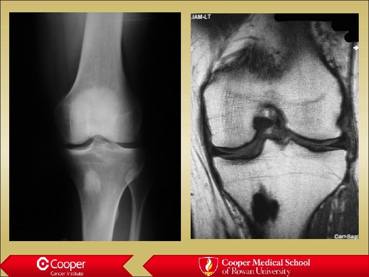
 Bone infarct • Well marginated • “Coast of Maine” contour with peripheral ossification • May have surrounding edema if acute

 Hemangioma • Maintenance of vertical trabeculae • “Jail house” vertebrae • Polka dots on axial CT
 50 y/o with Low Back Pain
 Intraosseous Lipoma • Sclerotic border • Fat density asymptomatic incidental finding


 Bone Island • Cortical bone in the medullary canal • Well marginated
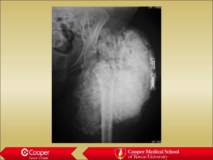
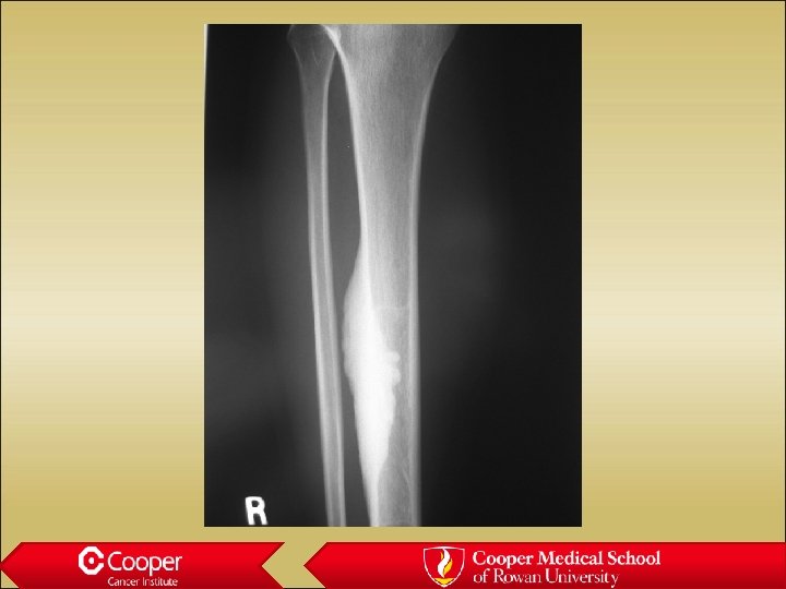
 Tumoral calcinosis • Amorphous calcium in soft tissues • Associated with renal failure
 Thank You


