
Puzzle of the day What is the link


- Размер: 2.6 Mегабайта
- Количество слайдов: 34
Описание презентации Puzzle of the day What is the link по слайдам
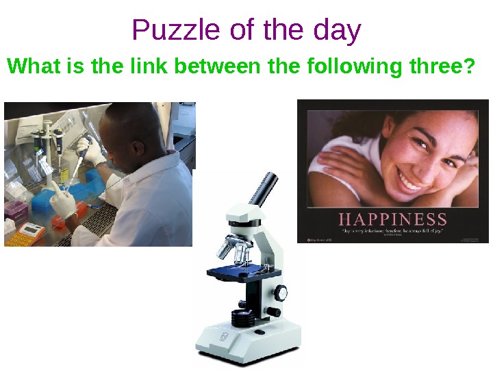
Puzzle of the day What is the link between the following three?
The study of cells Dr. Sophia Semerdjieva s. semerdjieva@ucl. ac. uk Office 2.
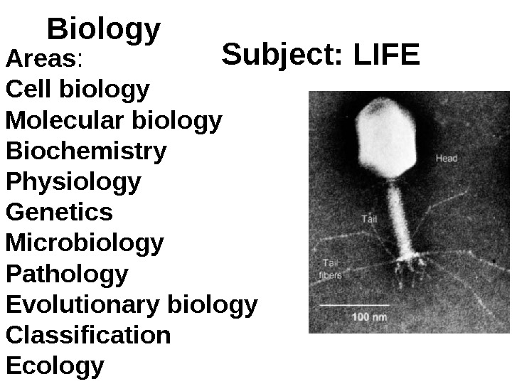
Biology Subject: LIFE Areas : Cell biology Molecular biology Biochemistry Physiology Genetics Microbiology Pathology Evolutionary biology Classification Ecology
At today’s lecture we will 1. Describe the main principles of light and electron microscopy. 2. Summarize the main points of the cell theory. 3. Outline the main features of animal and plant cells as seen under the light microscope.
By the end of today’s lecture you should: 1) Understand the main principles of both light and electron microscopy and be able to compare the two types of microscopes. 2) Be able to use confidently the standard SI units of measurement. 3) Understand be able to use appropriately the terms magnification and resolution. 4) Understand the cell theory. 5) Be able to compare the structure of animal and plant cells as seen under the light microscope. Learning outcomes
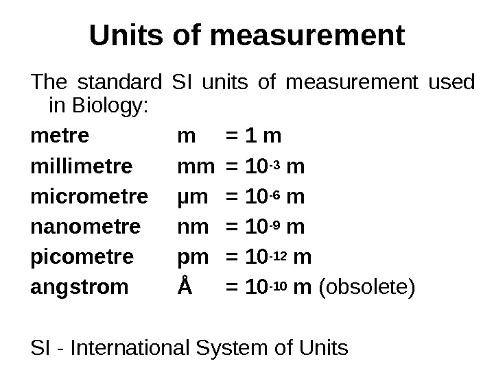
Units of measurement The standard SI units of measurement used in Biology: metre m = 1 m millimetre mm = 10 -3 m micrometre µm = 10 -6 m nanometre nm = 10 -9 m picometre pm = 10 -12 m angstrom Å = 10 -10 m (obsolete) SI — International System of Units
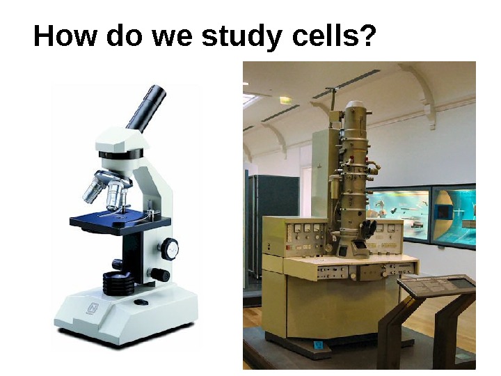
How do we study cells?
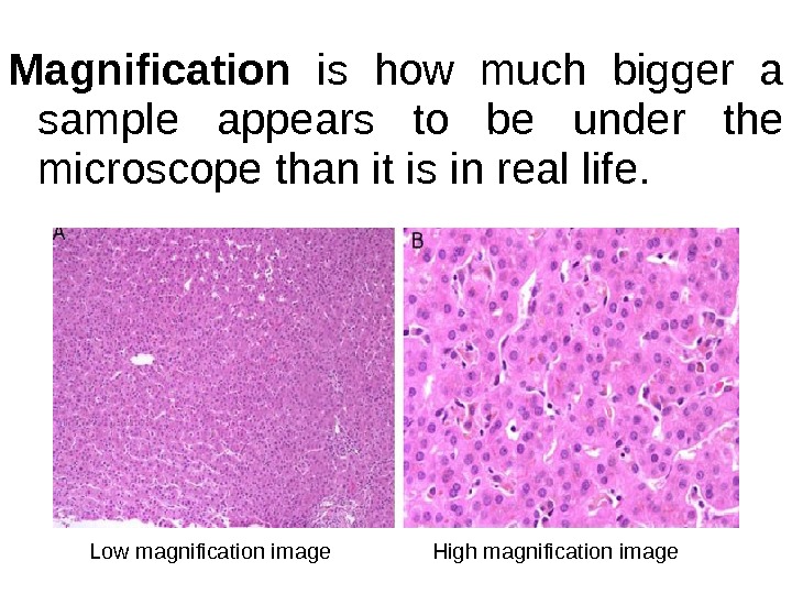
Magnification is how much bigger a sample appears to be under the microscope than it is in real life. Low magnification image High magnification image
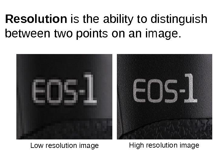
Low resolution image High resolution image. Resolution is the ability to distinguish between two points on an image.
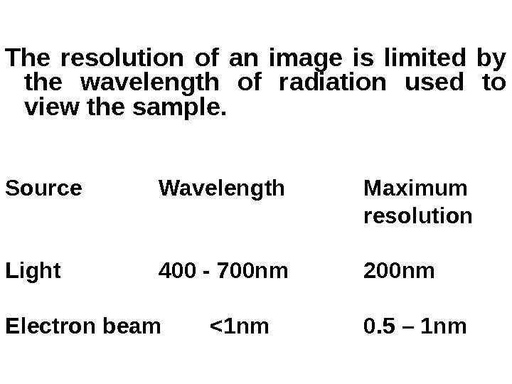
The resolution of an image is limited by the wavelength of radiation used to view the sample. Source Wavelength Maximum resolution Light 400 — 700 nm 200 nm Electron beam <1 nm 0. 5 – 1 nm
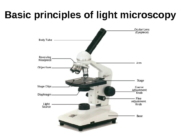
Basic principles of light microscopy
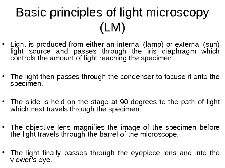
Basic principles of light microscopy (LM) • Light is produced from either an internal (lamp) or external (sun) light source and passes through the iris diaphragm which controls the amount of light reaching the specimen. • The light then passes through the condenser to focuse it onto the specimen. • The slide is held on the stage at 90 degrees to the path of light which next travels through the specimen. • The objective lens magnifies the image of the specimen before the light travels through the barrel of the microscope. • The light finally passes through the eyepiece lens and into the viewer’s eye.
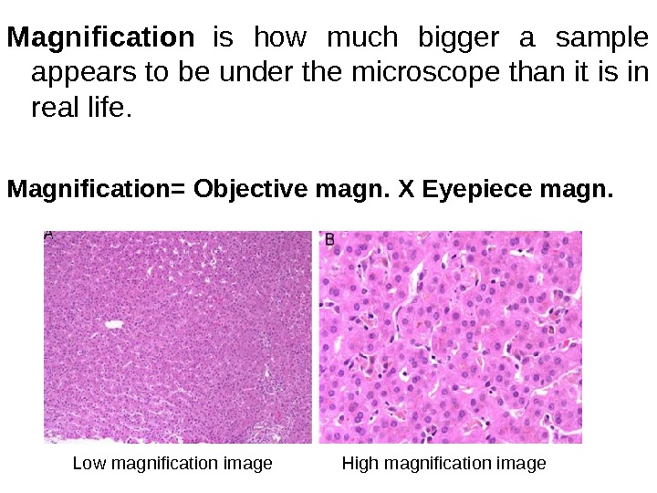
Magnification is how much bigger a sample appears to be under the microscope than it is in real life. Magnification= Objective magn. X Eyepiece magn. Low magnification image High magnification image
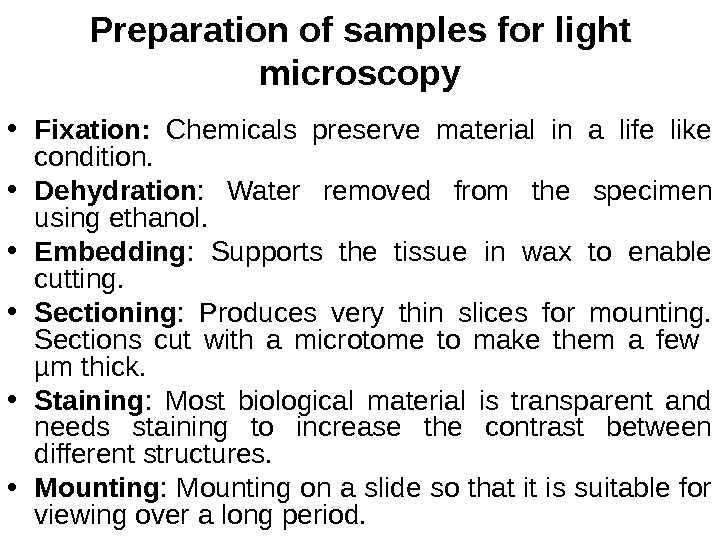
Preparation of samples for light microscopy • Fixation: Chemicals preserve material in a life like condition. • Dehydration : Water removed from the specimen using ethanol. • Embedding : Supports the tissue in wax to enable cutting. • Sectioning : Produces very thin slices for mounting. Sections cut with a microtome to make them a few µm thick. • Staining : Most biological material is transparent and needs staining to increase the contrast between different structures. • Mounting : Mounting on a slide so that it is suitable for viewing over a long period.
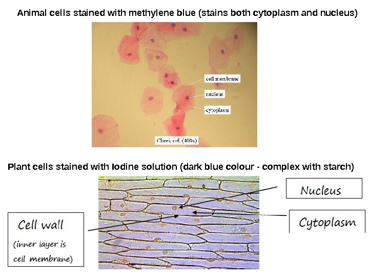
Animal cells stained with methylene blue (stains both cytoplasm and nucleus) Plant cells stained with Iodine solution (dark blue colour — complex with starch)
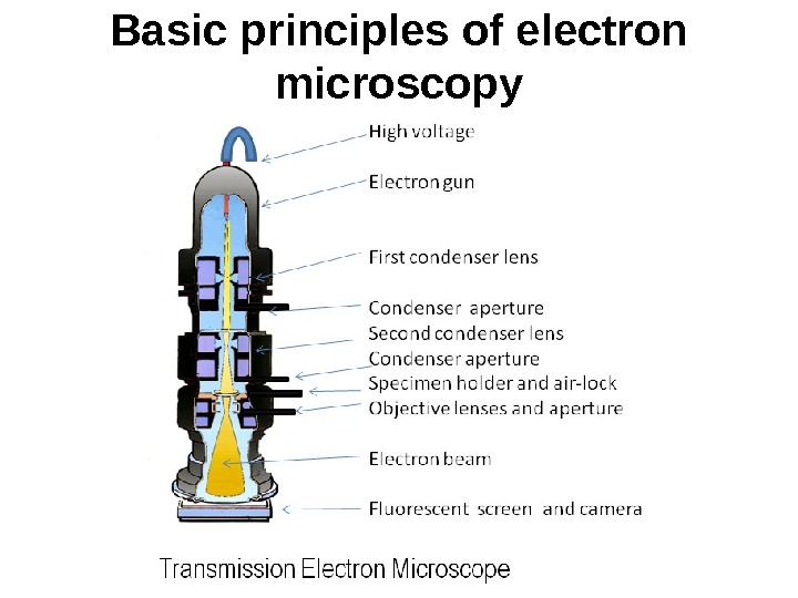
Basic principles of electron microscopy
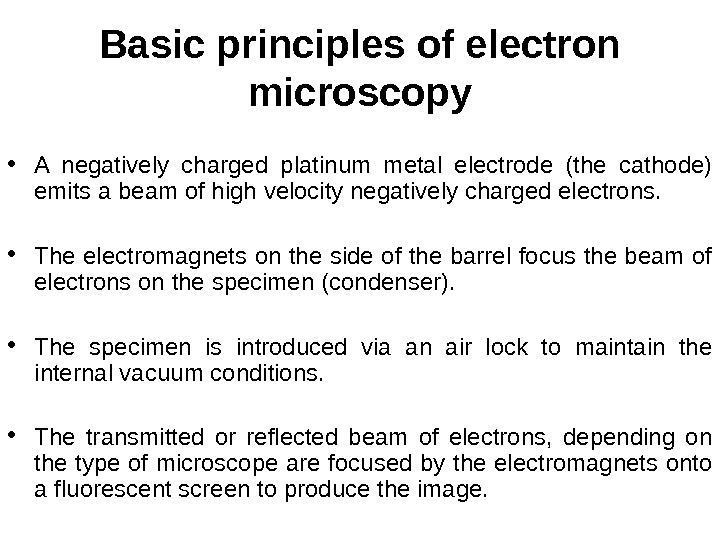
Basic principles of electron microscopy • A negatively charged platinum metal electrode (the cathode) emits a beam of high velocity negatively charged electrons. • The electromagnets on the side of the barrel focus the beam of electrons on the specimen (condenser). • The specimen is introduced via an air lock to maintain the internal vacuum conditions. • The transmitted or reflected beam of electrons, depending on the type of microscope are focused by the electromagnets onto a fluorescent screen to produce the image.
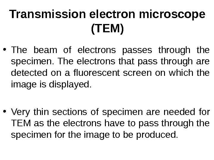
Transmission electron microscope (TEM) • The beam of electrons passes through the specimen. The electrons that pass through are detected on a fluorescent screen on which the image is displayed. • Very thin sections of specimen are needed for TEM as the electrons have to pass through the specimen for the image to be produced.
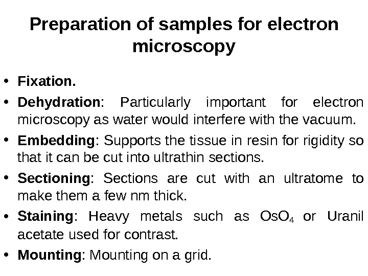
Preparation of samples for electron microscopy • Fixation. • Dehydration : Particularly important for electron microscopy as water would interfere with the vacuum. • Embedding : Supports the tissue in resin for rigidity so that it can be cut into ultrathin sections. • Sectioning : Sections are cut with an ultratome to make them a few nm thick. • Staining : Heavy metals such as Os. O 4 or Uranil acetate used for contrast. • Mounting : Mounting on a grid.
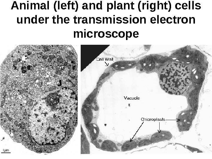
Animal (left) and plant (right) cells under the transmission electron microscope
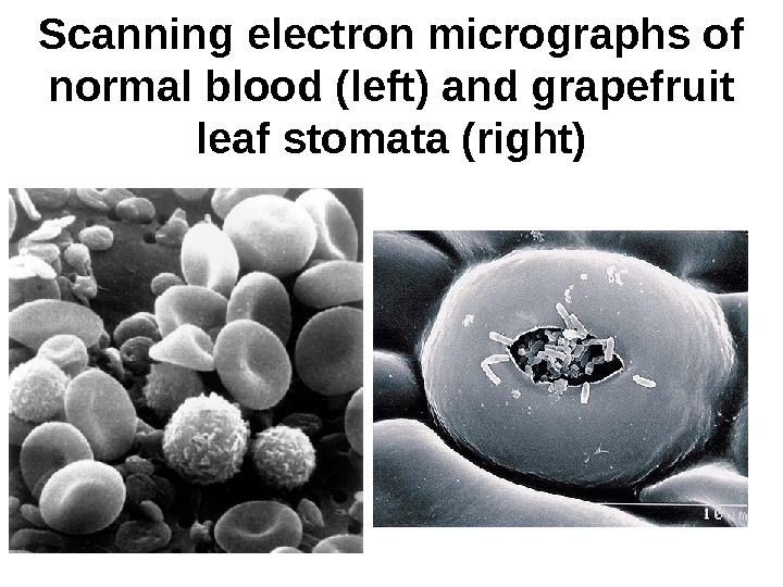
Scanning electron microscope (SEM) • The beam of electrons is passed over the surface of the specimen in the form of a ‘scanning’ beam. • Electrons are reflected off the surface of the specimen as it has been previously coated with heavy metals. • The reflected electrons are focussed on the fluorescent screen in order to produce the image. • Larger, thicker structures can be seen under the scanning electron microscope as the electrons do not have to pass through the sample in order to form the image. • The resolution of the SEM is lower than that of the TEM.
Scanning electron micrographs of normal blood (left) and grapefruit leaf stomata (right)
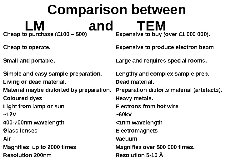
Cheap to purchase (£ 100 – 500) Expensive to buy (over £ 1 000). Cheap to operate. Expensive to produce electron beam Small and portable. Large and requires special rooms. Simple and easy sample preparation. Lengthy and complex sample prep. Living or dead material. Dead material. Material maybe distorted by preparation. Preparation distorts material (artefacts). Coloured dyes Heavy metals. Light from lamp or sun Electrons from hot wire ~12 V ~60 k. V 400 -700 nm wavelength <1 nm wavelength Glass lenses Electromagnets Air Vacuum Magnifies up to 2000 times Magnifies over 500 000 times. Resolution 200 nm Resolution 5 -10 ÅComparison between LM and TEM
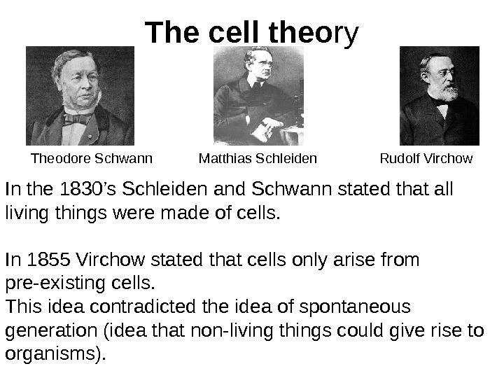
The cell theo ry Theodore Schwann Matthias Schleiden Rudolf Virchow In the 1830’s Schleiden and Schwann stated that all living things were made of cells. In 1855 Virchow stated that cells only arise from pre-existing cells. This idea contradicted the idea of spontaneous generation (idea that non-living things could give rise to organisms).
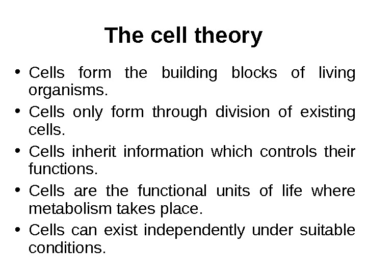
The cell theory • Cells form the building blocks of living organisms. • Cells only form through division of existing cells. • Cells inherit information which controls their functions. • Cells are the functional units of life where metabolism takes place. • Cells can exist independently under suitable conditions.
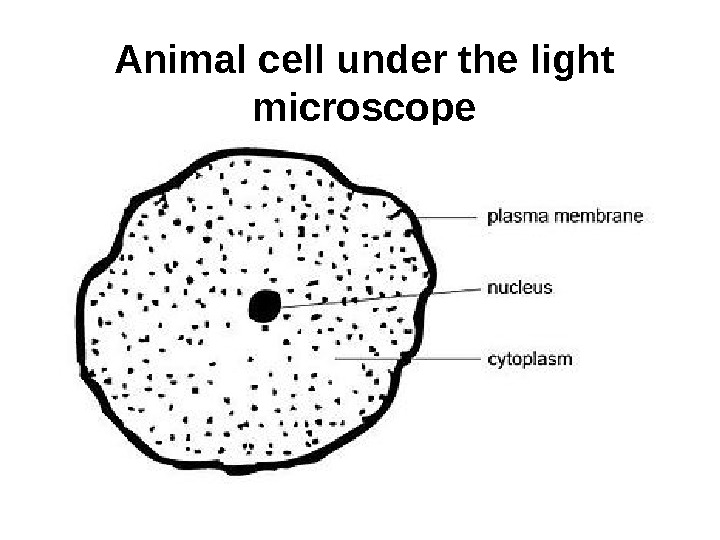
Animal cell under the light microscope
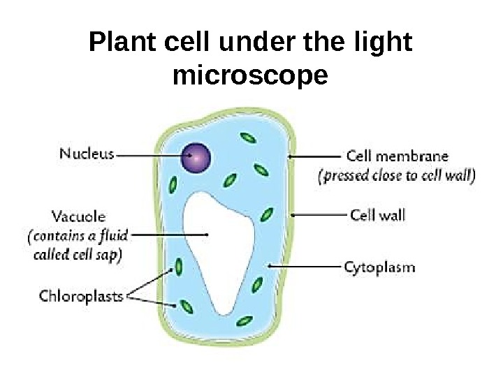
Plant cell under the light microscope
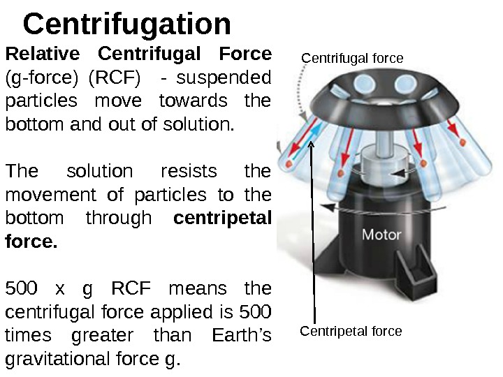
Centrifugation Relative Centrifugal Force (g-force) (RCF) — suspended particles move towards the bottom and out of solution. The solution resists the movement of particles to the bottom through centripetal force. 500 x g RCF means the centrifugal force applied is 500 times greater than Earth’s gravitational force g. Centrifugal force Centripetal force
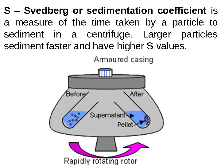
S – Svedberg or sedimentation coefficient is a measure of the time taken by a particle to sediment in a centrifuge. Larger particles sediment faster and have higher S values.
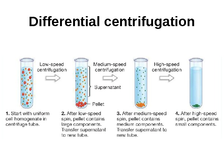
Differential centrifugation
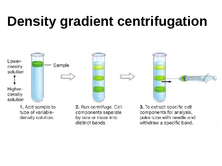
Density gradient centrifugation
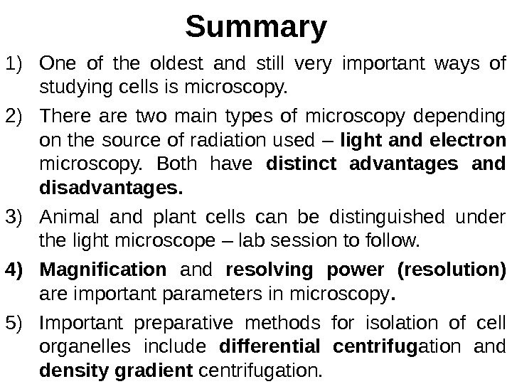
Summary 1) One of the oldest and still very important ways of studying cells is microscopy. 2) There are two main types of microscopy depending on the source of radiation used – light and electron microscopy. Both have distinct advantages and disadvantages. 3) Animal and plant cells can be distinguished under the light microscope – lab session to follow. 4) Magnification and resolving power (resolution) are important parameters in microscopy. 5) Important preparative methods for isolation of cell organelles include differential centrifug ation and density gradient centrifugation.
Puzzle of the day What is the link between the following three?
Dale Carnegie “ Are you bored with life? Then throw yourself into some work you believe in with all your heart, live for it, die for it, and you will find happiness that you had thought could never be yours. ”