Л.1,2 Микрофлора полости рта.ppt
- Количество слайдов: 42
 Проф. Чувилкин В. И. Микрофлора полости рта Москва – 2007
 Основные группы бактерий вегетирующих в полости рта. Родовые группы бактерий стрептококки вейлонеллы дифтероиды
 Типы дыхания бактерий • Анаэробное • Аэробное • Факультативно-анаэробное дыхание
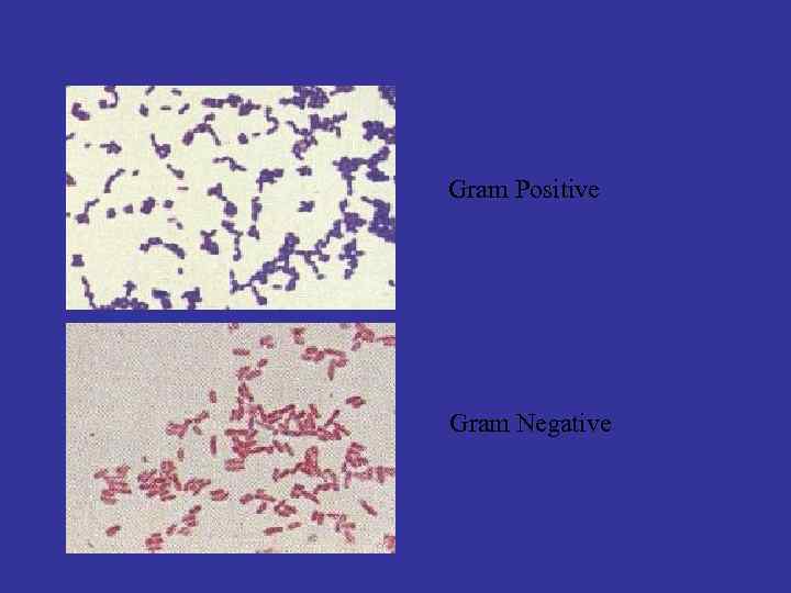 Gram Positive Gram Negative
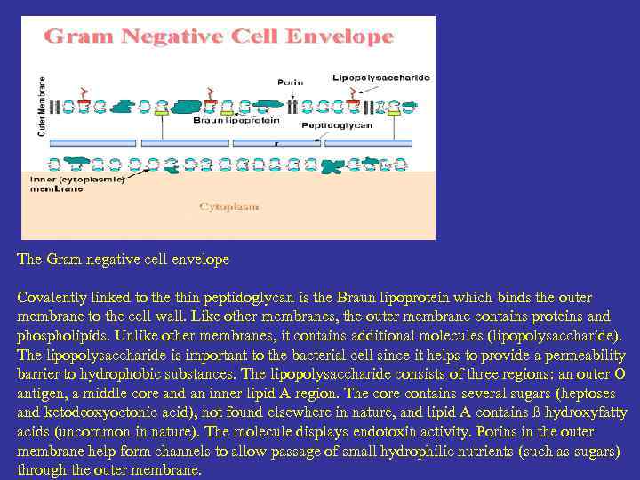 The Gram negative cell envelope Covalently linked to the thin peptidoglycan is the Braun lipoprotein which binds the outer membrane to the cell wall. Like other membranes, the outer membrane contains proteins and phospholipids. Unlike other membranes, it contains additional molecules (lipopolysaccharide). The lipopolysaccharide is important to the bacterial cell since it helps to provide a permeability barrier to hydrophobic substances. The lipopolysaccharide consists of three regions: an outer O antigen, a middle core and an inner lipid A region. The core contains several sugars (heptoses and ketodeoxyoctonic acid), not found elsewhere in nature, and lipid A contains ß hydroxyfatty acids (uncommon in nature). The molecule displays endotoxin activity. Porins in the outer membrane help form channels to allow passage of small hydrophilic nutrients (such as sugars) through the outer membrane.
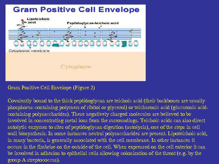 Gram Positive Cell Envelope (Figure 2) Covalently bound to the thick peptidoglycan are teichoic acid (their backbones are usually phosphorus-containing polymers of ribitol or glycerol) or teichuronic acid (glucuronic acidcontaining polysaccharides). These negatively charged molecules are believed to be involved in concentrating metal ions from the surroundings. Teichoic acids can also direct autolytic enzymes to sites of peptidoglycan digestion (autolysis), one of the steps in cell wall biosynthesis. In some instances neutral polysaccharides are present. Lipoteichoic acid, in many bacteria, is generally associated with the cell membrane. In other instances it occurs in the fimbriae on the outside of the cell. When expressed on the cell exterior it can be involved in adhesion to epithelial cells allowing colonization of the throat (e. g. by the group A streptococcus).
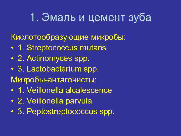 1. Эмаль и цемент зуба Кислотообразующие микробы: • 1. Streptococcus mutans • 2. Actinomyces spp. • 3. Lactobacterium spp. Микробы-антагонисты: • 1. Veillonella alcalescence • 2. Veillonella parvula • 3. Peptostreptococcus spp.
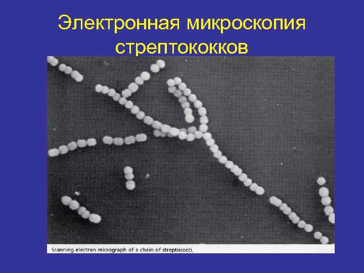 Электронная микроскопия стрептококков
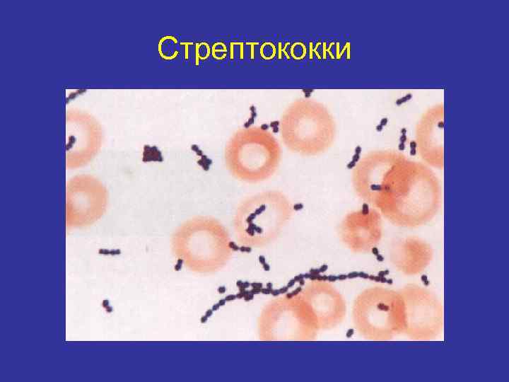 Стрептококки
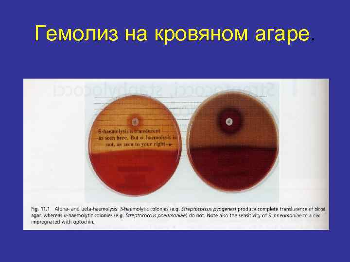 Гемолиз на кровяном агаре.
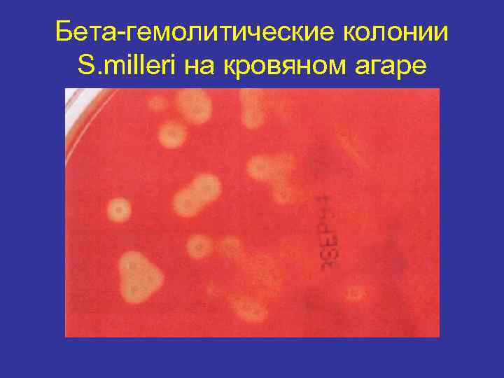 Бета-гемолитические колонии S. milleri на кровяном агаре
 2. Зубо-десневой желобок • Грам-отрицательные бактерии. • 1. Prevotella melaninogenica • 2. Fusobacterium spp. • • • Грам-положительные бактерии. 1. Peptostreptococcus spp. 2. Actinomyces spp. 3. Streptococcus sanguis 4. Streptococcus intermedius
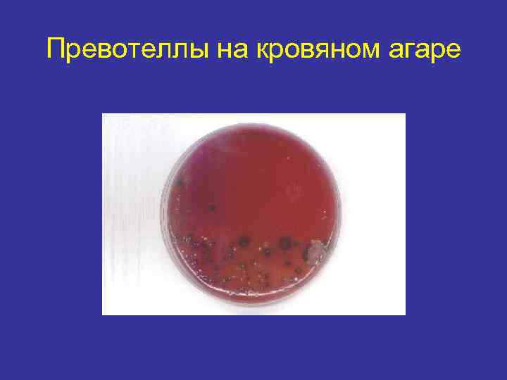 Превотеллы на кровяном агаре
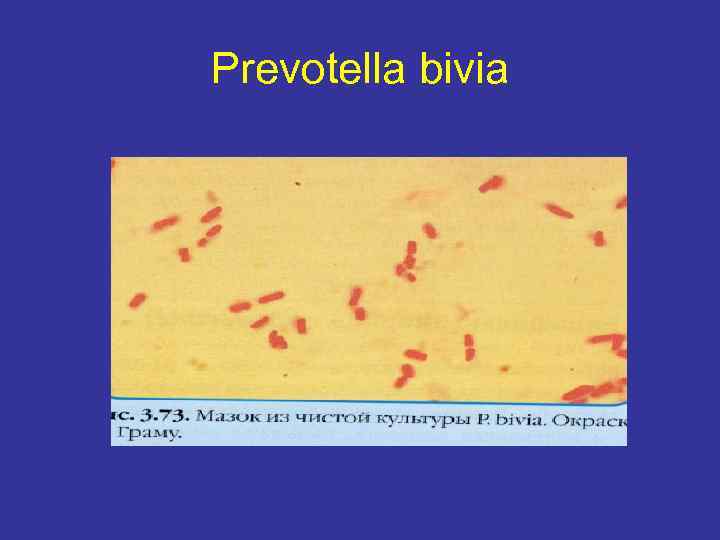 Prevotella bivia
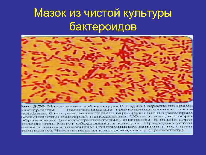 Мазок из чистой культуры бактероидов
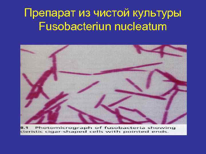 Препарат из чистой культуры Fusobacteriun nucleatum
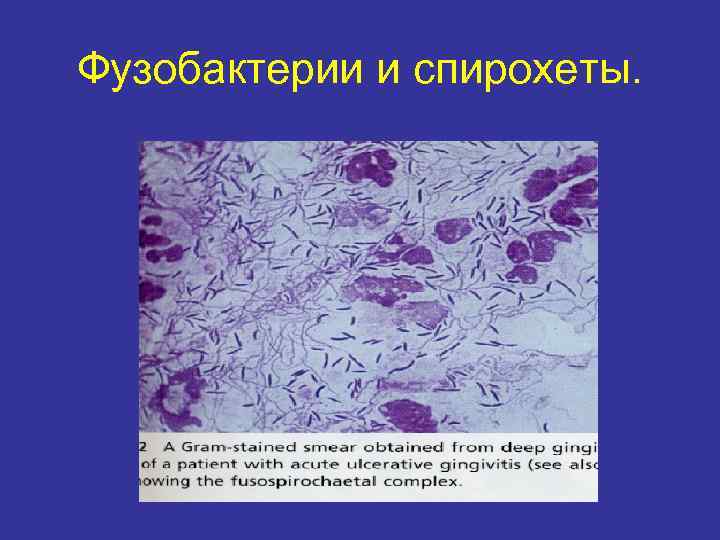 Фузобактерии и спирохеты.
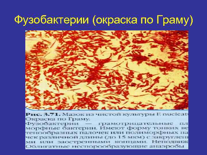 Фузобактерии (окраска по Граму)
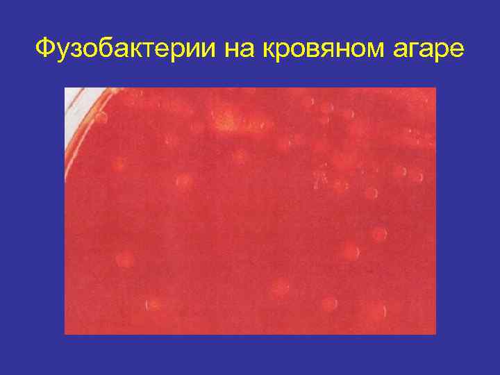 Фузобактерии на кровяном агаре
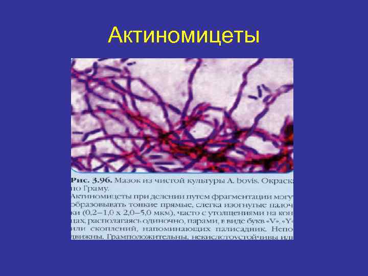 Актиномицеты
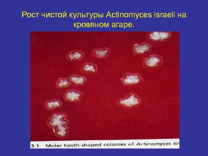 Рост чистой культуры Actinomyces israeli на кровяном агаре.
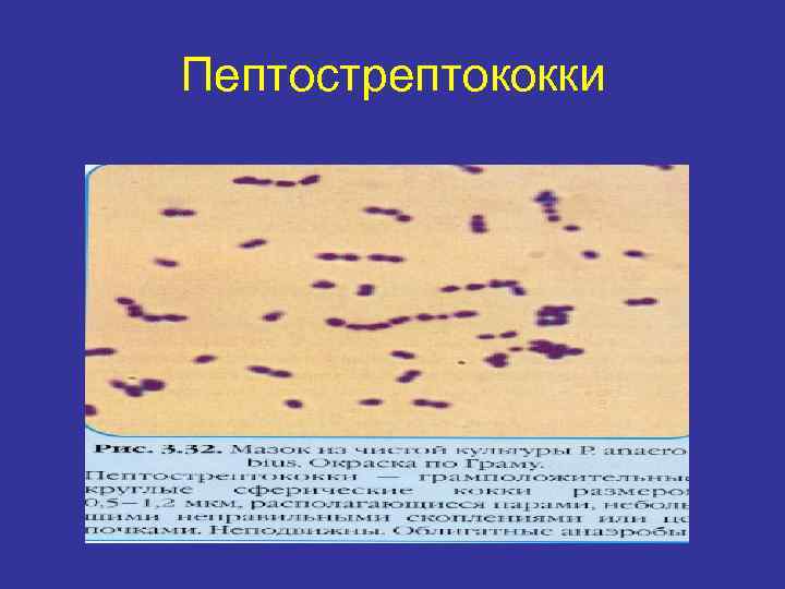 Пептострептококки
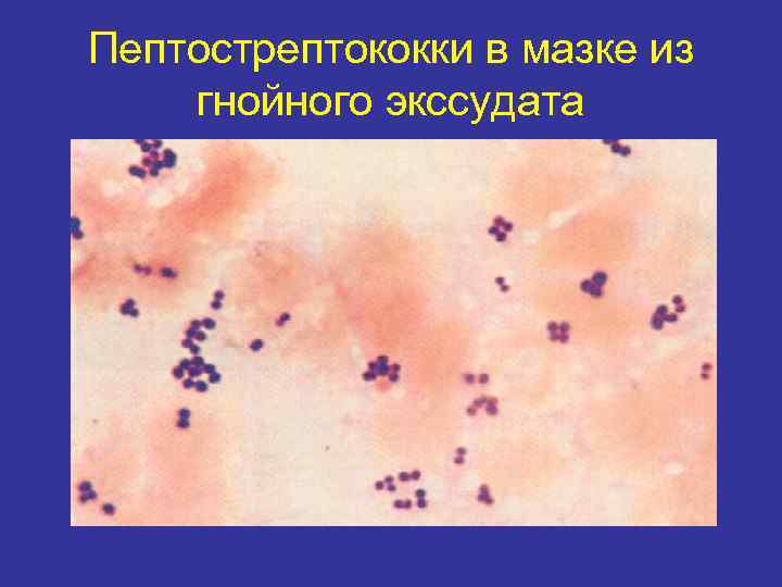 Пептострептококки в мазке из гнойного экссудата
 3. Спинка языка • • • 1. Streptococcus salivarius 2. Streptococcus sanguis 3. Lactobacillus spp. 4. Leptotrichia buccalis 5. Candida spp.
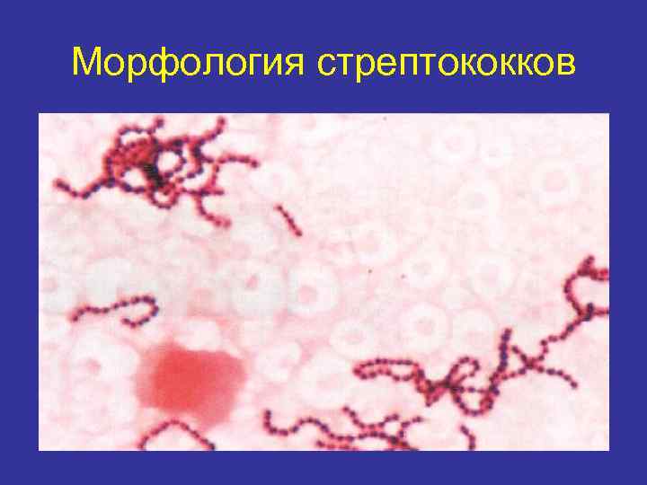 Морфология стрептококков
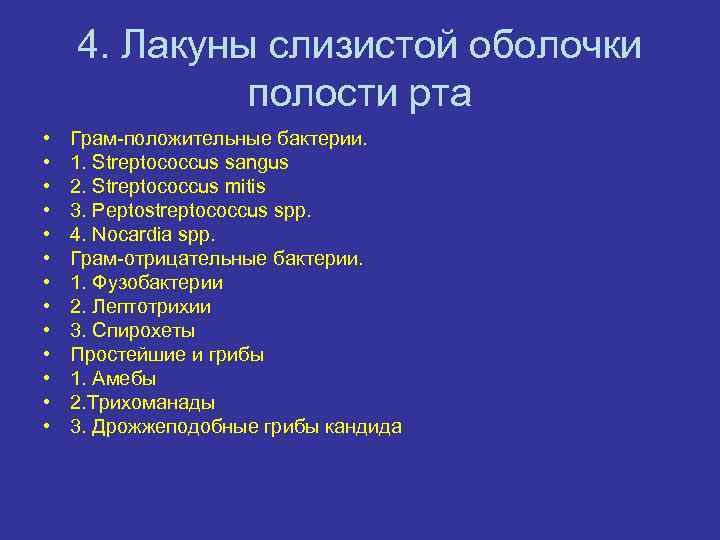 4. Лакуны слизистой оболочки полости рта • • • • Грам-положительные бактерии. 1. Streptococcus sangus 2. Streptococcus mitis 3. Peptostreptococcus spp. 4. Nocardia spp. Грам-отрицательные бактерии. 1. Фузобактерии 2. Лептотрихии 3. Спирохеты Простейшие и грибы 1. Амебы 2. Трихоманады 3. Дрожжеподобные грибы кандида
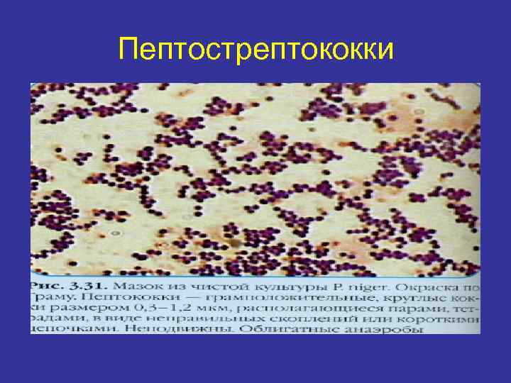 Пептострептококки
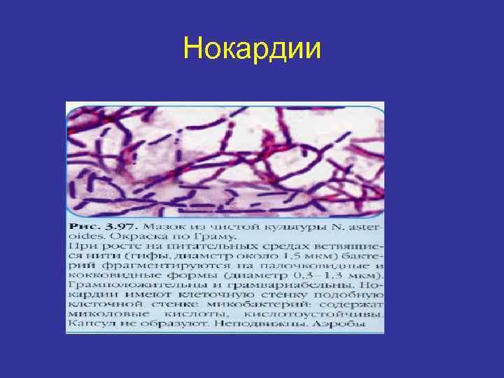 Нокардии
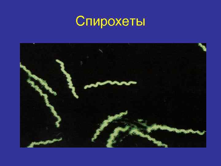 Спирохеты
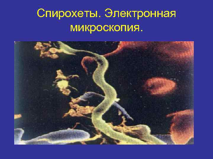 Спирохеты. Электронная микроскопия.
 5. Слизистая оболочка полости рта • • • Грам-положительные бактерии 1. S. sanguis 2. S. mitis 3. Corynebacterium spp. Грам-отрицательные бактерии 1. Veillonella spp. 2. Neiseria spp. 3. Propionibacterium spp. Дрожжеподобные грибы
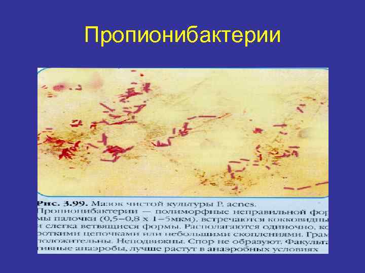 Пропионибактерии
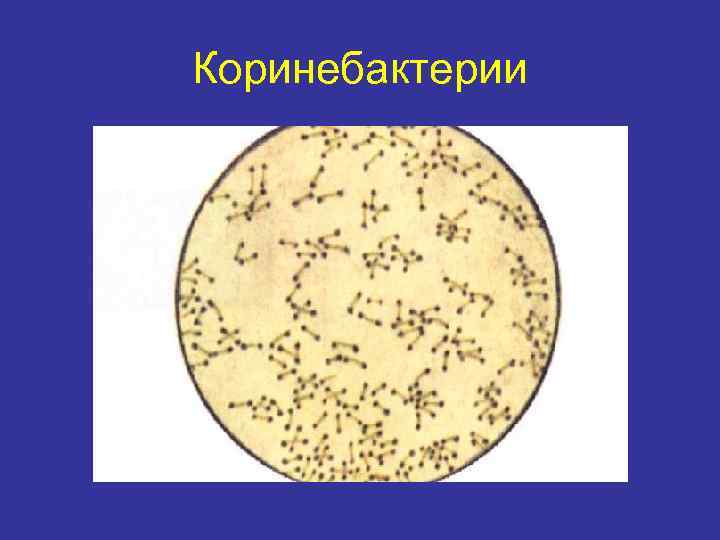 Коринебактерии
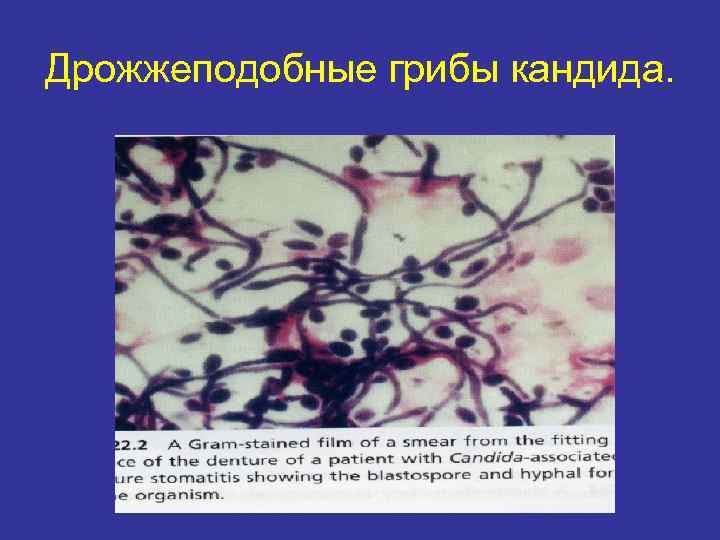 Дрожжеподобные грибы кандида.
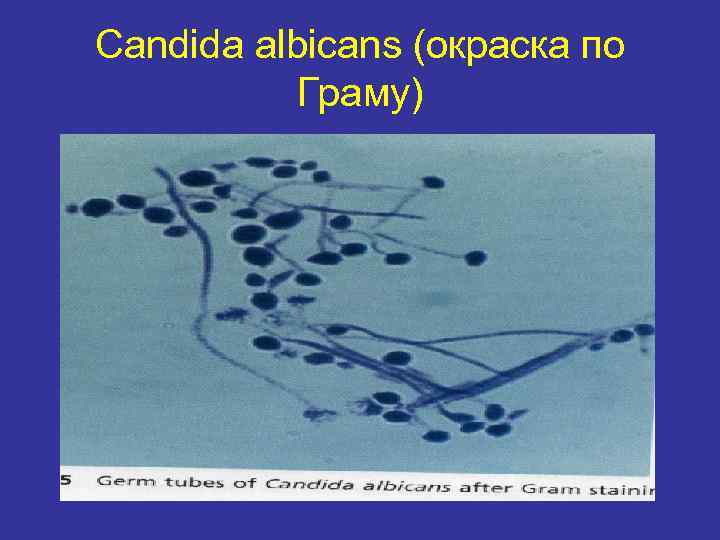 Candida albicans (окраска по Граму)
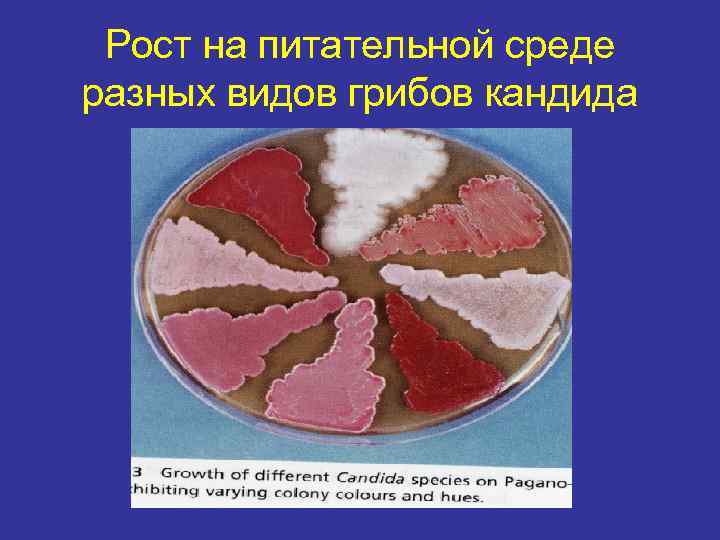 Рост на питательной среде разных видов грибов кандида
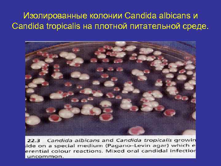 Изолированные колонии Candida albicans и Candida tropicalis на плотной питательной среде.
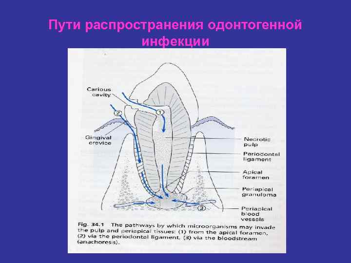 Пути распространения одонтогенной инфекции
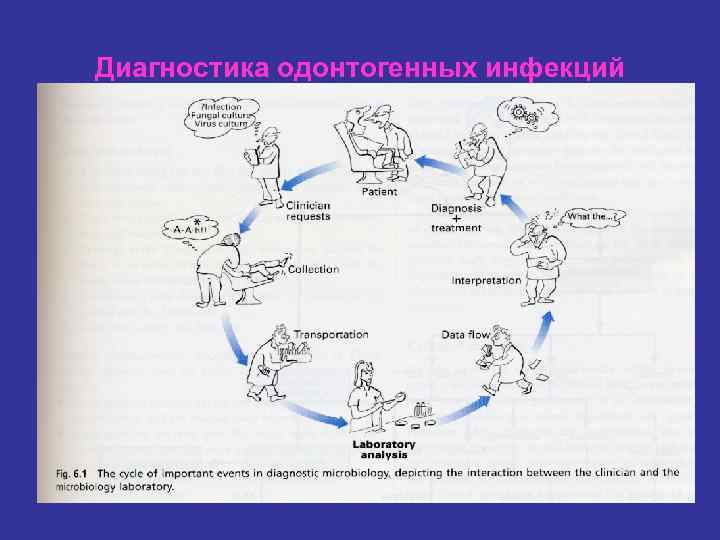 Диагностика одонтогенных инфекций


