Презентация wounds. wounds healing amp complications


wounds._wounds_healing_amp_complications.ppt
- Размер: 13.5 Mегабайта
- Количество слайдов: 82
Описание презентации Презентация wounds. wounds healing amp complications по слайдам
 Wounds, wound healing & complications


 Tidy incised wound on the finger


 Untidy avulsed wound on the hand
 Facial trauma – apparent tissue loss but none found after careful matching
 Degloving hand injury
 Dog bite in a child
 Degloving buttock injury
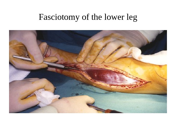
 Fasciotomy of the lower leg


 (a) Early inflammatory phase with platelet-enriched blood clot and dilated vessels. (b) Late inflammatory phase with increased vascularity and increase in polymorphonuclear lymphocytes (PMN) and lymphocytes (round cells). (c) Proliferative phase with capillary buds and fibroblasts. (d) Mature contracted scar.


 Multiple keloid scars


 Burn contractures showing hyperextended fingers and hyperflexed elbow.
 Post-traumatic (chainsaw) midline neck contracture
 Multiple Z-plasty release of finger contracture
 Treatment of hypertrophic and keloid scars • Pressure – local moulds or elasticated garments • Silicone gel sheeting (mechanism unknown) • Intralesional steroid injection (triamcinolone) • Excision and steroid injectiona • Excision and postoperative radiation (external beam or brachytherapy)a • Intralesional excision (keloids only) • Laser – to reduce redness (which may resolve in any event) • Vitamin E or palm oil massage (unproven)
 MANAGING THE ACUTE WOUND • The surgeon must remember to examine the whole patient • A bleeding wound should be elevated and a pressure • Clamps should not be put on vessels blindly as nerve damage is likely and vascular anastomosis is rendered impossible • Devitalised tissue must be excised until bleeding occurs with the obvious exception of nerves, vessels and tendons • Muscle viability is judged by the colour, bleeding pattern and contractility • In a tidy wound, repair of all damaged structures may be attempted
 Meshed split-skin graft
 Managing the acute wound • Cleansing • Exploration and diagnosis • Debridement • Repair of structures • Replacement of lost tissues where indicated • Skin cover if required • Skin closure without tension • All of the above with careful tissue handling and meticulous technique
 CHRONIC WOUNDS 1. Leg ulcers Aetiology of leg ulcers • Venous disease leading to local venous hypertension (e. g. varicose veins) • Arterial disease, either large vessel (atherosclerosis) or small vessel (diabetes) • Arteritis associated with autoimmune disease (rheumatoid • arthritis, lupus, etc. ) 2.
 THANK YOU!

