
Презентация projection of great vessels Tlemissova 350


projection_of_great_vessels_tlemissova_350.ppt
- Размер: 1 Mегабайта
- Количество слайдов: 17
Описание презентации Презентация projection of great vessels Tlemissova 350 по слайдам
The determination of the projection of large vessels on the surface of body: the determination of places for auscultation and pressing of vessels Tlemissova G 350 GM
Plan of work Heart(structure) The projection of the heart valves and large vessels The topography of the large vessels Point of auscultation Percussion
Heart(structure)and vessels The heart (Latin с or , Gr. Καρδιά) — fibromuscular organ that blood flow through blood vessels.
Human heart The human heart consists of four chambers separated by septa and valves. The blood from the upper and lower vena cava enters the right atrium, passes through the tricuspid valve (which is composed of three petals) in the right ventricle. Then through the pulmonary valve into the pulmonary artery enters, goes to the lungs where the exchange and returns to the left atrium. Then through the mitral (Bivalve), valve (it consists of two lobes) enters the left ventricle, then goes through the aortic valve into the aorta. In the right atrium are hollow to the left atrium — pulmonary veins. From the right and left ventricular out, respectively, the pulmonary artery (pulmonary trunk) and the ascending aorta. The right ventricle and left atrium close the pulmonary circulation, the left ventricle and right atrium — a large circle. The heart is located at the bottom of the anterior mediastinum, most of its front surface is covered with light. With flowing sections of hollow and the pulmonary veins, as well as facing the aorta and pulmonary trunk is covered with a shirt (pericardium). In the pericardial cavity contains a small amount of serous fluid. An adult man of his size and weight, on average for men 783 cm ³ and 332 g for women — 560 cm ³ and 253, the Through the heart of man during the day runs from 7000 to 10 000 liters of blood per year, about 3. 15 million liters. Adult length is 12 -15 cm of the heart, the transverse size of 8 -11 cm, 5. 8 cm anteroposterior size of the heart weight 220 — 300 g.
Human heart
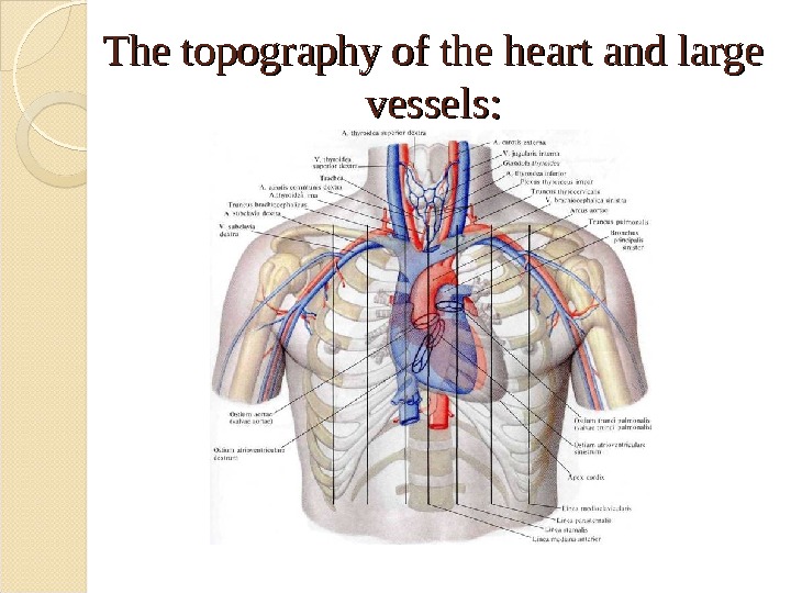
The projection of the heart valves and large vessels In Fig. 1. The projection of the heart valves and large vessels on the anterior chest wall (semi schematic): 1 — trachea, and 2 — the right common carotid artery, 3 — brachiocephalic trunk, 4 — subclavian artery, 5 — subclavian Vienna, 6 — opening of the aorta (aortic semilunar valves ) 7 — right atrio-ventricular orifice (tricuspid valve), 8 — Outside the carotid artery, 9 — internal jugular Vienna, 10 — thyroid, 11 — left brachiocephalic Vienna, 12 — aortic arch, 13 — pulmonary trunk, 14 — bronchi , 15 — hole pulmonary trunk (pulmonary valve), 16 — left atrioventricular opening (mitral valve), 17 — apex of the heart.
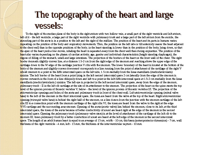
The topography of the heart and large vessels: The heart is located behind the lower half of the sternum and great vessels (aorta and pulmonary trunk) — behind its upper half. Occurring in the anterior mediastinum, heart toward the front of the median line, linea mediana anterior , located asymmetrically: almost one third of his lie to the left and about 1 / 2 — the right of the line. The longitudinal axis of the heart, running from the base to top, forming with the median and frontal planes of the body angle, reaching 40 °. The very same longitudinal axis goes downwards, left to right and back to front. Since the heart is, in addition, several rotated around its axis from right to left, a large part of the right half is more anteriorly, and most of the left half — backwards, so that the front surface of the right ventricle adjacent to the chest wall closest to the rest of the heart, right heart border which forms its lower boundary, reaches the angle between the chest wall and diaphragm of the right costophrenic sinus, sinus costodiaphrag-matica dexter; left atrium of all cavities of the heart is the most rearward position.
The topography of the heart and large vessels: To the right of the median plane of the body is the right atrium with two hollow vein, a small part of the right ventricle and left atrium, left of it — the left ventricle, a large part of the right ventricle with pulmonary trunk and a large part of the left atrium from the auricle, the ascending part of the aorta is in a position to the left and the right of the midline. The position of the heart and its parts in humans varies depending on the position of the body and respiratory movements. Thus, the position on the left side or tilt anteriorly nearer the heart adjacent to the chest wall than in the opposite positions of the body, in the heart standing is lower than in the position of the body, lying down, so that the apex of the heart push a few moves, inhaling the heart is separated away from the chest wall than during expiration. The position of the heart also varies depending on the phases of cardiac activity, age, gender and individual characteristics (height standing diaphragm), the degree of filling of the stomach, small and large intestines. The projection of the borders of the heart on the front wall of the chest. The right border descends slightly convex line, at a distance 1. 5 -2 cm from the right edge of the sternum and reaching above the upper edge of the cartilage down to the W edge of the cartilage junction V ribs with the sternum. The lower boundary of the heart is located at the bottom of the body of the sternum and slightly convex downward corresponds to a line running from the point of attachment of the cartilage of the right V rebrak sternum to a point in the fifth intercostal space on the left side, 1. 5 cm medially from the linea mamillaris (medioclavicularis) sinistra. The left border of the heart from a point lying in the left second intercostal space 2 cm laterally from the edge of the sternum is convex outwards in the form of a line obliquely down and left to a point in the left fifth intercostal space at 1. 5 -2 cm medially from the linea mamillaris (medioclavicularis ) sinistra. The left eye is projected in the left second intercostal space, away from the edge of the sternum, pulmonary trunk — II in the left rib cartilage at the site of its attachment to the sternum. The projection of the heart on the spine meets the top level of the spinous process of thoracic vertebra V, below — the level of the spinous process of thoracic vertebra IX. The projection of the atrioventricular openings and holes of the aorta and pulmonary trunk in front of the chest wall. Left atrioventricular opening (mitral valve base) to the left of the sternum in the third intercostal space, the tones are heard from the valve at the top of the heart. Right atrioventricular opening (tricuspid valve base) is behind the right half of the sternum, on a line drawn from the junction with the sternum cartilage of the left ribs III to a connection point with the sternum cartilage of the right ribs VI, the tones are heard from the valve to the right at the edge V-VI cartilage and the surrounding area sternum. Opening of the aorta (aortic valve) lies behind the sternum, close to its left, at the third intercostal space, the tones of the aorta because of better conductivity of sound are heard right at the edge of the sternum in the second intercostal space. Opening the pulmonary trunk (pulmonary valve stem) is at the level of attachment of the cartilage of the left rib to the sternum III; tones pulmonary trunk by a better conduction of sound are heard at the left edge of the sternum in the second intercostal space. The length of an adult human heart is equal to an average of 13 cm, width — 10 cm, thickness (anteroposterior dimension) — 7 cm, wall thickness of the right ventricle — 4 mm, left — 13 mm, the thickness of the interventricular septum — 10 mm.
The topography of the heart and large vessels:
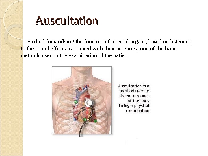
Auscultation Method for studying the function of internal organs, based on listening to the sound effects associated with their activities, one of the basic methods used in the examination of the patient
The 1 stst point of auscultation the apex of the heart, i. e, apex beat area or if it is not defined, then the left border of the heart at the V intercostal space (a point of listening to the mitral valve and left atrioventricular orifice ); during auscultation over the top woman at her need to pre-request to lift the left breast;
The 2 ndnd point of auscultation the second point — II intercostal space just at the right sternal border (the point of listening to the aortic valve and aorta mouth );
The 3 rdrd point of auscultation II intercostal space directly at the left sternal border (the point of listening to pulmonary artery valve and its mouth ); * second and third terms agreed to combine the concept of «base of the heart»
The 4 thth point of auscultation the base xiphoid process (the point of listening to the tricuspid valve and right atrioventricular orifice ).
The 5 thth point of auscultation the place of attachment IV edge to the left edge of the sternum (extra point of listening to the mitral valve, corresponding to its anatomical projections );
The 6 point of auscultation the point Botkin-Erb — III intercostal space at left sternal border (an additional point of listening to the aortic valve, corresponding to its anatomical projections. )
Percussion The boundaries of the vascular bundle are defined in II intercostal space as follows. Finger-plessimeter put on the right intercostal space II by mid-clavicular line parallel to the expected dullness and quiet percussion, gradually move it inwards towards the chest until the blunt sound. Boundaries mark on the outer edge of the finger, facing a clear percussion sound. Then, in the same way produce a silent percussion on the left. Also make a mark on the outer edge of the finger-plessimeter. The normal size diameter of the vascular bundle is 4, 5 -6 cm. Vascular bundle form on the right upper hollow Vienna and aortic arch, left — the pulmonary artery. Delimitation of the vascular bundle of the right (a) and left (b)