
Презентация nervous


- Размер: 3 Mегабайта
- Количество слайдов: 25
Описание презентации Презентация nervous по слайдам
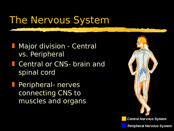
The Nervous System Major division — Central vs. Peripheral Central or CNS- brain and spinal cord Peripheral- nerves connecting CNS to muscles and organs Central Nervous System Peripheral Nervous System
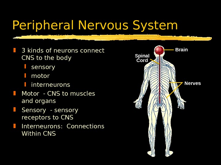
Peripheral Nervous System 3 kinds of neurons connect CNS to the body sensory motor interneurons Motor — CNS to muscles and organs Sensory — sensory receptors to CNS Interneurons: Connections Within CNS Spinal Cord Brain Nerves
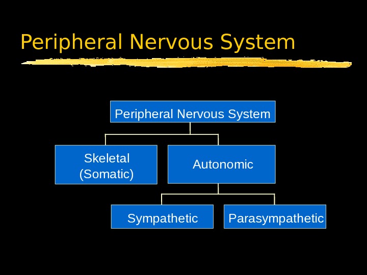
Peripheral Nervous System S k e l e t a l ( S o m a t i c ) S y m p a t h e t i c P a r a s y m p a t h e t i c. A u t o n o m i c. P e r i p h e r a l N e r v o u s S y s t e m
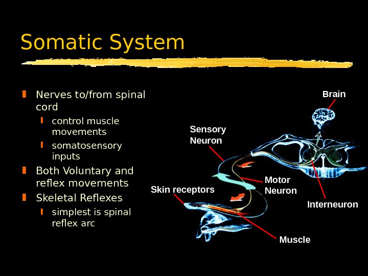
Somatic System Nerves to/from spinal cord control muscle movements somatosensory inputs Both Voluntary and reflex movements Skeletal Reflexes simplest is spinal reflex arc Muscle. Motor Neuron Interneuron. Skin receptors Sensory Neuron Brain
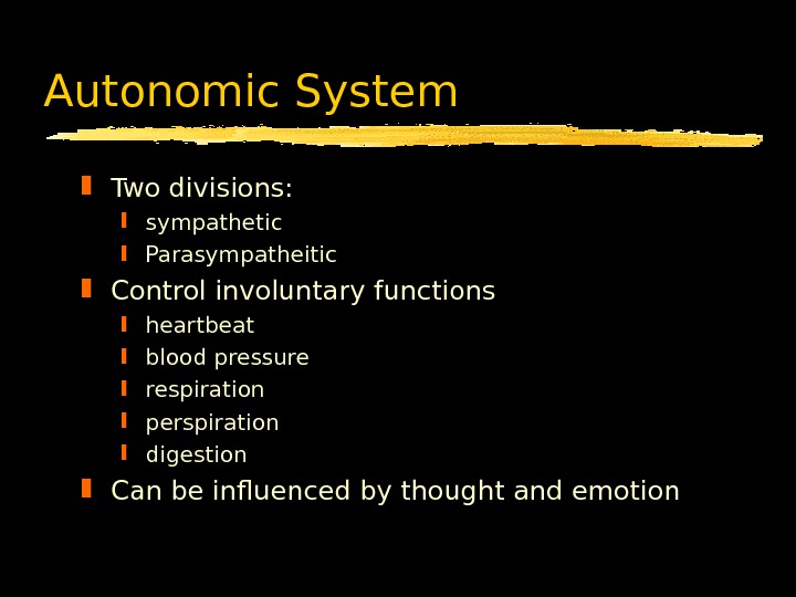
Autonomic System Two divisions: sympathetic Parasympatheitic Control involuntary functions heartbeat blood pressure respiration perspiration digestion Can be influenced by thought and emotion
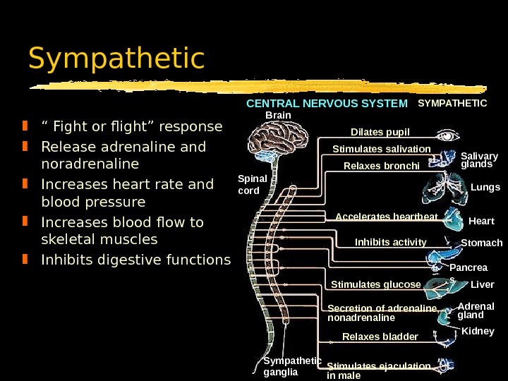
Sympathetic “ Fight or flight” response Release adrenaline and noradrenaline Increases heart rate and blood pressure Increases blood flow to skeletal muscles Inhibits digestive functions CENTRAL NERVOUS SYSTEM Brain Spinal cord SYMPATHETIC Dilates pupil Stimulates salivation Relaxes bronchi Accelerates heartbeat Inhibits activity Stimulates glucose Secretion of adrenaline, nonadrenaline Relaxes bladder Stimulates ejaculation in male. Sympathetic ganglia Salivary glands Lungs Heart Stomach Pancrea s Liver Adrenal gland Kidney
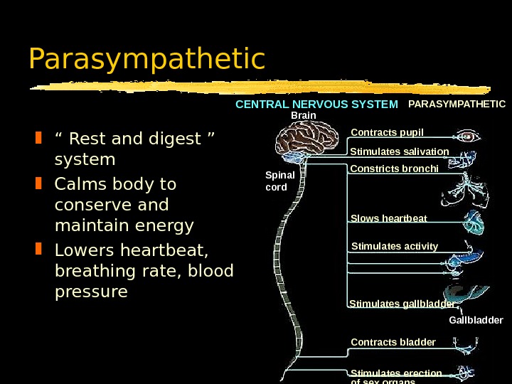
Parasympathetic “ Rest and digest ” system Calms body to conserve and maintain energy Lowers heartbeat, breathing rate, blood pressure CENTRAL NERVOUS SYSTEM Brain PARASYMPATHETIC Spinal cord Stimulates salivation Constricts bronchi Slows heartbeat Stimulates activity Contracts bladder Stimulates erection of sex organs. Stimulates gallbladder Gallbladder. Contracts pupil
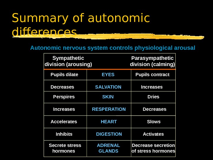
Summary of autonomic differences Autonomic nervous system controls physiological arousal Sympathetic division (arousing) Parasympathetic division (calming) Pupils dilate EYES Pupils contract Decreases SALVATION Increases Perspires SKIN Dries Increases RESPERATION Decreases Accelerates HEART Slows Inhibits DIGESTION Activates Secrete stress hormones ADRENAL GLANDS Decrease secretion of stress hormones
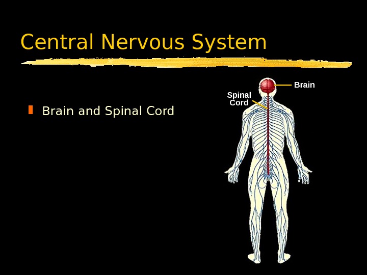
Central Nervous System Brain and Spinal Cord Brain
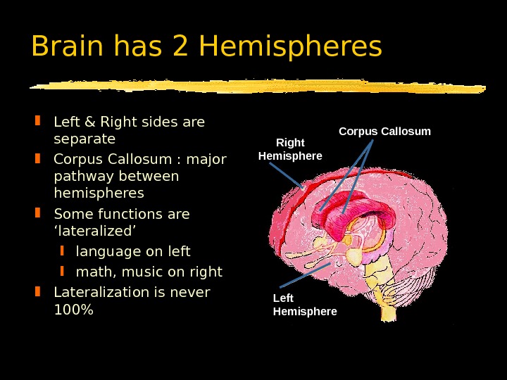
Left & Right sides are separate Corpus Callosum : major pathway between hemispheres Some functions are ‘lateralized’ language on left math, music on right Lateralization is never 100% Brain has 2 Hemispheres Left Hemisphere Corpus Callosum Right Hemisphere
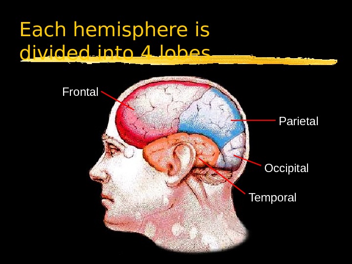
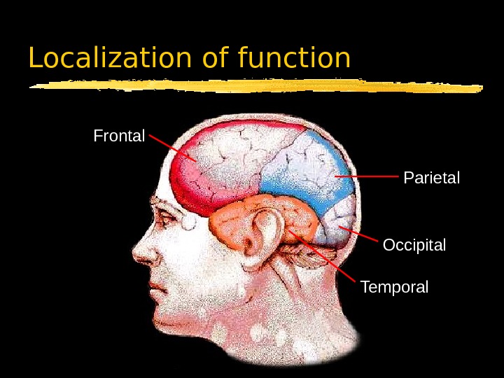
Each hemisphere is divided into 4 lobes Frontal Parietal Occipital Temporal
Sensory Information sent to opposite hemisphere Principle is Contralateral Organization Sensory data crosses over in pathways leading to the cortex Visual Crossover left visual field to right hemisphere right field to left Other senses similar Left visual field Right visual field Optic nerves Corpus Callosum. Left Visual Cortex Right Visual Cortex
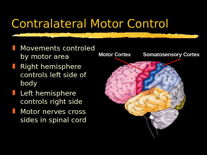
Contralateral Motor Control Movements controled by motor area Right hemisphere controls left side of body Left hemisphere controls right side Motor nerves cross sides in spinal cord Somatosensory Cortex. Motor Cortex
Corpus Callosum Major ( but not only) pathway between sides Connects comparable structures on each side Permits data received on one side to be processed in both hemispheres Aids motor coordination of left and right side Corpus Callosum Medial surface of right hemisphere
Corpus Callosum What happens when the corpus callosum is cut? Sensory inputs are still crossed Motor outputs are still crossed Hemispheres can’t exchange data
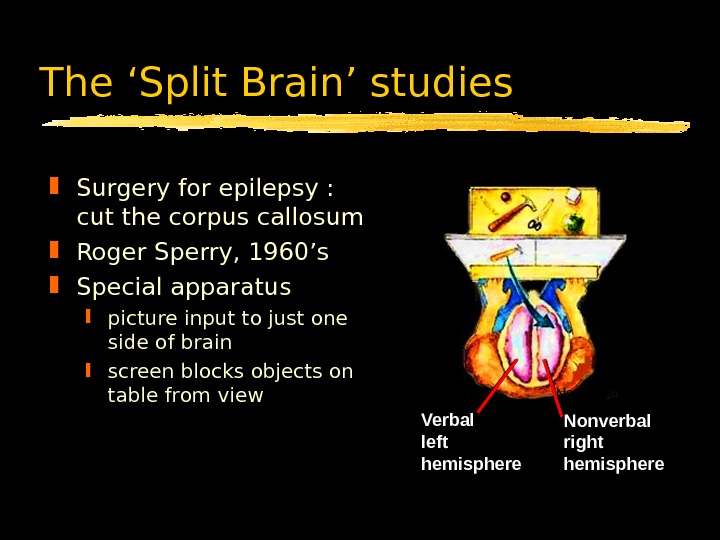
The ‘Split Brain’ studies Surgery for epilepsy : cut the corpus callosum Roger Sperry, 1960’s Special apparatus picture input to just one side of brain screen blocks objects on table from view Nonverbal right hemisphere. Verbal left hemisphere
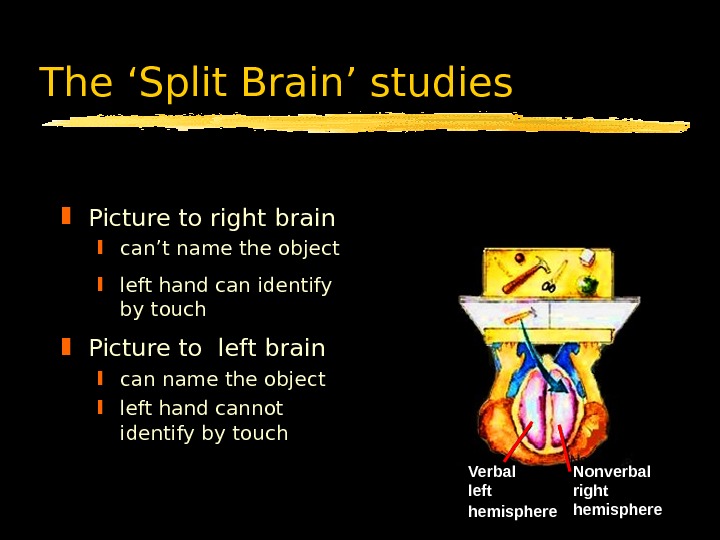
Nonverbal right hemisphere. Verbal left hemisphere ? ? “ What did you see? ” Picture to left brain can name the object left hand cannot identify by touch Picture to right brain can’t name the object left hand can identify by touch “ Using your left hand, Pick up what you saw. ” The ‘Split Brain’ studies I saw an apple. “ What did you see? ” Nonverbal right hemisphere. Verbal left hemisphere
Localization of function Frontal Parietal Occipital Temporal
Occipital Lobe Input from Optic nerve Contains primary visual cortex most is on surface inside central fissure Outputs to parietal and temporal lobes Occipital Lobe Visual Lobe
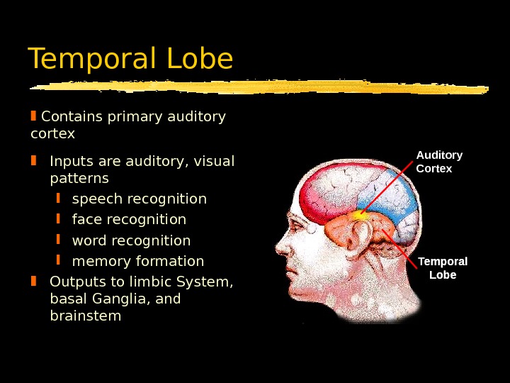
Temporal Lobe Inputs are auditory, visual patterns speech recognition face recognition word recognition memory formation Outputs to limbic System, basal Ganglia, and brainstem Contains primary auditory cortex Temporal Lobe. Auditory Cortex
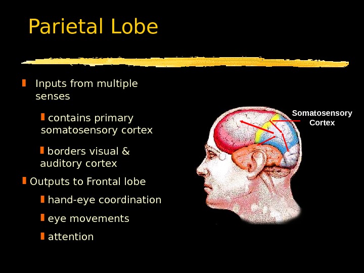
Parietal Lobe. Somatosensory Cortex. Parietal Lobe Inputs from multiple senses contains primary somatosensory cortex borders visual & auditory cortex Outputs to Frontal lobe hand-eye coordination eye movements attention
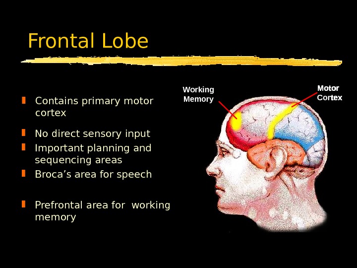
Frontal Lobe Contains primary motor cortex Motor Cortex. Broca’s Area Motor Cortex. Working Memory No direct sensory input Important planning and sequencing areas Broca’s area for speech Prefrontal area for working memory
Frontal Lobe Disorders Broca’s area productive aphasia Prefrontal area lose track of ongoing context fail to inhibit inappropriate responses Often measured with the Wisconsin Card Sorting Task
Wisconsin Card Sorting Task Patient is given a deck of 64 different cards Told to place each card under the one it best matches Told correct or incorrect after each card Row of 4 example cards set out Must deduce what the underlying rule is. Correct!
The Nervous System: Summary Major structures of the nervous CNS, Somatic, Autonomic Two hemispheres & 4 lobes Organization contralateral input & output primary sensory areas motor areas Commissure Localization of functions Central Nervous System Peripheral Nervous System