
Презентация murmur


- Размер: 7.3 Mегабайта
- Количество слайдов: 41
Описание презентации Презентация murmur по слайдам
Cardiac Murmurs Lubna Piracha, D. O. Assistant Professor of Medicine Department of Cardiology
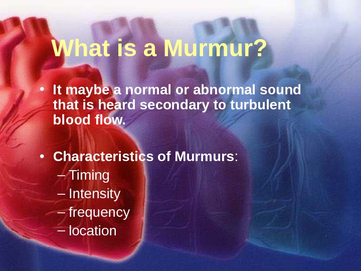
11/12/02 Lubna Piracha, D. O. 2 What is a Murmur? • It maybe a normal or abnormal sound that is heard secondary to turbulent blood flow. • Characteristics of Murmurs : – Timing – Intensity – frequency – location
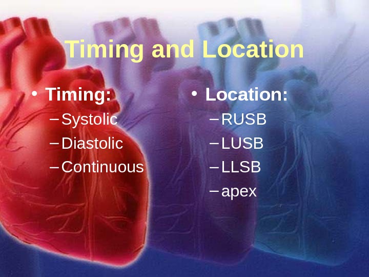
11/12/02 Lubna Piracha, D. O. 3 Timing and Location • Timing: – Systolic – Diastolic – Continuous • Location: – RUSB – LLSB – apex
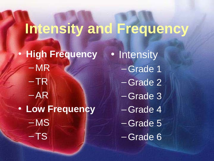
11/12/02 Lubna Piracha, D. O. 4 Intensity and Frequency • High Frequency – MR – TR – AR • Low Frequency – MS – TS • Intensity – Grade 1 – Grade 2 – Grade 3 – Grade 4 – Grade 5 – Grade
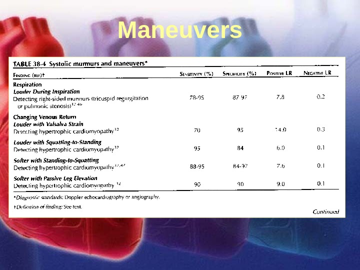
11/12/02 Lubna Piracha, D. O. 5 Maneuvers
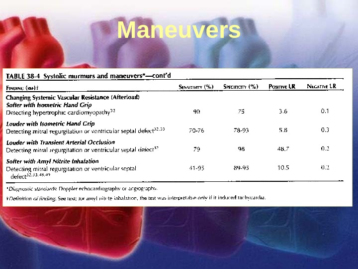
11/12/02 Lubna Piracha, D. O. 6 Maneuvers
11/12/02 Lubna Piracha, D. O. 7 Case Studies • A 50 year old male with a known heart murmur presents with complaints of substernal chest pain, which increases with exertion, and shortness of breath which is starting to limit his lifestyle. No risk factors for coronary artery disease. – On Physical Exam you find the following: • Delayed carotid upstroke • A sustained apical pulse • Prominent A wave in the neck • PMI is sustained but not displaced laterally • and you hear
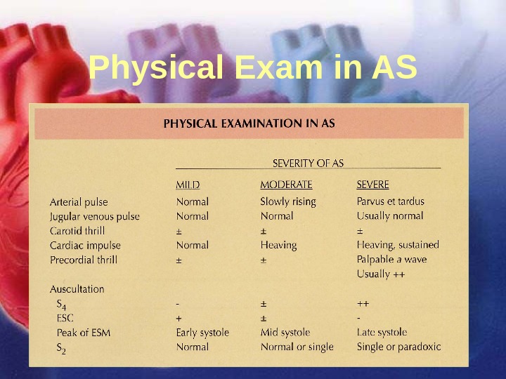
11/12/02 Lubna Piracha, D. O. 8 Physical Exam in AS
11/12/02 Lubna Piracha, D. O. 9 EKG shows
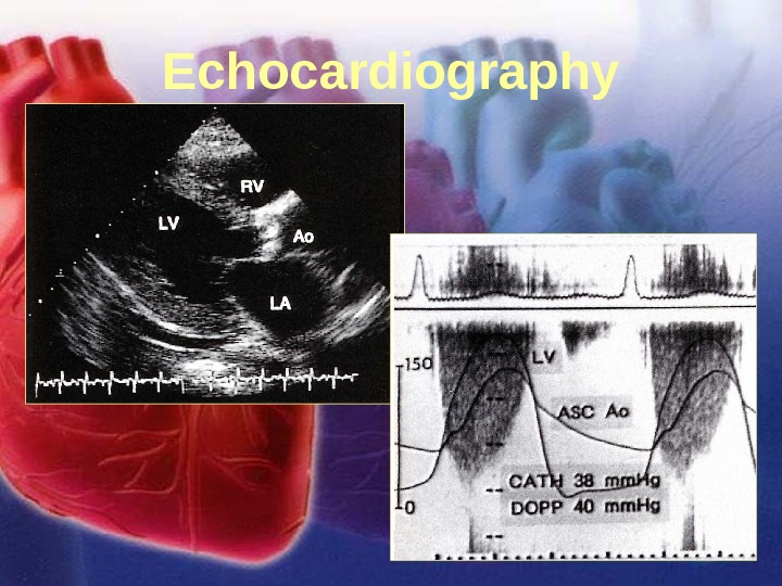
11/12/02 Lubna Piracha, D. O. 10 Echocardiography
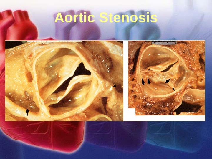
11/12/02 Lubna Piracha, D. O. 11 Aortic Stenosis
11/12/02 Lubna Piracha, D. O. 12 Aortic Stenosis There is little hemodynamic disturbance that occurs as the valve area is reduced from 3 to 4 cm 2 to 1. 5 to 2 cm 2. However, an additional reduction in t he valve area from half its normal size to a quarter of it’s normal size produces severe obstruction to flow and progressive pressure overload on the left ventricle.
11/12/02 Lubna Piracha, D. O. 13 Aortic Stenosis continued: • Concentric hypertrophy develops in response to this overload. The increased muscle mass allows the ventricle to generate the increased force necessary to propel blood past the obstruction. The hypertrophied myocardium has decreased coronary blood flow reserve and can cause systolic and diastolic failure. • Patients may present with symptoms: – Angina: 35% of patients with severe AS present with chest pain and half will die in 5 years. – Syncope: 15% of patients with severe AS present with syncope and half will die in 3 years. – CHF: 50% of patients with severe AS present with CHF and half will die in 2 years.
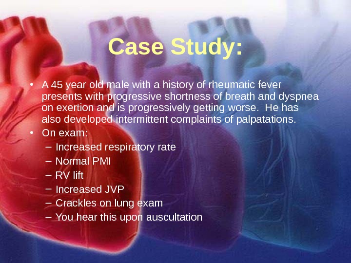
11/12/02 Lubna Piracha, D. O. 14 Case Study: • A 45 year old male with a history of rheumatic fever presents with progressive shortness of breath and dyspnea on exertion and is progressively getting worse. He has also developed intermittent complaints of palpatations. • On exam: – Increased respiratory rate – Normal PMI – RV lift – Increased JVP – Crackles on lung exam – You hear this upon auscultation
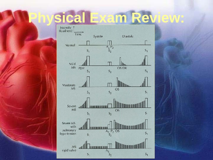
11/12/02 Lubna Piracha, D. O. 15 Physical Exam Review:
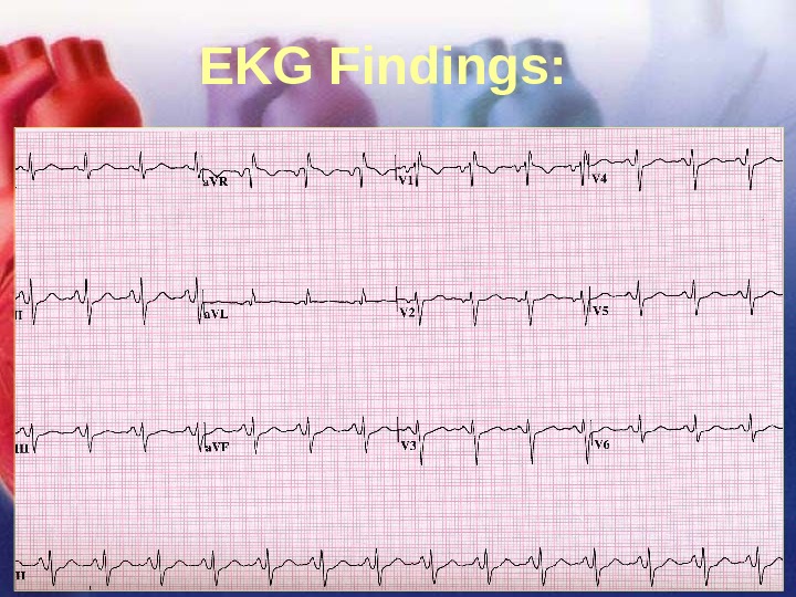
11/12/02 Lubna Piracha, D. O. 16 EKG Findings:
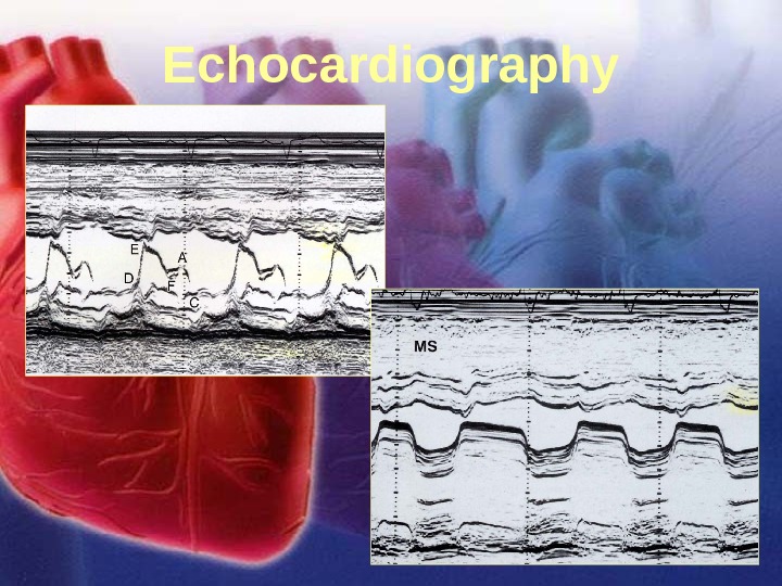
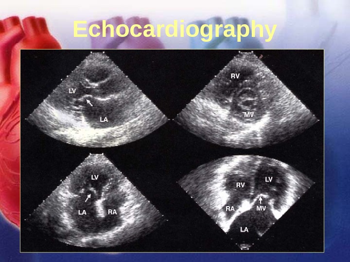
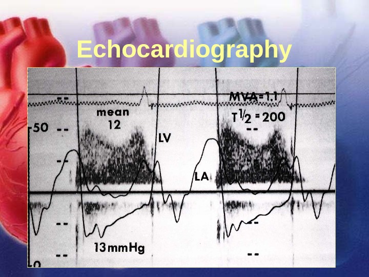
11/12/02 Lubna Piracha, D. O. 17 Echocardiography
11/12/02 Lubna Piracha, D. O. 18 Echocardiography
11/12/02 Lubna Piracha, D. O. 19 Echocardiography
11/12/02 Lubna Piracha, D. O. 20 Mitral Stenosis In severe mitral stenosis the left ventricle is spared and tends to be small and under filled. There is significant elevation in the left atrial pressures leading to left atrial enlargement which then gets transmitted to the pulmonary circulation leading to pulmonary edema and pulmonary hypertension. The left atrial enlargement can lead to atrial fibrillation and loss of atrial kick and decreased filling of the left ventricle. Systemic embolic events are seen in approximately one-third of patients with atrial fibrillation and mitral stenosis and maybe the presenting event before the diagnosis of mitral stenosis is made.
11/12/02 Lubna Piracha, D. O. 21 Case Studies: A 52 year old female presents with complaints of slowly progressive dyspnea on exertion and an uncomfortable awareness of pulsations in the neck and chest. On Exam you find the following: -Abnormal brisk pulses -Wide pulse pressures -Quincke’s pulse -Head bobbing -Pistol shot sounds On auscultation you hear this:
11/12/02 Lubna Piracha, D. O. 22 Physical Exam Review • Early diastolic murmur of regurgitation – blowing, and high frequency, and decrescendo in shape. • Systolic aortic flow murmur • Austin flint murmur
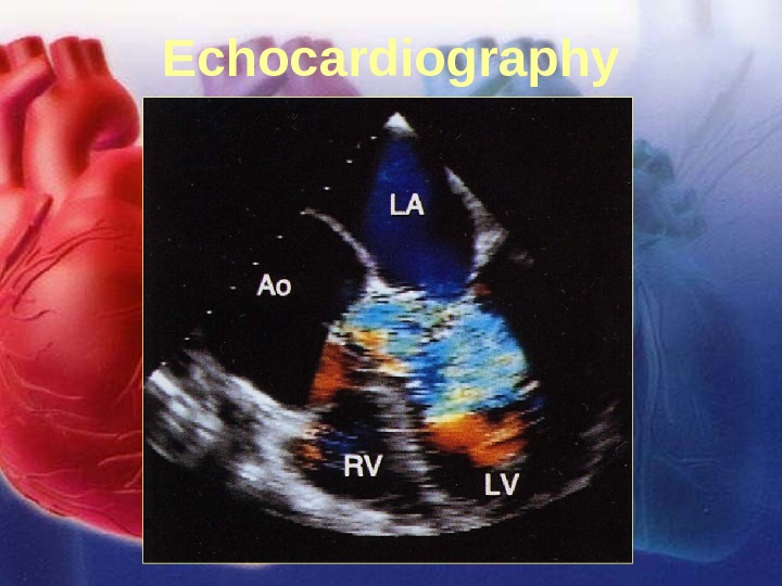
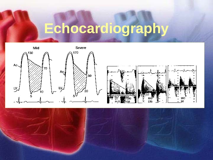
11/12/02 Lubna Piracha, D. O. 23 Echocardiography
11/12/02 Lubna Piracha, D. O. 24 Echocardiography
11/12/02 Lubna Piracha, D. O. 25 Aortic Insufficiency – Acute aortic insufficiency usually due to acute aortic dissection or aortic valve endocarditis usually presents with significant shortness of breath and the murmur maybe minimal and peripheral manifestations maybe diminished. This causes the abrupt introduction of a large volume of blood into a non-compliant ventricle increasing the LV end diastolic and pulmonary venous pressures leading to significant dyspnea. A murmur maybe minimal because the abrupt increase LV diastolic pressure rapidly diminishes the aortic to LV diastolic gradient.
11/12/02 Lubna Piracha, D. O. 26 Aortic Insufficiency – In chronic aortic insufficiency, compensatory left ventricular changes occur over time. The chronic volume overload causes stretching and elongation of myocardial fibers (eccentric hypertrophy). Eventually, the LV cannot compensate and you have LV dilatation and congestive heart failure.
11/12/02 Lubna Piracha, D. O. 27 Case Study • A 75 year old male present to the emergency room with complaints of severe chest tightness (10/10) and acutely short of breath. He has PND and orthopnea. He is hypotensive, tachycardic and in respiratory distress. His EKG reveals an inferior and posterior wall myocardial infarction. – On Exam: • Vital signs are unstable • Crackles are noted bilaterally • PMI is still relatively normal • Ausculatory findings reveal this:
11/12/02 Lubna Piracha, D. O. 28 Physical Exam Review • In acute MR, there is tachycardia, the murmur maybe short and confined to early systole, because the LA pressures are elevated. • In chronic MR, the murmur is typically holosystolic starting after S 1.
11/12/02 Lubna Piracha, D. O. 29 EKG Findings:
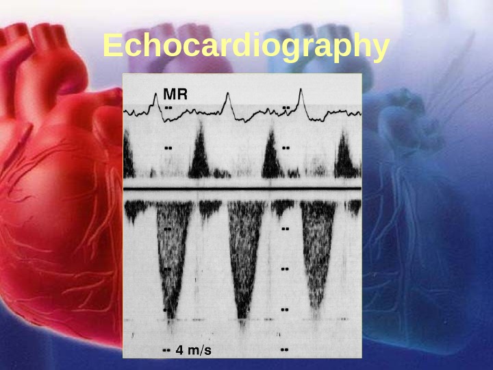
11/12/02 Lubna Piracha, D. O. 30 Echocardiography
11/12/02 Lubna Piracha, D. O. 31 Echocardiography
11/12/02 Lubna Piracha, D. O. 32 Mitral Regurgitation • There is acute volume overload on left ventricle with an increase in end diastolic volume. At the same time, there is new pathway for LV ejection into a low pressure system into the LA. The left ventricle initially is hypercontractile because it can eject blood back into the LA and out the aortic valve. Forward stroke volume is actually decreased. • In acute MR, the LA cannot accommodate the increased volume and builds up in the lungs leading to respiratory distress.
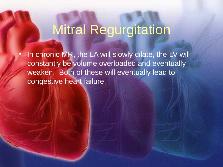
11/12/02 Lubna Piracha, D. O. 33 Mitral Regurgitation • In chronic MR, the LA will slowly dilate, the LV will constantly be volume overloaded and eventually weaken. Both of these will eventually lead to congestive heart failure.
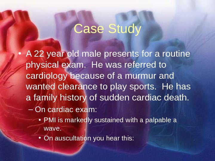
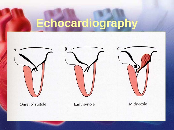
11/12/02 Lubna Piracha, D. O. 34 Case Study • A 22 year old male presents for a routine physical exam. He was referred to cardiology because of a murmur and wanted clearance to play sports. He has a family history of sudden cardiac death. – On cardiac exam: • PMI is markedly sustained with a palpable a wave. • On auscultation you hear this:
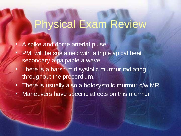
11/12/02 Lubna Piracha, D. O. 35 Physical Exam Review • A spike and dome arterial pulse • PMI will be sustained with a triple apical beat secondary a palpable a wave • There is a harsh mid systolic murmur radiating throughout the precordium. • There is usually also a holosystolic murmur c/w MR • Maneuvers have specific affects on this murmur
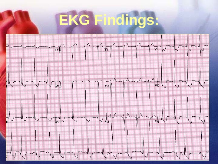
11/12/02 Lubna Piracha, D. O. 36 EKG Findings:
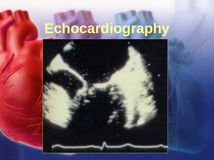
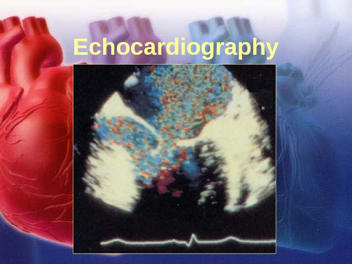
11/12/02 Lubna Piracha, D. O. 37 Echocardiography
11/12/02 Lubna Piracha, D. O. 38 Echocardiography
11/12/02 Lubna Piracha, D. O. 39 Echocardiography
11/12/02 Lubna Piracha, D. O. 40 Hypertrophic Cardiomyopathy • HCM is frequently a hereditary disorder, with transmission to first-degree relatives in 50% of cases. The most common location of ventricular hypertrophy is subaortic, septal, and anterior wall hypertrophy. • Traditionally, dynamic left ventricular outflow tract obstruction has been considered as the cause of symptoms in patients, but it should be remembered that diastolic dysfunction, ischemia, MR, and arrhythmia’s are also important in producing symptoms.
11/12/02 Lubna Piracha, D. O. 41 Hypertrophic Cardiomyopathy • Atrial arrhythmia’s are common. Ventricular ectopy is a common finding on Holter monitoring. Sustained ventricular tachycardia and fibrillation are the most likely mechanisms of syncope and sudden death in these patients. • Cardiac output may decrease as much as 40% if atrial fibrillation occurs, and these patients tend to rely on their atrial kick.