
Презентация human-digestion


- Размер: 23.3 Mегабайта
- Количество слайдов: 118
Описание презентации Презентация human-digestion по слайдам
Human digestion
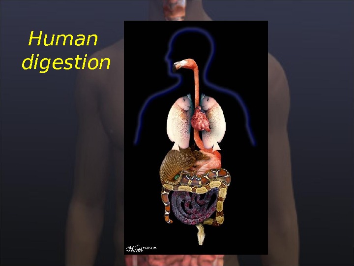
Human digestion
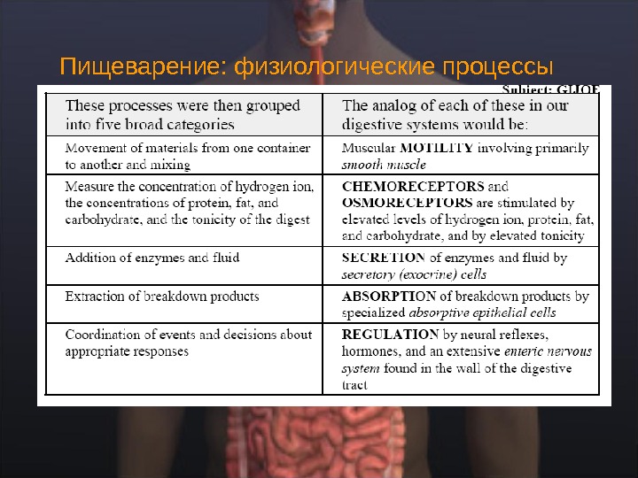
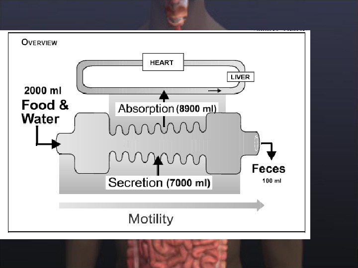
Пищеварение: физиологические процессы
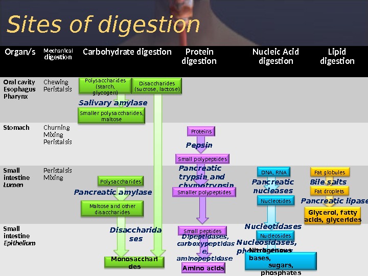
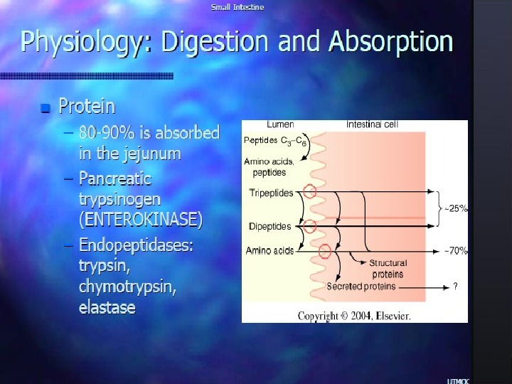
Organ/s Mechanical digestion Carbohydrate digestion Protein digestion Nucleic Acid digestion Lipid digestion Oral cavity Esophagus Pharynx Chewing Peristalsis Stomach Churning Mixing Peristalsis Small intestine Lumen Peristalsis Mixing Small intestine Epithelium Polysaccharides (starch, glycogen) Disaccharides (sucrose, lactose) Salivary amylase Smaller polysaccharides, maltose Polysaccharides Pancreatic amylase Maltose and other disaccharides Disaccharida ses Monosacchari des Proteins Pepsin Small polypeptides Pancreatic trypsin and chymotrypsin Smaller polypeptides Small peptides Dipeptidases, carboxypeptidas e, aminopeptidase Amino acids DNA, RNA Pancreatic nucleases Nucleotidases Nucleosidases, phosphatases Nitrogenous bases, sugars, phosphates Fat globules Bile salts Fat droplets Pancreatic lipase Glycerol, fatty acids, glycerides. Sites of digestion
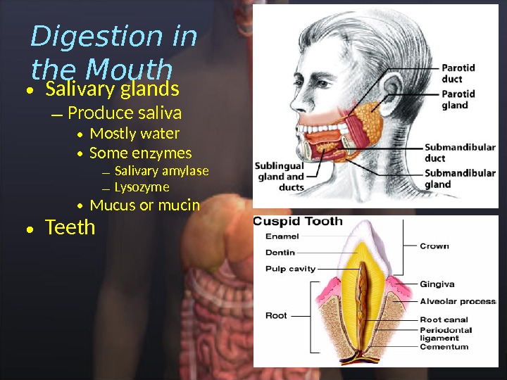
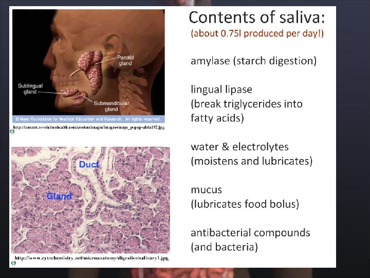
• Salivary glands – Produce saliva • Mostly water • Some enzymes – Salivary amylase – Lysozyme • Mucus or mucin • Teeth. Digestion in the Mouth
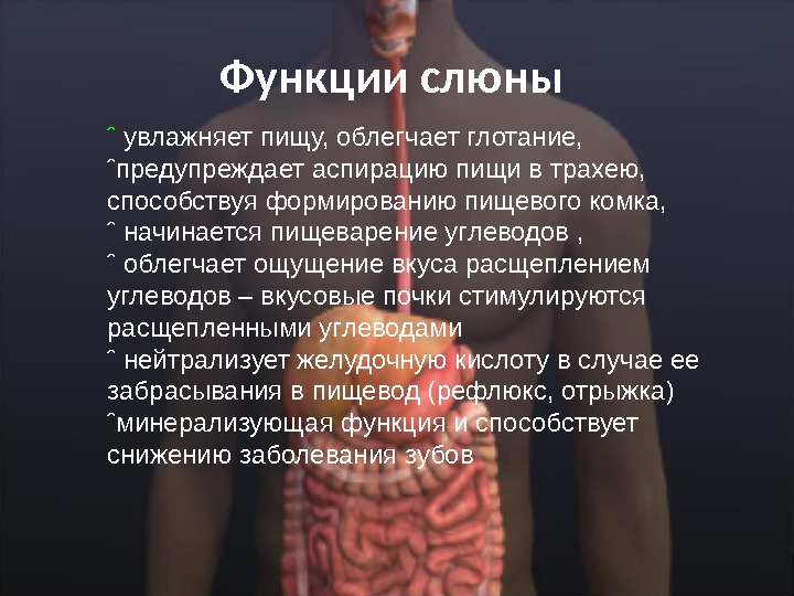
Функции слюны ˆ увлажняет пищу, облегчает глотание, ˆпредупреждает аспирацию пищи в трахею, способствуя формированию пищевого комка, ˆ начинается пищеварение углеводов , ˆ облегчает ощущение вкуса расщеплением углеводов – вкусовые почки стимулируются расщепленными углеводами ˆ нейтрализует желудочную кислоту в случае ее забрасывания в пищевод (рефлюкс, отрыжка) ˆминерализующая функция и способствует снижению заболевания зубов
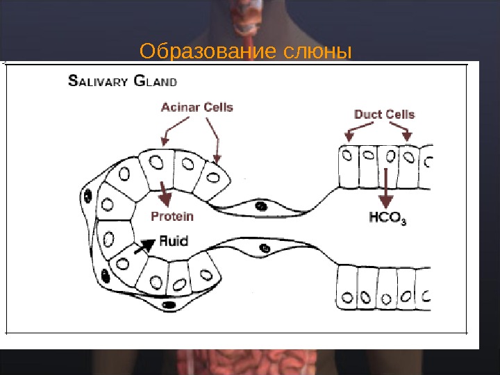
Образование слюны
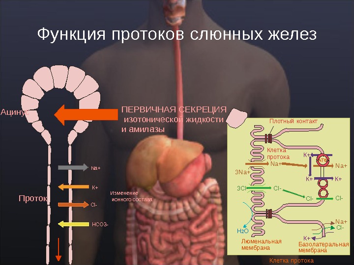
Функция протоков слюнных желез. Проток Ацинус. ПЕРВИЧНАЯ СЕКРЕЦИЯ изотонической жидкости и амилазы Изменение ионного состава. Na+ К+ Cl- НСО 3 — АТФПлотный контакт Базолатеральная мембрана. Люменальная мембрана. Н О 2 К+ N a+ К+ К+3 N a+ С l-3 С l-N a+ К+Клетка протока
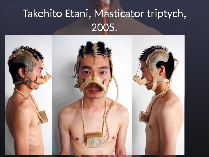
Takehito Etani, Masticator triptych, 2005.
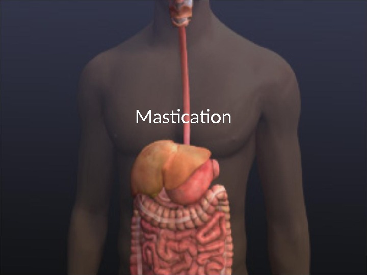
Mastication. masticatory cycle ( chewing cycle ) the complete pathway of the mandible
Mastication
The Muscles of Mastication (Chewing ) • Перетирание или жевание, выполняется скоординированной функцией четырех важных мышц, которые сжимают зубы вместе в различных направлениях. • These are: • The masseter • The lateral pterygoid • The medial pterygoid • The temporalis • Каждая из этих мышц — парная и ими управляет мозг через нервные волокна тройничного нерва (cranial nerve V).
M ain force vectors of masticatory muscles
Swallowing: from mouth to stomach The Oral Phase The Pharyngeal Phase The Esophageal Phase
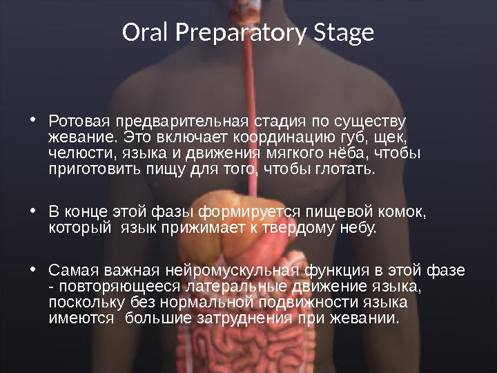
Oral Preparatory Stage • Ротовая предварительная стадия по существу жевание. Это включает координацию губ, щек, челюсти, языка и движения мягкого нёба, чтобы приготовить пищу для того, чтобы глотать. • В конце этой фазы формируется пищевой комок, который язык прижимает к твердому небу. • Самая важная нейромускульная функция в этой фазе — повторяющееся латеральные движение языка, поскольку без нормальной подвижности языка имеются большие затруднения при жевании.
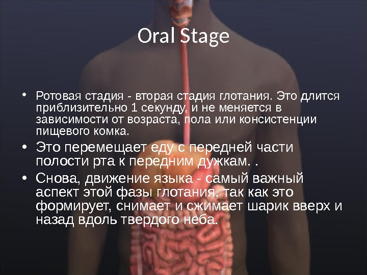
Oral Stage • Ротовая стадия — вторая стадия глотания. Это длится приблизительно 1 секунду, и не меняется в зависимости от возраста, пола или консистенции пищевого комка. • Это перемещает еду с передней части полости рта к передним дужкам. . • Снова, движение языка — самый важный аспект этой фазы глотания, так как это формирует, снимает и сжимает шарик вверх и назад вдоль твердого неба.
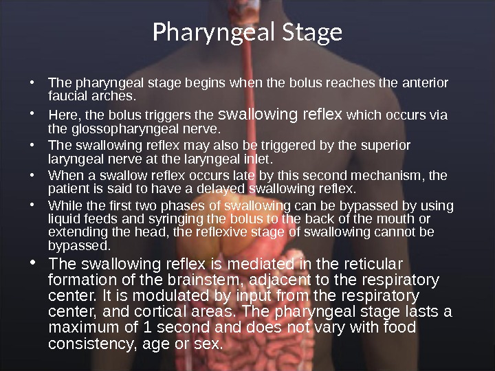
Pharyngeal Stage • The pharyngeal stage begins when the bolus reaches the anterior faucial arches. • Here, the bolus triggers the swallowing reflex which occurs via the glossopharyngeal nerve. • The swallowing reflex may also be triggered by the superior laryngeal nerve at the laryngeal inlet. • When a swallow reflex occurs late by this second mechanism, the patient is said to have a delayed swallowing reflex. • While the first two phases of swallowing can be bypassed by using liquid feeds and syringing the bolus to the back of the mouth or extending the head, the reflexive stage of swallowing cannot be bypassed. • The swallowing reflex is mediated in the reticular formation of the brainstem, adjacent to the respiratory center. It is modulated by input from the respiratory center, and cortical areas. The pharyngeal stage lasts a maximum of 1 second and does not vary with food consistency, age or sex.
Swallowing Reflex (Pharyngeal Stage) • When triggered, the swallowing reflex results in four neuromuscular functions, which occur in rapid sequence in the following order: • 1) Velopharyngeal closure (to prevent nasopharyngeal reflux) • 2) Pharyngeal peristalsis (to propel the bolus into the pharynx) Pharyngeal peristalsis occurs in a sequential fashion, from superior to inferior constrictor. It functions to clear material from the pharyngeal recesses. Vallecular or pyriform sinus pooling on modified barium swallow is an indication of reduced pharyngeal peristalsis. • 3) Elevation and closure of the larynx (for airway protection Laryngeal elevation occurs by contraction of the strap muscles, which positions the larynx anterosuperiorly (under the tongue base).
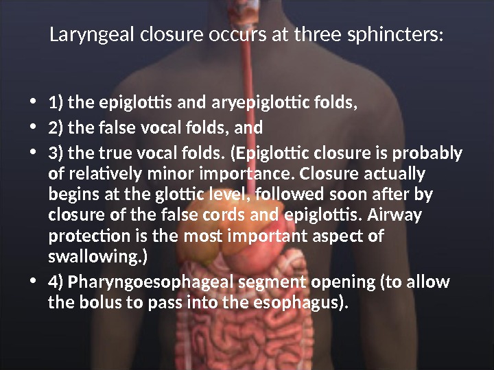
Laryngeal closure occurs at three sphincters: • 1) the epiglottis and aryepiglottic folds, • 2) the false vocal folds, and • 3) the true vocal folds. (Epiglottic closure is probably of relatively minor importance. Closure actually begins at the glottic level, followed soon after by closure of the false cords and epiglottis. Airway protection is the most important aspect of swallowing. ) • 4) Pharyngoesophageal segment opening (to allow the bolus to pass into the esophagus).
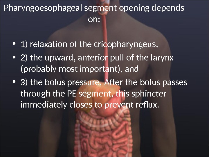
Pharyngoesophageal segment opening depends on: • 1) relaxation of the cricopharyngeus, • 2) the upward, anterior pull of the larynx (probably most important), and • 3) the bolus pressure. After the bolus passes through the PE segment, this sphincter immediately closes to prevent reflux.
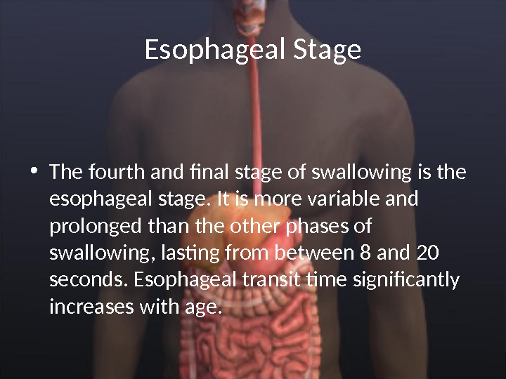
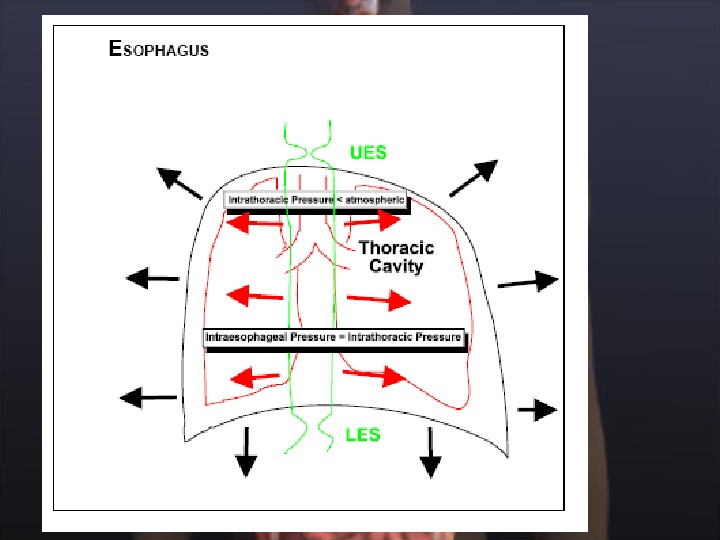
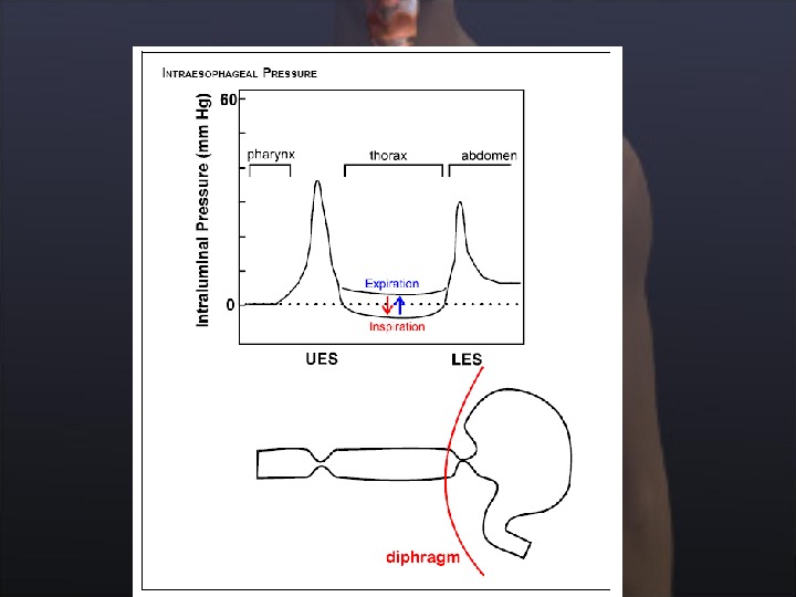
Esophageal Stage • The fourth and final stage of swallowing is the esophageal stage. It is more variable and prolonged than the other phases of swallowing, lasting from between 8 and 20 seconds. Esophageal transit time significantly increases with age.
Swallowing Centers • In humans, the development of • functional magnetic resonance imaging (f. MRI) has allowed the identification of the cortical • regions involved in voluntary swallowing. • The primary motor and sensory areas are • consistently active in healthy adults during swallowing. The anterior cingular cortex is also • activated during swallowing (Hamdy, Mikulis et al. 1999; Humbert and Robbins 2007).
Swallowing Centers • There is convincing evidence that the sequential and rhythmic patterns of swallowing are • formed and organized by a central pattern generator (CPG) located within the medulla • oblongata (Jean 2001). The CPG was first described as a swallowing center that can be • subdivided into three systems: an afferent system composed of the central and peripheral • inputs to the center; an efferent system composed of outputs from the center to various • motor neuron pools involved in swallowing; and an organizing system composed of • interneuronal networks that program the motor pattern.
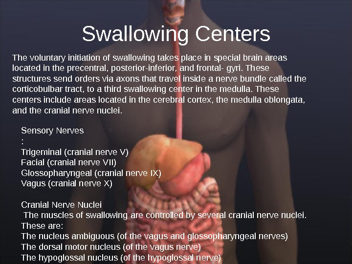
Swallowing Centers The voluntary initiation of swallowing takes place in special brain areas located in the precentral, posterior-inferior, and frontal- gyri. These structures send orders via axons that travel inside a nerve bundle called the corticobulbar tract, to a third swallowing center in the medulla. These centers include areas located in the cerebral cortex, the medulla oblongata, and the cranial nerve nuclei. Sensory Nerves : Trigeminal (cranial nerve V) Facial (cranial nerve VII) Glossopharyngeal (cranial nerve IX) Vagus (cranial nerve X) Cranial Nerve Nuclei The muscles of swallowing are controlled by several cranial nerve nuclei. These are: The nucleus ambiguous (of the vagus and glossopharyngeal nerves) The dorsal motor nucleus (of the vagus nerve) The hypoglossal nucleus (of the hypoglossal nerve)
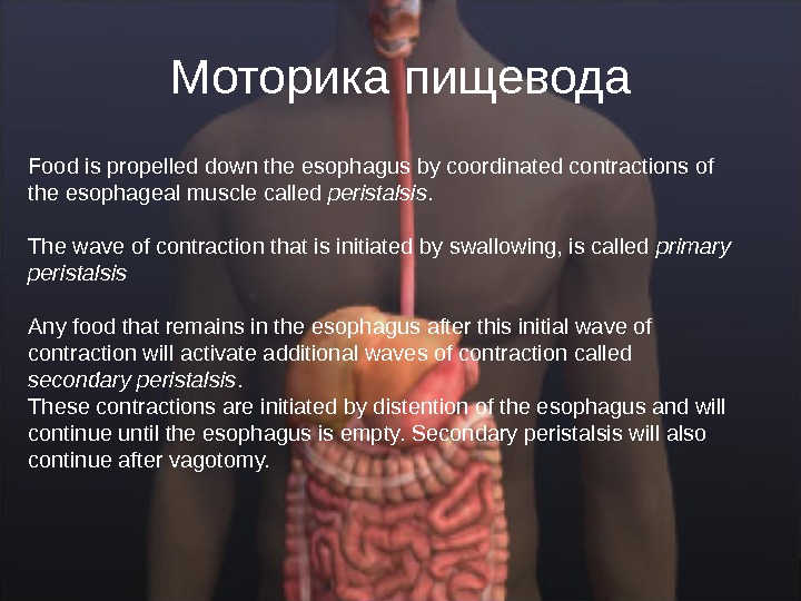
Моторика пищевода Food is propelled down the esophagus by coordinated contractions of the esophageal muscle called peristalsis. The wave of contraction that is initiated by swallowing, is called primary peristalsis Any food that remains in the esophagus after this initial wave of contraction will activate additional waves of contraction called secondary peristalsis. These contractions are initiated by distention of the esophagus and will continue until the esophagus is empty. Secondary peristalsis will also continue after vagotomy.
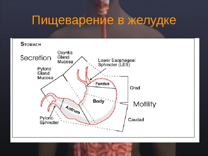
Пищеварение в желудке
Muscular sac ◦ Churns & mixes food Gastric glands ◦ Parietal cells HCl, intrinsic factor ◦ Goblet cells mucus ◦ Chief cells pepsinogen, weak gastric lipase ◦ Gastrin Hormone Controls gastric juices Comes out as chyme (2 -6 hours)Digestion in the Stomach
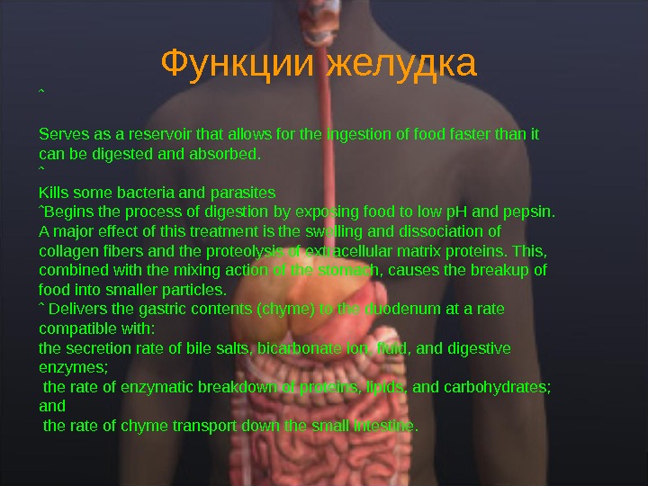
Функции желудка ˆ Serves as a reservoir that allows for the ingestion of food faster than it can be digested and absorbed. ˆ Kills some bacteria and parasites ˆBegins the process of digestion by exposing food to low p. H and pepsin. A major effect of this treatment is the swelling and dissociation of collagen fibers and the proteolysis of extracellular matrix proteins. This, combined with the mixing action of the stomach, causes the breakup of food into smaller particles. ˆ Delivers the gastric contents (chyme) to the duodenum at a rate compatible with: the secretion rate of bile salts, bicarbonate ion, fluid, and digestive enzymes; the rate of enzymatic breakdown of proteins, lipids, and carbohydrates; and the rate of chyme transport down the small intestine.
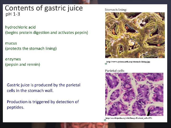
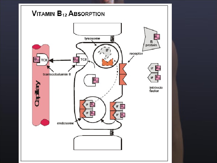
Секреция в желудке The major components of gastric juice are: ˆ Hydrochloric acid (HCl) — denatures proteins ˆ Pepsinogen — proenzyme that is converted to pepsin (protease) ˆ Intrinsic factor — vitamin B 12 -binding protein ˆ Mucous — protects stomach wall from HCl, pepsin, and mechanical trauma
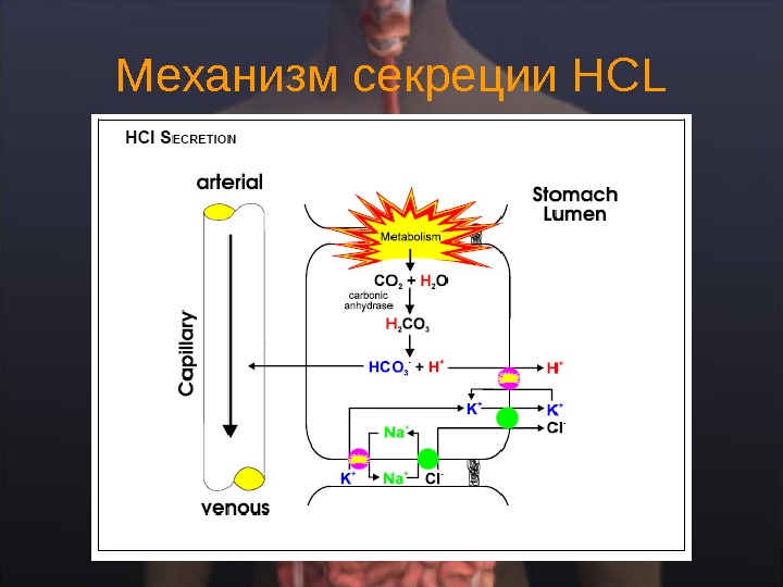
Механизм секреции НС L
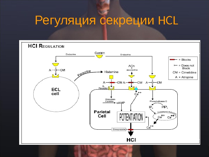
Регуляция секреции HCL
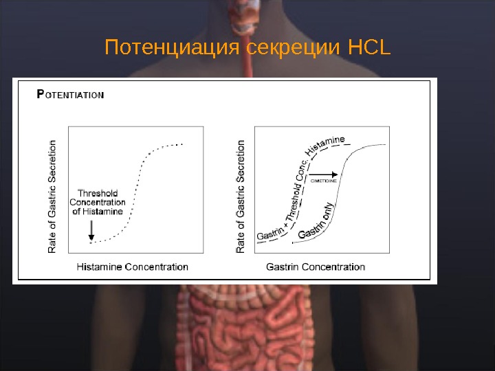
Потенциация секреции HCL
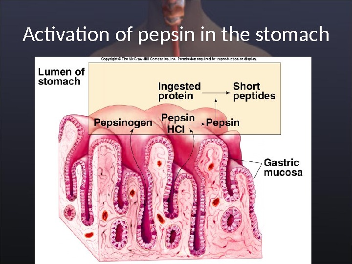
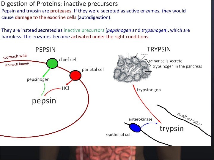
Activation of pepsin in the stomach
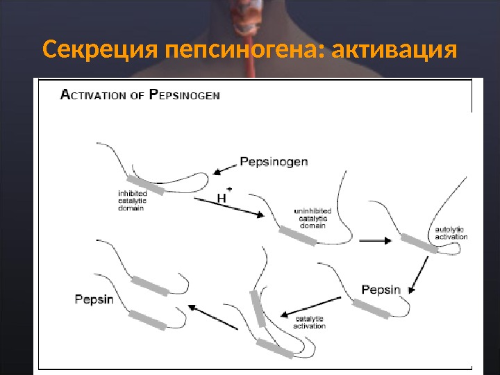
Секреция пепсиногена: активация
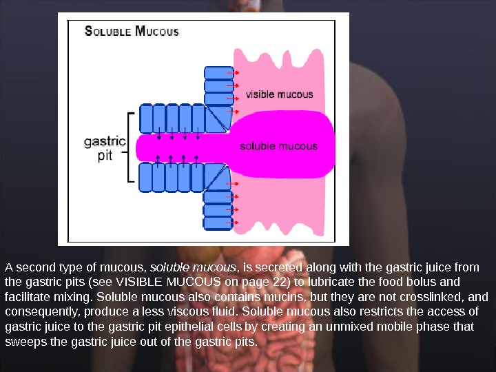
Секреция слизи в желудке: типы слизи The surface epithelial cells of the stomach secrete visible mucous (slightly opaque) that lines the surface of the stomach. Visible mucous contains crosslinked mucins (glycoproteins) that form an almost gelatinous coating which contains relatively high concentrations of bicarbonate ion
A second type of mucous, soluble mucous , is secreted along with the gastric juice from the gastric pits (see VISIBLE MUCOUS on page 22) to lubricate the food bolus and facilitate mixing. Soluble mucous also contains mucins, but they are not crosslinked, and consequently, produce a less viscous fluid. Soluble mucous also restricts the access of gastric juice to the gastric pit epithelial cells by creating an unmixed mobile phase that sweeps the gastric juice out of the gastric pits.
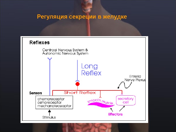
Регуляция секреции в желудке
Регуляция секреции HCL
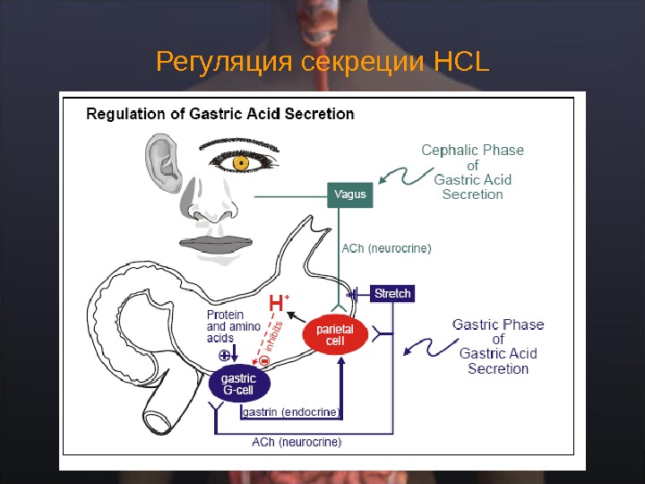
Фазы желудочной секреции Аnticipation of a meal, or the sight of food activates a long reflex that stimulates secretion of gastric juice ( cephalic phase of gastric digestion ) proteins and amino acids in the stomach directly stimulate the release of gastrin from G-cells in the gastric mucosa, which then travels by way of the general circulation back to the stomach (endocrine) to stimulate gastric juice secretion ( gastric phase of gastric digestion ) ˆ Between meals, the secretion of gastric juice is very low. In the absence of protein, which is a good p. H buffer, a small amount of gastric juice can reduce the p. H to 1. Hydrogen ions inhibit secretion by directly inhibiting the release of gastrin from gastric mucosal G-cells ( gastric phase of gastric digestion ). This negative feedback process halts the secretion of gastric juice and prevents the hydrogen ion concentration from reaching levels that would be toxic to the gastric mucosal cells. Pepsinogen secretion closely parallels the secretion of HCl. This is in part due to a vagal reflex activated by low p. H in the stomach that directly stimulates chief cells. ACh released from nerve fibers is a strong stimulant of pepsinogen secretion.
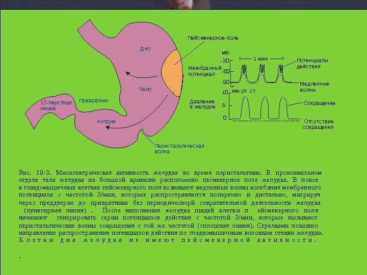
Дно Тело Перис тальтичес кая волна. Антрум 12 -перс тная кишка Привратник -30 -60 -90 1 мин 10 5 0 мм рт. с т. М ембранный потенциал С окращение. Потенциалы дейс твия. Пейсмекерное поле Рис. 10 -3. Мио электрическая активность желудка во время перистальтики. В проксимальном отделе тела желудка на большой кривизне расположено песмекерное поле желудка. В покое в глакдомышечных клетках пейсмекерного поля возникают медленны е волны колебания мембранного потенциала с частотой 3/мин, которы е распространяются поперечно и дистально, мигрируя через преддверие до привратника без периодическорй сократительной деятельности желудка (пунк тирная л иния) . После наполнения желудк а пищей к летк и п ейсмек ерного поля начинают генерировать серии потенциалов действия с частотой 3/мин, которые вы зывают перистальтические волны сокращения с той же частотой (сплошная линия). Стрелками показано направление распространения потенциалов действия по гладкомы шечным волокнам стенки желудка. К л е т к и д н а ж е л у д к а н е и м е ю т п е й с м е к е р н о й а к т и в н о с т и. . Давление в желудке М едленные волны Отс у тс твие сокращениям. В
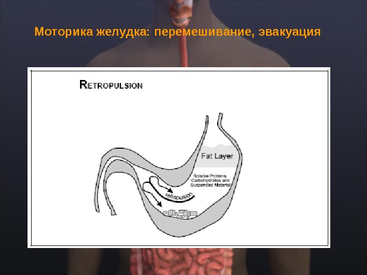
Моторика желудка: перемешивание, эвакуация
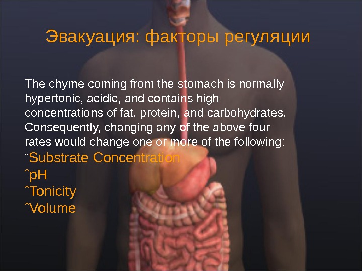
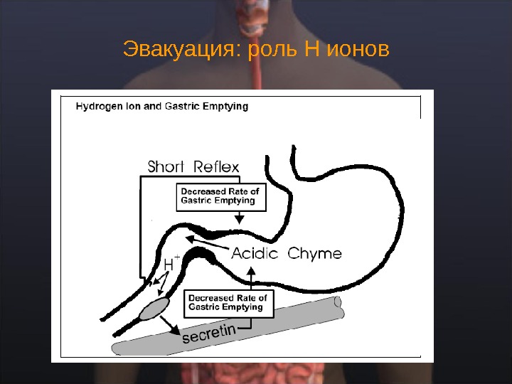
Эвакуация: факторы регуляции The chyme coming from the stomach is normally hypertonic, acidic, and contains high concentrations of fat, protein, and carbohydrates. Consequently, changing any of the above four rates would change one or more of the following: ˆ Substrate Concentration ˆp. H ˆTonicity ˆVolume
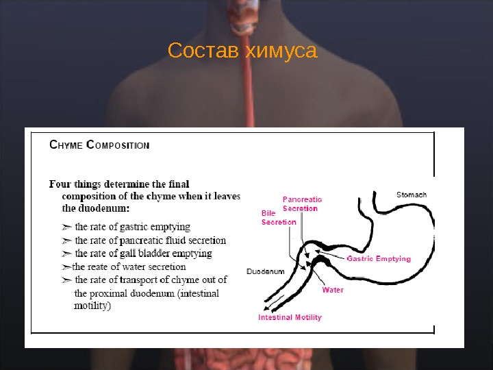
Состав химуса
Эвакуация: роль Н ионов
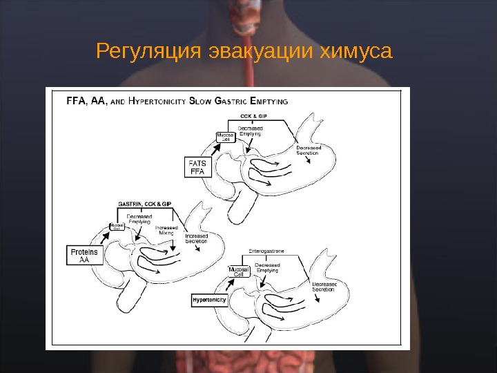
Регуляция эвакуации химуса
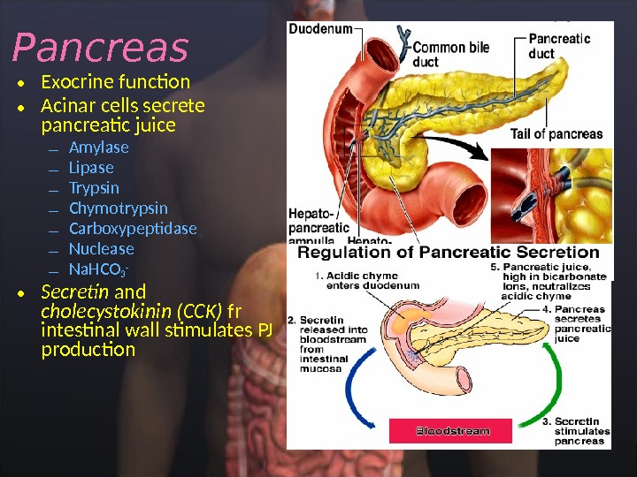
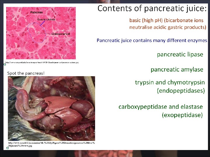
• Exocrine function • Acinar cells secrete pancreatic juice – Amylase – Lipase – Trypsin – Chymotrypsin – Carboxypeptidase – Nuclease – Na. HCO 3 — • Secretin and cholecystokinin (CCK) fr intestinal wall stimulates PJ production. Pancreas
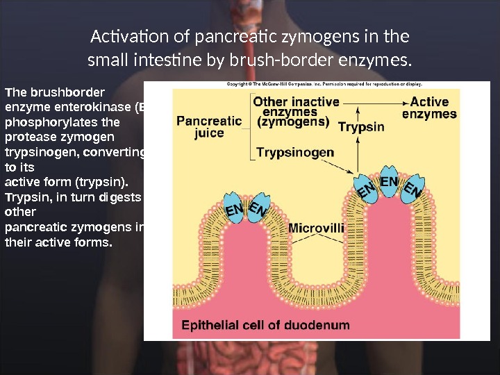
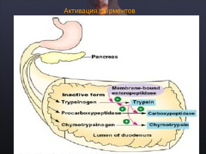
Activation of pancreatic zymogens in the small intestine by brush-border enzymes. The brushborder enzyme enterokinase (EN) phosphorylates the protease zymogen trypsinogen, converting it to its active form (trypsin). Trypsin, in turn digests other pancreatic zymogens into their active forms.
Активация ферментов
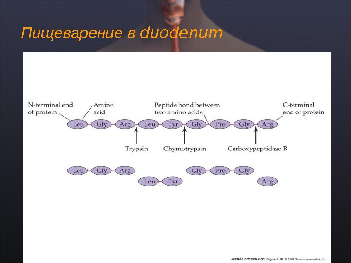
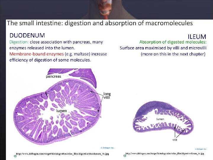
Пищеварение в duodenum
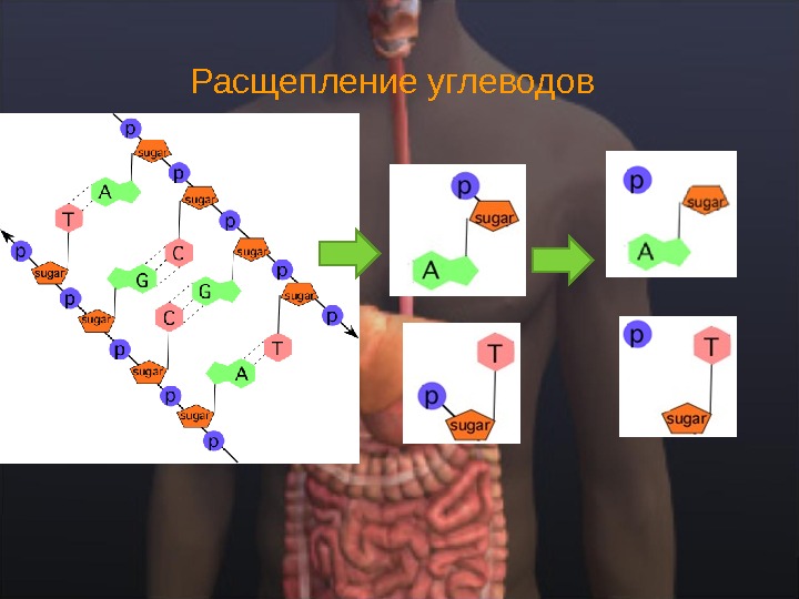
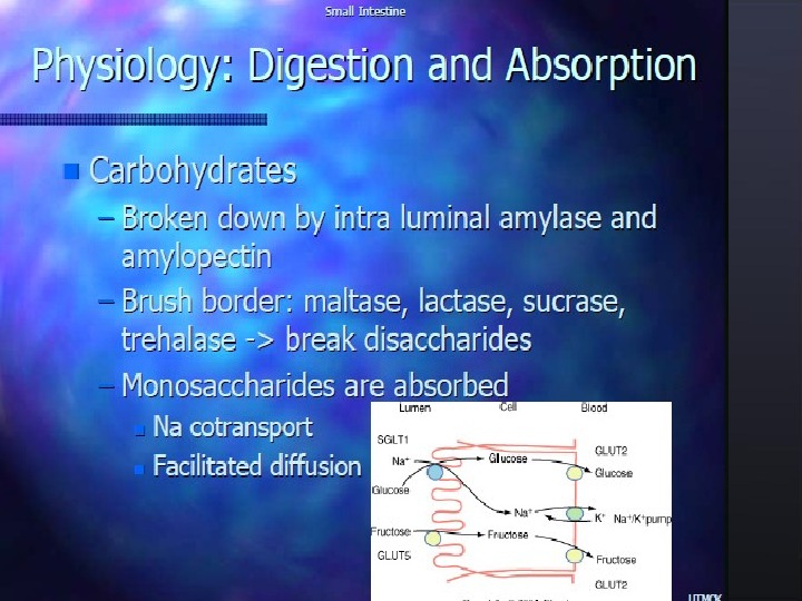
Расщепление углеводов
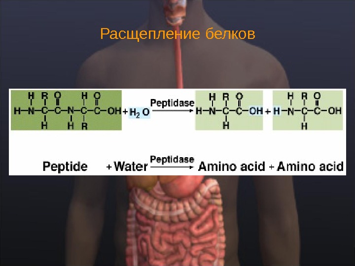
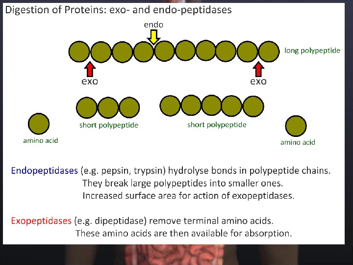
Расщепление белков
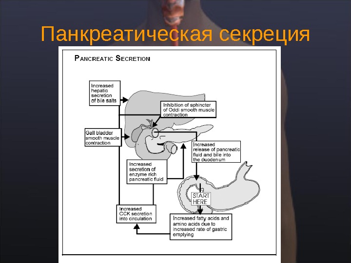
Панкреатическая секреция
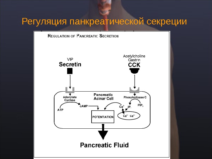
Регуляция панкреатической секреции
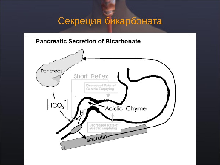
Секреция бикарбоната
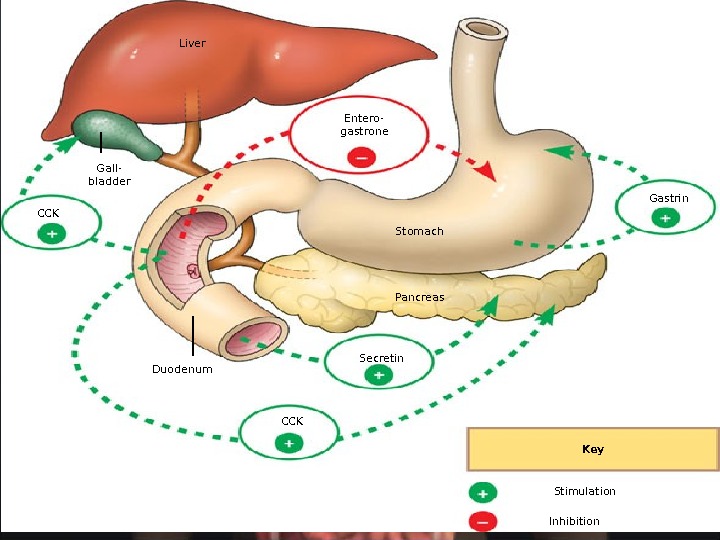
Liver Gall- bladder CCK Entero- gastrone Gastrin Stomach Pancreas Secretin CCKDuodenum Key Stimulation Inhibition
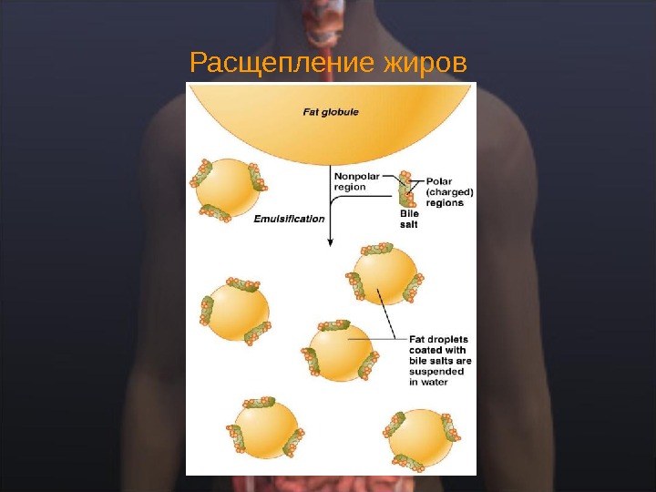
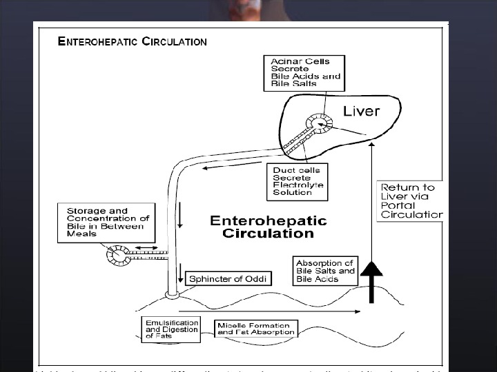
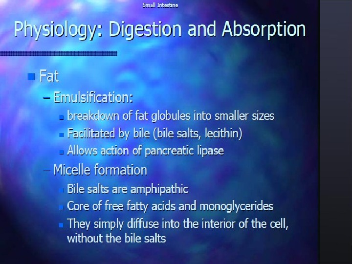
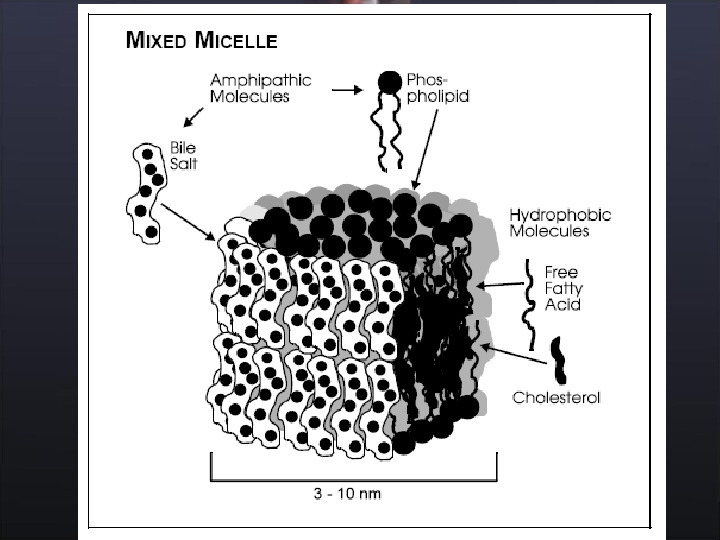
Emulsification and digestion of fat in the small intestine.
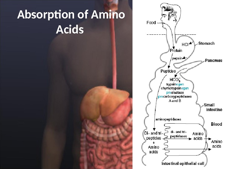
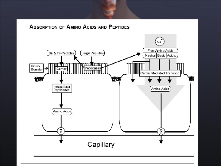
Пищеварение и всасывание углеводов и белков
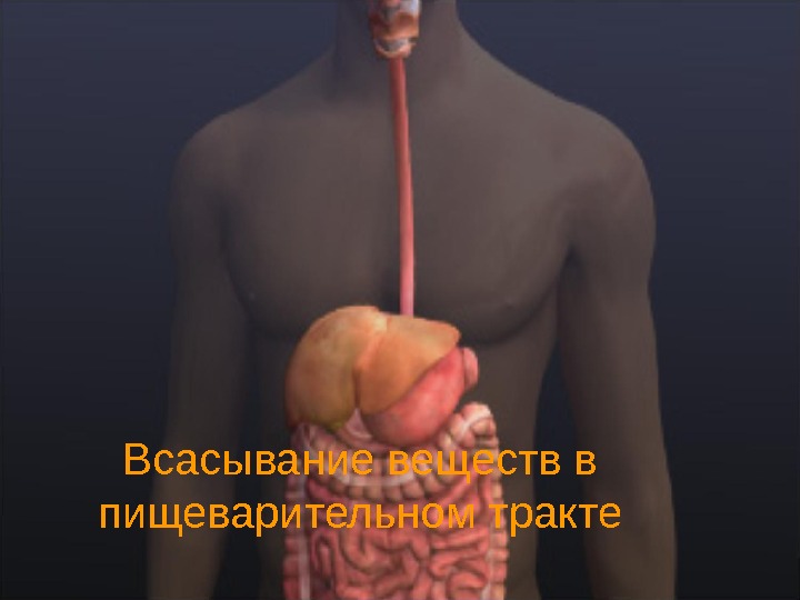
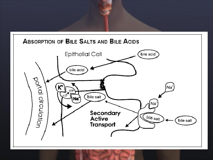
Всасывание веществ в пищеварительном тракте
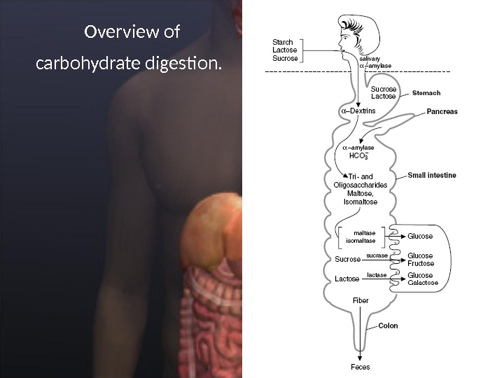
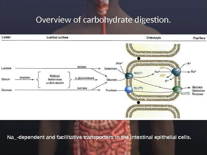
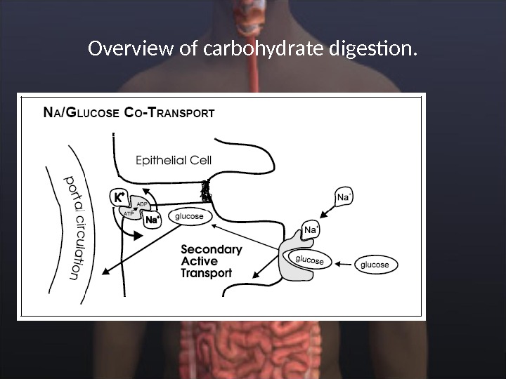
Overview of carbohydrate digestion.
Overview of carbohydrate digestion. Na_-dependent and facilitative transporters in the intestinal epithelial cells.
Overview of carbohydrate digestion.
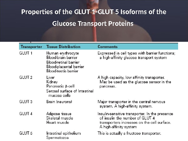
Properties of the GLUT 1 -GLUT 5 Isoforms of the Glucose Transport Proteins
CLINICAL CORRELATION Disaccharidase Deficiency Intestinal disaccharidase deficiencies are encountered relatively frequently in humans. Deficiency can be present in one enzyme or several enzymes for a variety of reasons (genetic defect, physiological decline with age, or the result of «injuries» to the mucosa). Of the disaccharidases, lactase is the most common enzyme with an absolute or relative deficiency, which is experienced as milk intolerance. The consequences of an inability to hydrolyze lactose in the upper small intestine are inability to absorb lactose and bacterial fermentation of ingested lactose in the lower small intestine.
Absorption of Amino Acids
CLINICAL CORRELATION Neutral Amino Aciduria (Hartnup Disease) Transport functions, like enzymatic functions, are subject to modification by mutations. An example of a genetic lesion in epithelial amino acid transport is Hartnup disease, named after the family in which the disease entity resulting from the defect was first recognized. The disease is characterized by the inability of renal and intestinal epithelial cells to absorb neutral amino acids from the lumen. In the kidney, in which plasma amino acids reach the lumen of the proximal tubule through the ultrafiltrate, the inability to reabsorb amino acids manifests itself as excretion of amino acids in the urine (amino aciduria). The intestinal defect results in malabsorption of free amino acids from the diet.
Kwashiorkor , A common problem of children in Third World countries, is caused by a deficiency of protein in a diet that is adequate in calories. Children with kwashiorkor suffer from muscle wasting and a decreased concentration of plasma proteins, particularly albumin. The result is an increase in interstitial fluid that causes edema and a distended abdomen that make the children appear “plump”.
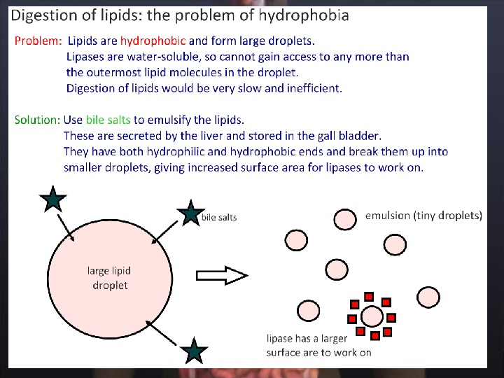
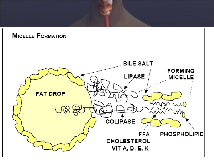
Расщепление жиров
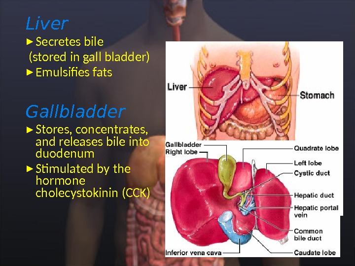
Liver Secretes bile (stored in gall bladder) Emulsifies fats Gallbladder Stores, concentrates, and releases bile into duodenum Stimulated by the hormone cholecystokinin (CCK)
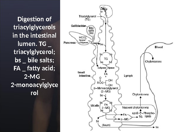
Digestion of triacylglycerols in the intestinal lumen. TG _ triacylglycerol; bs _ bile salts; FA _ fatty acid; 2 -MG _ 2 -monoacylglyce rol
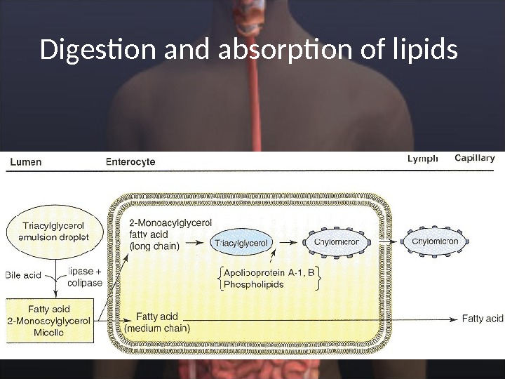
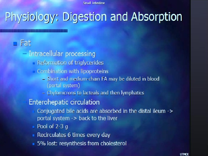
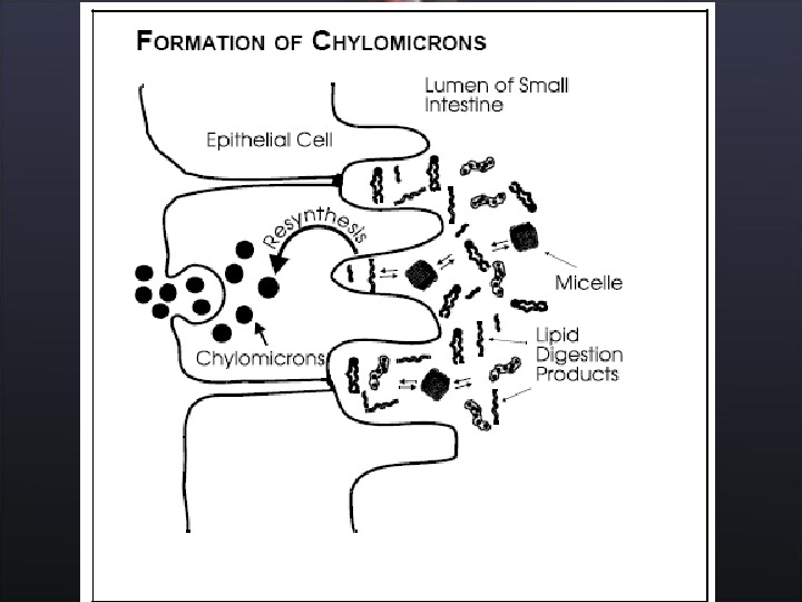
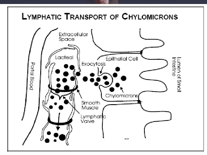
Digestion and absorption of lipids
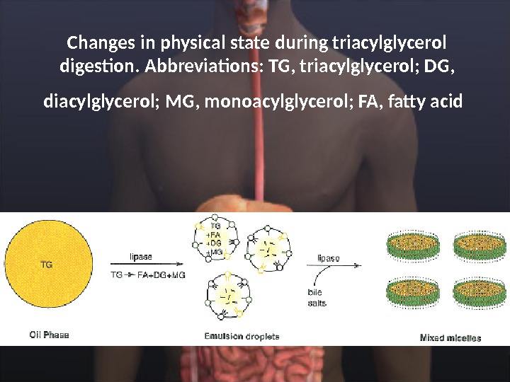
Changes in physical state during triacylglycerol digestion. Abbreviations: TG, triacylglycerol; DG, diacylglycerol; MG, monoacylglycerol; FA, fatty acid
CLINICAL CORRELATION A- β -Lipoproteinemia A- b -lipoproteinemia is an autosomal recessive disorder characterized by the absence of all lipoproteins containing apo-β-lipoprotein, that is, chylomicrons, very low density lipoproteins (VLDLs), and low density lipoproteins (LDLs). Serum cholesterol is extremely low. This defect is associated with severe malabsorption of triacylglycerol and lipid-soluble vitamins (especially tocopherol and vitamin E) and accumulation of apo B in enterocytes and hepatocytes.
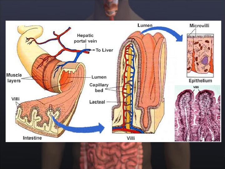
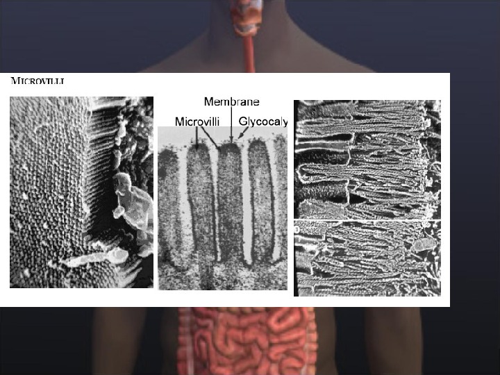
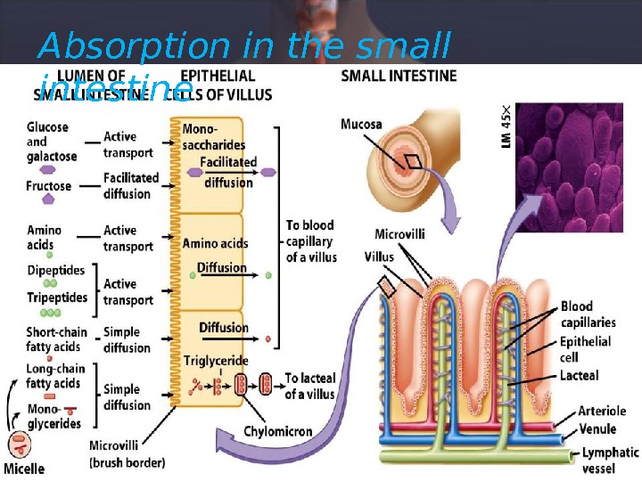
Absorption in the small intestine
Absorption of Minerals and Metals Calcium Active, transcellular absorption occurs only in the duodenum when calcium intake has been low. This process involves import of calcium into the enterocyte, transport across the cell, and export into extracellular fluid and blood. The rate limiting step in transcellular calcium absorption is transport across the epithelial cell, which is greatly enhanced by the carrier protein calbindin, the synthesis of which is totally dependent on vitamin D.
Calcium • Passive, paracellular absorption occurs in the jejunum and ileum, and, to a much lesser extent, in the colon when dietary calcium levels have been moderate or high. In this case, ionized calcium diffuses through tight junctions into the basolateral spaces around enterocytes, and hence into blood. Such transport depends on having higher concentrations of free calcium in the intestinal lumen than in blood.
Phosphorus is predominantly absorbed as inorganic phosphate in the upper small intestine. Phosphate is transported into the epithelial cells by cotransport with sodium, and expression of this (or these) transporters is enhanced by vitamin D.
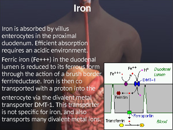
Iron is absorbed by villus enterocytes in the proximal duodenum. Efficient absorption requires an acidic environment. Ferric iron (Fe+++) in the duodenal lumen is reduced to its ferrous form through the action of a brush border ferrireductase. Iron is then co transported with a proton into the enterocyte via the divalent metal transporter DMT-1. This transporter is not specific for iron, and also transports many divalent metal ions.
Iron • Once inside the enterocyte, iron follows one of two major pathways: • • Iron abundance states : iron within the enterocyte is trapped by incorporation into ferritin and hence, not transported into blood. When the enterocyte dies and is shed, this iron is lost. • • Iron limiting states : iron is exported out of the enterocyte via a transporter (ferroportin) located in the basolateral membrane. It then binds to the iron-carrier transferrin for transport throughout the body.
Copper • There appear to be two processes responsible for copper absorption: • i) a rapid, low capacity system and • ii) a slower, high capacity system, which may be similar to the two processes seen with calcium absorption. • Many of the molecular details of copper absorption remain to be elucidated. • A number of dietary factors have been shown to influence copper absorption. For example, excessive dietary intake of either zinc or molybdenum can induce secondary copper deficiency states.
Zinc • Zinc homeostasis is largely regulated by its uptake and loss through the small intestine. Although a number of zinc transporters and binding proteins have been identified in villus epithelial cells, a detailed picture of the molecules involved in zinc absorption is not yet in hand. • A number of nutritional factors have been identified that modulate zinc absorption. Certain animal proteins in the diet enhance zinc absorption. Phytates from dietary plant material (including cereal grains, corn, rice) chelate zinc and inhibit its absorption. Subsistence on phytate-rich diets is thought responsible for a considerable fraction of human zinc deficiencies.
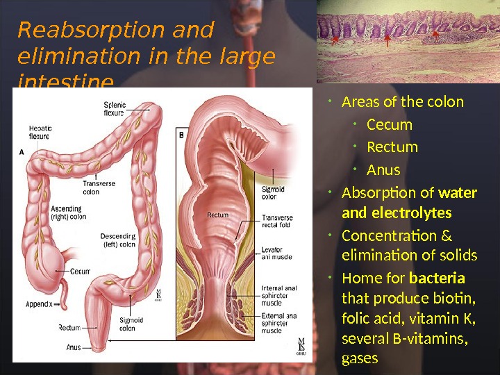
Reabsorption and elimination in the large intestine • Areas of the colon • Cecum • Rectum • Anus • Absorption of water and electrolytes • Concentration & elimination of solids • Home for bacteria that produce biotin, folic acid, vitamin K, several B-vitamins, gases
The Large Intestine: Introduction • Recovery of water and electrolytes from ingesta: By the time ingesta reaches the terminal ileum, roughly 90% of its water has been absorbed, but considerable water and electrolytes like sodium and chloride remain and must be recovered by absorption in the large gut. • Formation and storage of feces: As ingesta is moved through the large intestine, it is dehydrated, mixed with bacteria and mucus, and formed into feces. The craftsmanship (for want of a better term) with which this is carried out varies among species. • Microbial fermentation: The large intestine of all species teems with microbial life. Those microbes produce enzymes capable of digesting many of molecules that to vertebrates are indigestible, cellulose being a premier example. The extent and benefit of fermentation also varies greatly among species.
Large Intestinal Motility • Segmentation contractions which chop and mix the ingesta, presenting it to the mucosa where absorption occurs. These contractions are quite prominent in some species, forming sacculations in the colon known as hausta. • Antiperistaltic contractions propagate toward the ileum, which serves to retard the movement of ingesta through the colon, allowing additional opportunity for absorption of water and electrolytes. Peristaltic contractions, in addition to influx from the small intestine, facilitate movement of ingesta through the colon. • Mass movements constitute a type of motility not seen elsewhere in the digestive tube. Known also as giant migrating contractions, this pattern of motility is like a very intense and prolonged peristaltic contraction which strips an area of large intestine clear of contents.
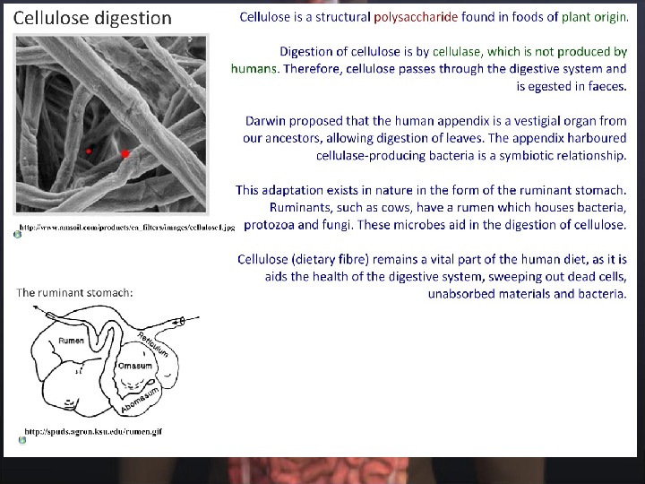
Fermentation • Fermentation is the enzymatic decomposition and utililization of foodstuffs, particularly carbohydrates, by microbes. • Large intestinal epithelial cells do not produce digestive enzymes, but contain huge numbers of bacteria which have the enzymes to digest and utilize many substrates. In all animals, two processes are attributed to the microbial flora of the large intestine: • Digestion and metabolism of carbohydrates not digested in the small intestine (e. g. cellulose, residual starch) • Synthesis of vitamin K and certain B vitamins • Synthesis of vitamin K by colonic bacteria provides a valuable supplement to dietary sources and makes clinical vitamin K deficiency rare. Similarly, formation of B vitamins by the microbial flora in the large intestine is useful to many animals.