
Презентация heart murmurs


- Размер: 101.5 Кб
- Количество слайдов: 18
Описание презентации Презентация heart murmurs по слайдам
Heart Murmurs David Leder
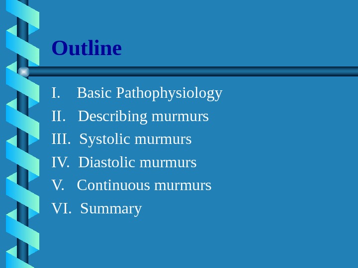
Outline I. Basic Pathophysiology II. Describing murmurs III. Systolic murmurs IV. Diastolic murmurs V. Continuous murmurs VI. Summary
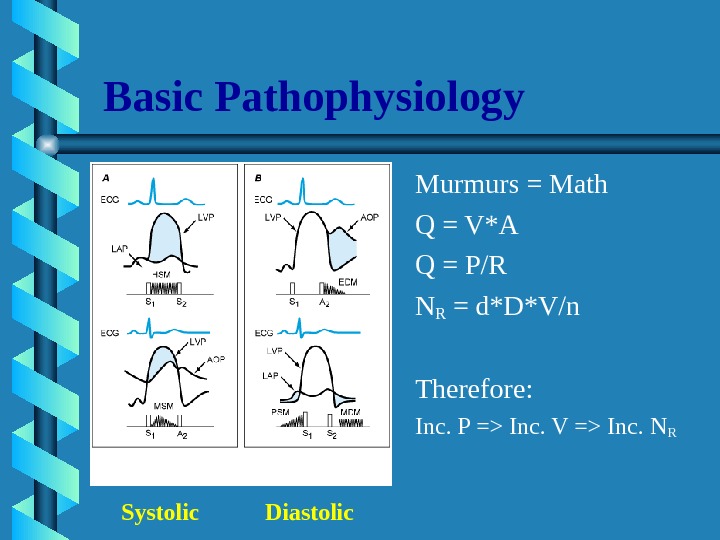
Basic Pathophysiology Murmurs = Math Q = V*A Q = P/R NR = d*D*V/n Therefore: Inc. P => Inc. V => Inc. N R Systolic Diastolic
Describing a heart murmur 1. Timing • murmurs are longer than heart sounds • HS can distinguished by simultaneous palpation of the carotid arterial pulse • systolic, diastolic, continuous 2. Shape • crescendo (grows louder), decrescendo, crescendo-decrescendo, plateau 3. Location of maximum intensity • is determined by the site where the murmur originates • e. g. A, P, T, M listening areas
Describing a heart murmur con’t: 4. Radiation • reflects the intensity of the murmur and the direction of blood flow 5. Intensity • graded on a 6 point scale – Grade 1 = very faint – Grade 2 = quiet but heard immediately – Grade 3 = moderately loud – Grade 4 = loud – Grade 5 = heard with stethoscope partly off the chest – Grade 6 = no stethoscope needed *Note: Thrills are assoc. with murmurs of grades 4 —
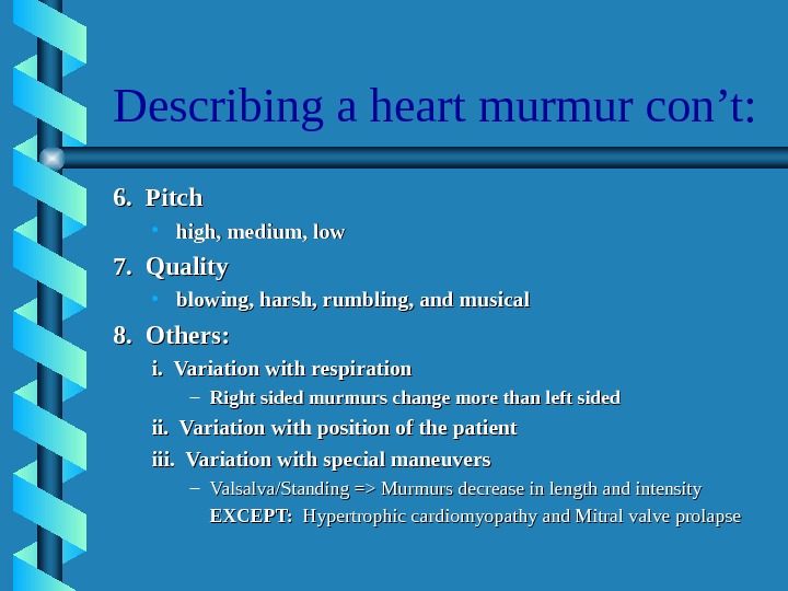
Describing a heart murmur con’t: 6. Pitch • high, medium, low 7. Quality • blowing, harsh, rumbling, and musical 8. Others: i. Variation with respiration – Right sided murmurs change more than left sided ii. Variation with position of the patient iii. Variation with special maneuvers – Valsalva/Standing => Murmurs decrease in length and intensity EXCEPT: Hypertrophic cardiomyopathy and Mitral valve prolapse
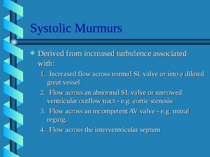
Systolic Murmurs Derived from increased turbulence associated with: 1. Increased flow across normal SL valve or into a dilated great vessel 2. Flow across an abnormal SL valve or narrowed ventricular outflow tract — e. g. aortic stenosis 3. Flow across an incompetent AV valve — e. g. mitral regurg. 4. Flow across the interventricular septum
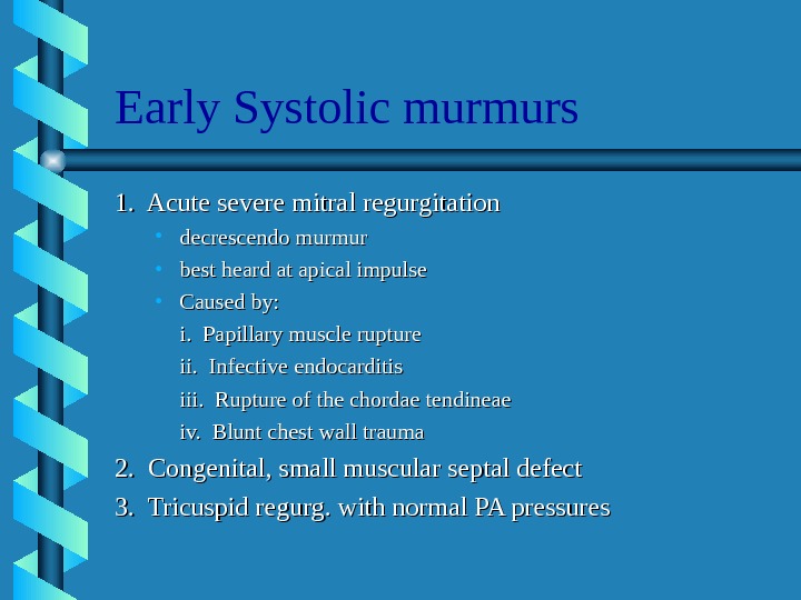
Early Systolic murmurs 1. Acute severe mitral regurgitation • decrescendo murmur • best heard at apical impulse • Caused by: i. Papillary muscle rupture ii. Infective endocarditis iii. Rupture of the chordae tendineae iv. Blunt chest wall trauma 2. Congenital, small muscular septal defect 3. Tricuspid regurg. with normal PA pressures
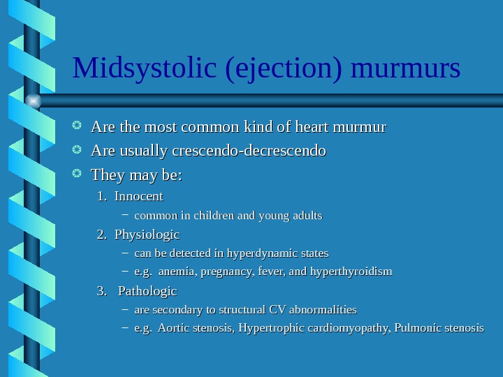
Midsystolic (ejection) murmurs Are the most common kind of heart murmur Are usually crescendo-decrescendo They may be: 1. Innocent – common in children and young adults 2. Physiologic – can be detected in hyperdynamic states – e. g. anemia, pregnancy, fever, and hyperthyroidism 3. Pathologic – are secondary to structural CV abnormalities – e. g. Aortic stenosis, Hypertrophic cardiomyopathy, Pulmonic stenosis
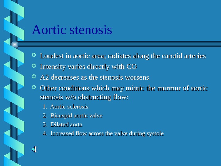
Aortic stenosis Loudest in aortic area; radiates along the carotid arteries Intensity varies directly with CO A 2 decreases as the stenosis worsens Other conditions which may mimic the murmur of aortic stenosis w/o obstructing flow: 1. Aortic sclerosis 2. Bicuspid aortic valve 3. Dilated aorta 4. Increased flow across the valve during systole
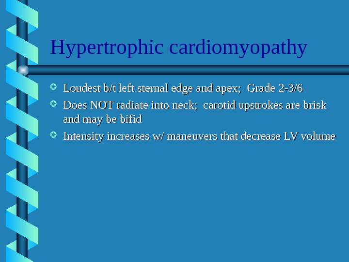
Hypertrophic cardiomyopathy Loudest b/t left sternal edge and apex; Grade 2 -3/6 Does NOT radiate into neck; carotid upstrokes are brisk and may be bifid Intensity increases w/ maneuvers that decrease LV volume
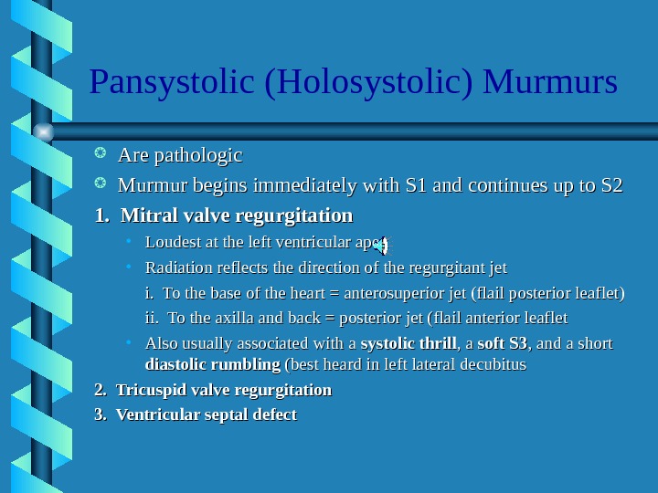
Pansystolic (Holosystolic) Murmurs Are pathologic Murmur begins immediately with S 1 and continues up to S 2 1. Mitral valve regurgitation • Loudest at the left ventricular apex • Radiation reflects the direction of the regurgitant jet i. To the base of the heart = anterosuperior jet (flail posterior leaflet) ii. To the axilla and back = posterior jet (flail anterior leaflet • Also usually associated with a systolic thrill , a soft S 3 , and a short diastolic rumbling (best heard in left lateral decubitus 2. Tricuspid valve regurgitation 3. Ventricular septal defect
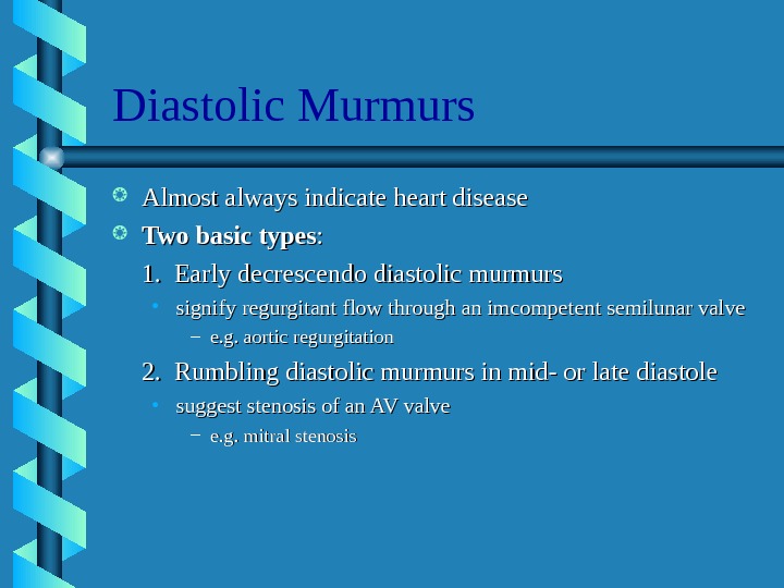
Diastolic Murmurs Almost always indicate heart disease Two basic types : : 1. Early decrescendo diastolic murmurs • signify regurgitant flow through an imcompetent semilunar valve – e. g. aortic regurgitation 2. Rumbling diastolic murmurs in mid- or late diastole • suggest stenosis of an AV valve – e. g. mitral stenosis
Aortic Regurgitation Best heard in the 2 nd ICS at the left sternal edge High pitched, decrescendo Blowing quality => may be mistaken for breath sounds Radiation: i. Left sternal border = assoc. with primary valvular pathology; ii. Right sternal edge = assoc. w/ primary aortic root pathology Other associated murmurs: i. Midsystolic murmur ii. Austin Flint murmur
Mitral Stenosis Two components: 1. Middiastolic — during rapid ventricular filling 2. Presystolic — during atrial contraction; therefore, it disappears if atrial fibrillation develops Is low-pitched and best heard over the apex (w/ the bell) Little or no radiation Murmur begins after an Opening Snap; S 1 is accentuated
Continuous Murmurs Begin in systole, peak near s 2, and continue into all or part of diastole. 1. Cervical venous hum • Audible in kids; can be abolished by compression over the IJV 2. Mammary souffle • Represents augmented arterial flow through engorged breasts • Becomes audible during late 3 rd trimester and lactation 3. Patent Ductus Arteriosus • Has a harsh, machinery-like quality 4. Pericardial friction rub • Has scratchy, scraping quality
Back to the Basics 1. When does it occur — systole or diastole 2. Where is it loudest — A, P, T, M I. Systolic Murmurs: 1. Aortic stenosis — ejection type 2. Mitral regurgitation — holosystolic 3. Mitral valve prolapse — late systole II. Diastolic Murmurs: 1. Aortic regurgitation — early diastole 2. Mitral stenosis — mid to late diastole
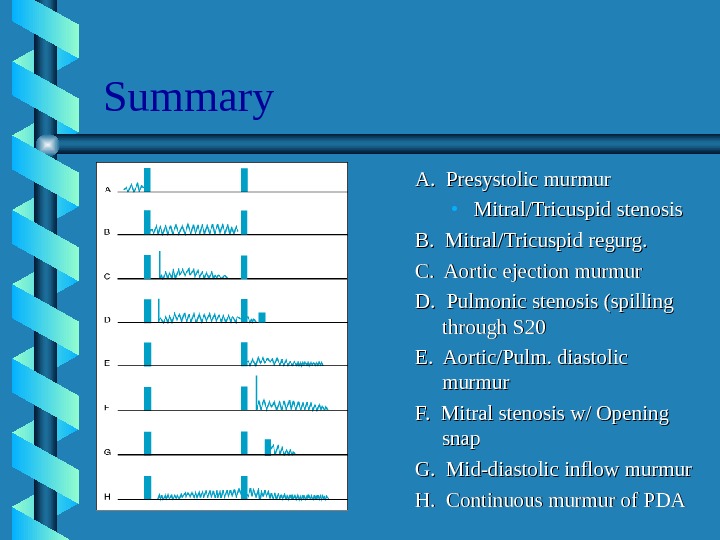
Summary A. Presystolic murmur • Mitral/Tricuspid stenosis B. Mitral/Tricuspid regurg. C. Aortic ejection murmur D. Pulmonic stenosis (spilling through S 20 E. Aortic/Pulm. diastolic murmur F. Mitral stenosis w/ Opening snap G. Mid-diastolic inflow murmur H. Continuous murmur of P