Презентация heart-auscultation4181



- Размер: 196.5 Кб
- Количество слайдов: 18
Описание презентации Презентация heart-auscultation4181 по слайдам
 CARDIOVASCULAR SYSTEM Heart auscultation Lecture 2/
 The Goals of Auscultation • The intensity of S 1 in all areas • The intensity of S 2 in all areas • The characterization of any systolic sounds • The characterization of any diastolic sounds

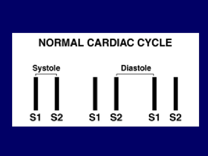
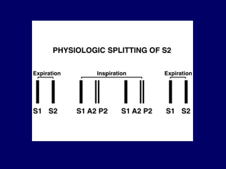
 Normal heart sounds • In younger patients physiologic splitting of S 2. is possible – S 2 is made up of 2 components, aortic (A 2) and pulmonic (P 2) valve closure. – On inspiration, venous return to the heart is augmented and pulmonic valve closure is delayed, allowing you to hear first A 2 and then P 2. – On expiration, the two sounds occur closer together and are detected as a single S 2. • The two components of S 1 (mitral and tricuspid valve closure) occur so close together that splitting is not appreciated.

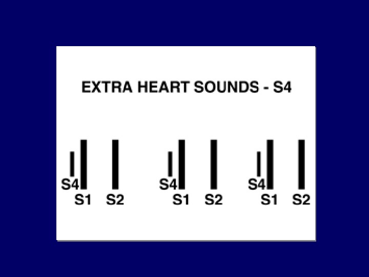
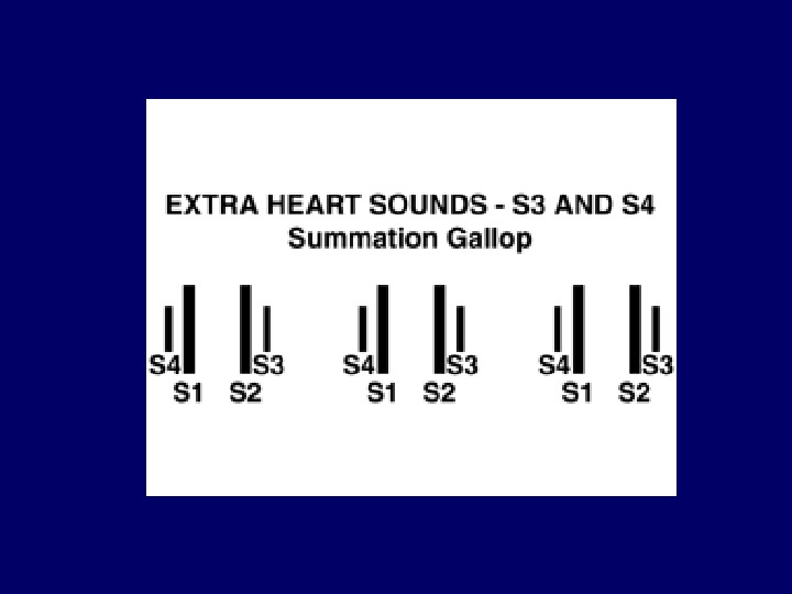
 Extra heart sounds • While present in normal subjects up to the ages of 20 -30, they represent pathology in older patients. • An S 3 is most commonly associated with left ventricular failure and is caused by blood from the left atrium slamming into an already overfilled ventricle during early diastolic filling. • The S 4 is a sound created by blood trying to enter a stiff, non-compliant left ventricle during atrial contraction. It’s most frequently associated with left ventricular hypertrophy that is the result of long standing hypertension. • Positioning the patient on their left side while you listen may improve the yield of this exam. • The presence of both an S 3 and S 4 simultaneously is referred to as a summation gallop.


 Factors that may influence the intensity of the heart sounds: first sound Loud first sound • Hyperdinamic circulation • Mitral stenosis • Atril myxoma (rare) Soft first sound • Low cardiac output (rest, heart failure) • Tachycardia • Severe mitral reflux (caused by destruction of valve) Variable intensity of first sound • Atrial fillibration • Complete hart block
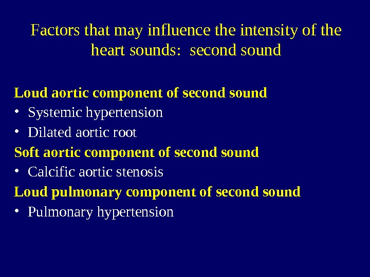
 Factors that may influence the intensity of the heart sounds: second sound Loud aortic component of second sound • Systemic hypertension • Dilated aortic root Soft aortic component of second sound • Calcific aortic stenosis Loud pulmonary component of second sound • Pulmonary hypertension
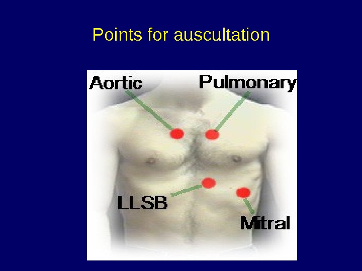
 Points for auscultation
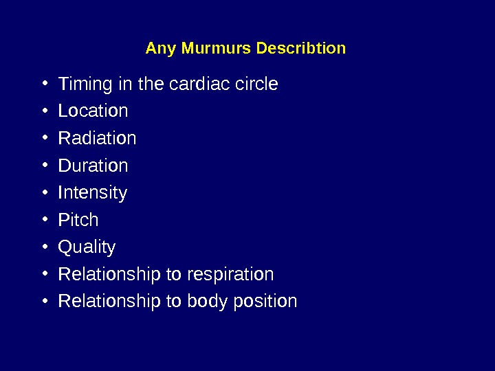
 Any Murmurs Describtion • Timing in the cardiac circle • Location • Radiation • Duration • Intensity • Pitch • Quality • Relationship to respiration • Relationship to body position
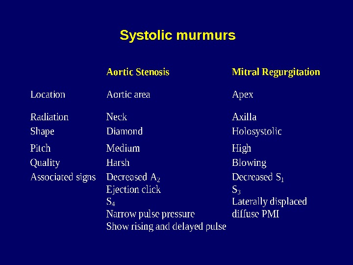
 Systolic murmurs
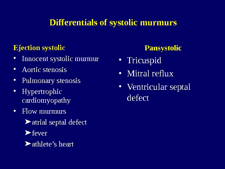
 Differentials of systolic murmurs Ejection systolic • Innocent systolic murmur • Aortic stenosis • Pulmonary stenosis • Hypertrophic cardiomyopathy • Flow murmurs atrial septal defect fever athlete’s heart Pansystolic • Tricuspid • Mitral reflux • Ventricular septal defect
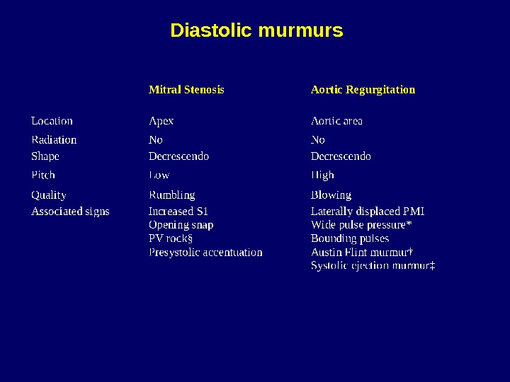
 Diastolic murmurs
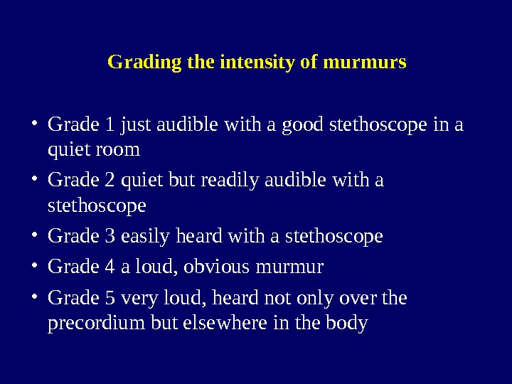
 Grading the intensity of murmurs • Grade 1 just audible with a good stethoscope in a quiet room • Grade 2 quiet but readily audible with a stethoscope • Grade 3 easily heard with a stethoscope • Grade 4 a loud, obvious murmur • Grade 5 very loud, heard not only over the precordium but elsewhere in the body
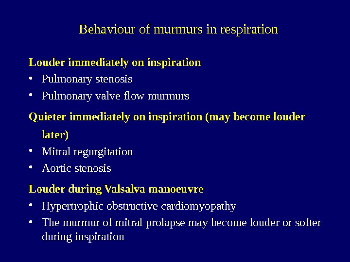
 Behaviour of murmurs in respiration Louder immediately on inspiration • Pulmonary stenosis • Pulmonary valve flow murmurs Quieter immediately on inspiration (may become louder later) • Mitral regurgitation • Aortic stenosis Louder during Valsalva manoeuvre • Hypertrophic obstructive cardiomyopathy • The murmur of mitral prolapse may become louder or softer during inspiration

