
Презентация cervix and uteri cancer during pregnancy


cervix_and_uteri_cancer_during_pregnancy.ppt
- Размер: 1.6 Mегабайта
- Количество слайдов: 27
Описание презентации Презентация cervix and uteri cancer during pregnancy по слайдам
С ervix and uteri cancer during pregnancy Prepared: Nurmanbetova G. Nurzhauova A. Course: 7 Faculty: General medicine Group: 705 Teacher: Igisinova G. S. Р ДЕНСАУЛЫ СА ТАУ МИНИСТРЛІГІҚ Қ Қ С. Д. АСФЕНДИЯРОВ АТЫНДА Ы Ғ АЗА ЛТТЫ МЕДИЦИНА Қ Қ Ұ Қ УНИВЕРСИТЕТІ МИНИС Т ЕРСТВО ЗДРАВООХРАНЕНИЯ РК КАЗАХСКИЙ НАЦИОНАЛЬНЫЙ МЕДИЦИНСКИЙ УНИВЕРСИТЕТ ИМЕНИ С. Д. АСФЕНДИЯРОВА Department of internship and residency in oncology
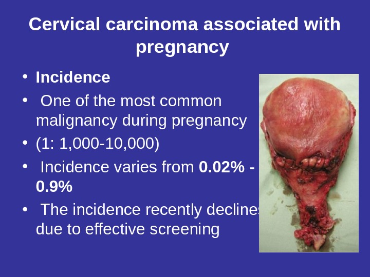
Cervical carcinoma associated with pregnancy • Incidence • One of the most common malignancy during pregnancy • (1: 1, 000 -10, 000) • Incidence varies from 0. 02% — 0. 9% • The incidence recently declines due to effective screening
Physiological changes during pregnancy in the mucous membrane of the cervix • In the I trimester of the cervix characterized by cyanosis and colposcopic noted the presence of whitish spot elevations due to physiological hypertrophy, and an extensive network of blood vessels. • In the II and III trimesters can be determined visually physiological ectopia prismatic epithelium and colposcopy — white spots on the background metaplastic epithelium vascular pattern. Changes regress within 2 -4 months after birth
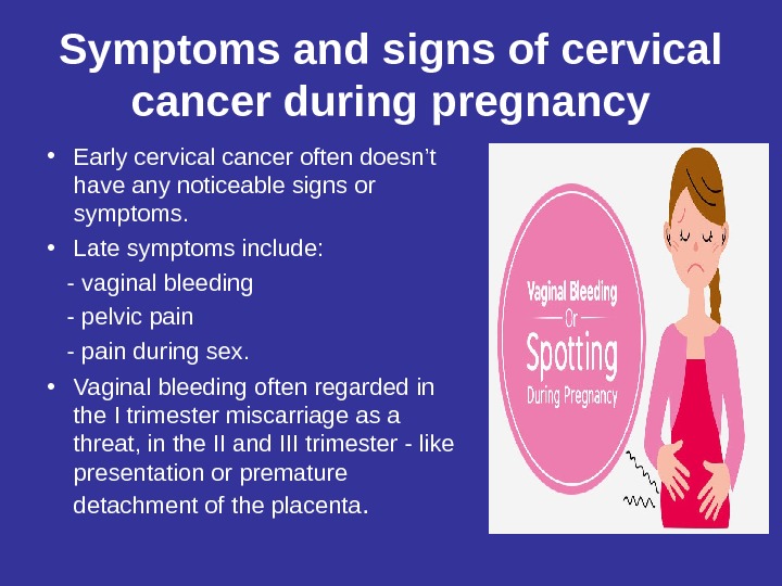
Symptoms and signs of cervical cancer during pregnancy • Early cervical cancer often doesn’t have any noticeable signs or symptoms. • Late symptoms include: — vaginal bleeding — pelvic pain — pain during sex. • Vaginal bleeding often regarded in the I trimester miscarriage as a threat, in the II and III trimester — like presentation or premature detachment of the placenta.
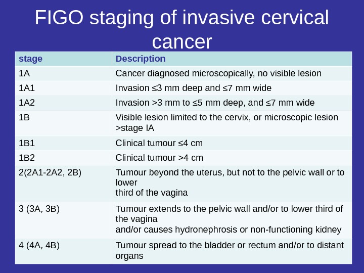
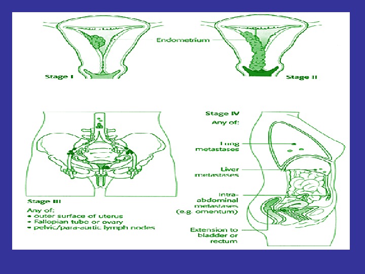
FIGO staging of invasive cervical cancer stage Description 1 A Cancer diagnosed microscopically, no visible lesion 1 A 1 Invasion ≤ 3 mm deep and ≤ 7 mm wide 1 A 2 Invasion >3 mm to ≤ 5 mm deep, and ≤ 7 mm wide 1 B Visible lesion limited to the cervix, or microscopic lesion >stage IA 1 B 1 Clinical tumour ≤ 4 cm 1 B 2 Clinical tumour >4 cm 2(2 A 1 -2 A 2, 2 B) Tumour beyond the uterus, but not to the pelvic wall or to lower third of the vagina 3 (3 A, 3 B) Tumour extends to the pelvic wall and/or to lower third of the vagina and/or causes hydronephrosis or non-functioning kidney 4 (4 A, 4 B) Tumour spread to the bladder or rectum and/or to distant organs
FIGO staging of invasive cervical cancer
Treatment of cervical cancer during pregnancy • The decision to continue the pregnancy should be based on careful discussion of the cancer prognosis, treatment and future fertility with the woman and her partner and multidisciplinary team
Treatment of intraepithelial cervical cancer (in situ) • I trimester • If patient don’t want to continue her pregnancy — medical abortion after 4 -8 weeks – conization cervix. • If patient wish to continue her pregnancy — 4 -8 weeks after vaginal delivery — cone biopsy of the cervix. • If invasive growth — a medical abortion after 4 -8 weeks – conization cervix; when strong desire to continue the pregnancy — atypical cone biopsy or wedge biopsy 6 -8 weeks after vaginal delivery — cone biopsy of the cervix. • II, III trimesters • 1. Preservation of pregnancy, 6 -8 weeks • after vaginal childbirth — cervical conization
Treatment of microinvasive cervical cancer I trimester • If patient wish to continue the pregnancy, but if patient want to preserve fertility — a medical abortion after 4 -8 weeks – cervix conization. • If patient would not keep the pregnancy and at the decision to complete the childbirth — a simple hysterectomy. • If patient wish to continue the pregnancy, and Fertility — continuation of the pregnancy through 6 -8 weeks after term birth (abdominal or vaginal) — cone biopsy of the cervix. • If you wish to continue the pregnancy, but then the completion of childbearing – Caesar section at term with simultaneous simple hysterectomy.
Treatment of cervical IA 2, II stages • In the case of diagnosis before 20 weeks of gestation, treatment should begin immediately. The method of choice is radical hysterectomy with fetus in situ. During the chemoradiotherapy, as a rule, the termination of pregnancy is not carried out, as after the start of treatment there is a spontaneous miscarriage.
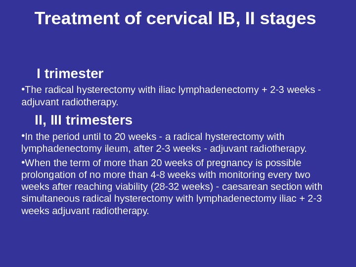
Treatment of cervical IB, II stages I trimester • The radical hysterectomy with iliac lymphadenectomy + 2 -3 weeks — adjuvant radiotherapy. II, III trimesters • In the period until to 20 weeks — a radical hysterectomy with lymphadenectomy ileum, after 2 -3 weeks — adjuvant radiotherapy. • When the term of more than 20 weeks of pregnancy is possible prolongation of no more than 4 -8 weeks with monitoring every two weeks after reaching viability (28 -32 weeks) — caesarean section with simultaneous radical hysterectomy with lymphadenectomy iliac + 2 -3 weeks adjuvant radiotherapy.
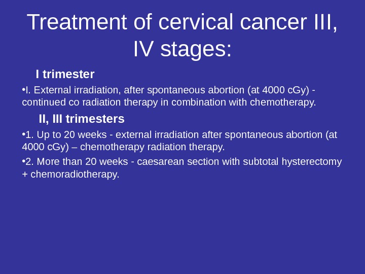
Treatment of cervical cancer III, IV stages: I trimester • I. External irradiation, after spontaneous abortion (at 4000 c. Gy) — continued co radiation therapy in combination with chemotherapy. II, III trimesters • 1. Up to 20 weeks — external irradiation after spontaneous abortion (at 4000 c. Gy) – chemotherapy radiation therapy. • 2. More than 20 weeks — caesarean section with subtotal hysterectomy + chemoradiotherapy.
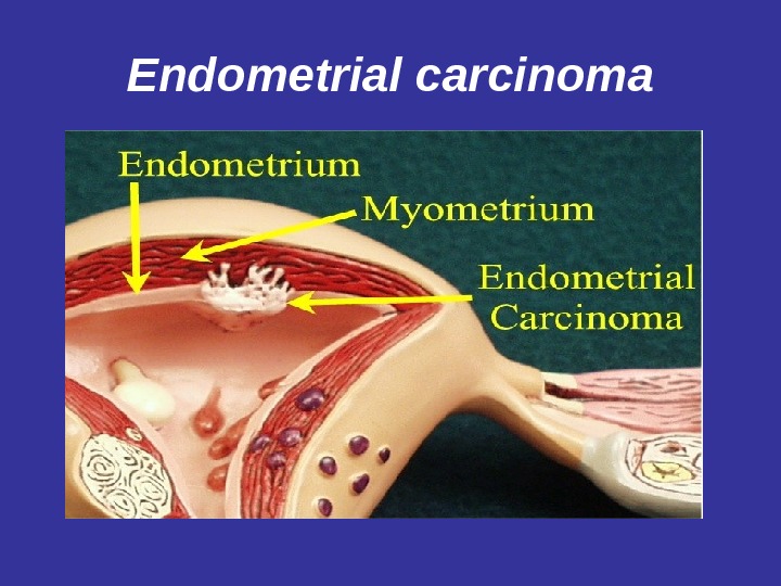
Endometrial carcinoma
Cancer During Pregnancy • Cancer during pregnancy is uncommon. It occurs in only about one out of every 1, 000 pregnancies. For many years, both doctors and women were often unsure about how to deal with cancer during pregnancy. But now more women with cancer and their doctors are starting or continuing treatment while pregnant. This means more information about treating and living with cancer during pregnancy is available than ever before.
Diagnosing cancer during pregnancy • Being pregnant may delay a cancer diagnosis. This is because some cancer symptoms, such as bloating, headaches, breast changes, or rectal bleeding, are also common during pregnancy. On the other hand, pregnancy can sometimes uncover cancer. For example, a Pap test done as part of standard pregnancy care can detect cervical cancer. And an ultrasound performed during pregnancy could find ovarian cancer.
Cancer treatment during pregnancy • When making treatment decisions for cancer during pregnancy, it is important to consider the best treatment options for the pregnant woman balanced against the possible risks to the growing baby.
Treatment recommendations are based on many factors, including the following • Stage of the pregnancy • The type, location, size, and stage of the cancer • The woman’s and her family’s wishes
• Treatment must be carefully planned to ensure the woman and unborn baby are safe. In general, treatment during pregnancy requires close teamwork with a multidisciplinary team, including both cancer doctors as well as high-risk obstetricians. These professionals can closely monitor the woman during treatment and make sure the baby is healthy.
Treatments that may be used during pregnancy • Surgery is the removal of the tumor and some of the surrounding healthy tissue. It poses little risk to the growing baby and is considered the safest cancer treatment during pregnancy.
Chemotherapy. • There is a risk of harm to the fetus if chemotherapy is given in the first three months of pregnancy. This is when the fetus’s organs are still growing. Chemotherapy during the first trimester carries risk of birth defects or pregnancy loss.
• During the second and third trimesters, doctors can give several types of chemotherapy without apparent risk to the fetus. Because the placenta acts as a barrier between the women and the baby, some drugs cannot pass through, or they pass through in very small amounts. Studies have suggested babies exposed to chemotherapy while in the mother’s uterus do not show any abnormalities either immediately after delivery, or during their future growth and development, when compared with children not exposed to chemotherapy.
Radiation therapy. • Radiation therapy is the use of high-energy x-rays to destroy cancer cells. Because radiation therapy can harm the fetus in all trimesters, doctors generally avoid using this treatment during pregnancy. The risks to the developing baby depend on the dose of radiation and the area of the body being treated.
Breastfeeding during treatment • Doctors advise women who are receiving chemotherapy after a pregnancy not to breastfeed. Chemotherapy can transfer to the infant through breast milk.
Reference List • Barber H. R. K. , Brunschwig A: Am. J. OB/GYN, 85. 156, 1963. • Baltzer J. , Regenfrecht M. , Kopche W. , Carcinoma of the Cervix and Pregnancy Int. J. Gyneco Obstet. 31: 317, 1990. • Zemlickis D. , Lishner M. Degendorfer P. et. el. Maternal and fetal outcome after breast cancer pregnancy. Am. J. Obstet. Gynecol. 9: 1956, 1991. • Karlen J. R. et. al. Dysgermenoma associated with pregnancy. OB/GYN 53: 330, 1979. • P. Struyk, P. S. Ovarian Masses in Pregnancy Acta. Scand. 63: 421, 1984. • Aviles, A. et. al. Growth and Development of Children of Mothers Treated with Chemotherapy during pregnancy: Current status of 43 children. Am. J. Hematology 36: 243, 1991. • Brodsky et. al. Am. J. Obstet, Gynecol. 138: 1165, 1980.