
Презентация bleeding disordes in children


- Размер: 6.5 Mегабайта
- Количество слайдов: 90
Описание презентации Презентация bleeding disordes in children по слайдам
Bleeding (hemorrhagic) disorders in children. Assistant of pediatric department Tatyana Golovko
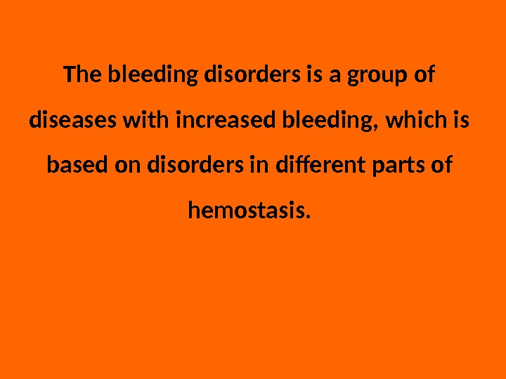
The bleeding disorders is a group of diseases with increased bleeding, which is based on disorders in different parts of hemostasis.
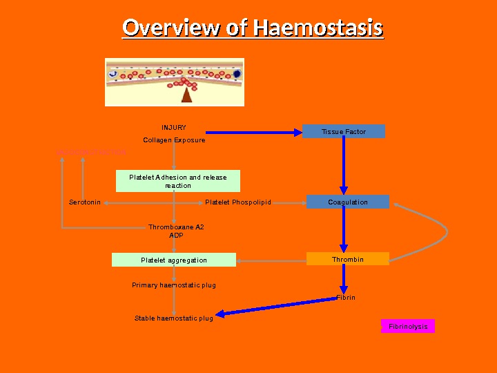
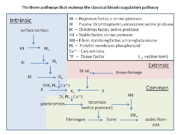
Overview of Haemostasis INJURY Collagen Exposure Platelet Adhesion and release reaction Platelet aggregation. VASOCONSTRICTION Serotonin Platelet Phospolipid Thromboxane A 2 ADP Primary haemostatic plug Stable haemostatic plug Tissue Factor Coagulation Thrombin Fibrinolysis
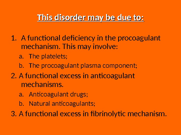
This disorder may be due to: 1. A functional deficiency in the procoagulant mechanism. This may involve: a. The platelets; b. The procoagulant plasma component; 2. A functional excess in anticoagulant mechanisms. a. Anticoagulant drugs; b. Natural anticoagulants; 3. A functional excess in fibrinolytic mechanism.
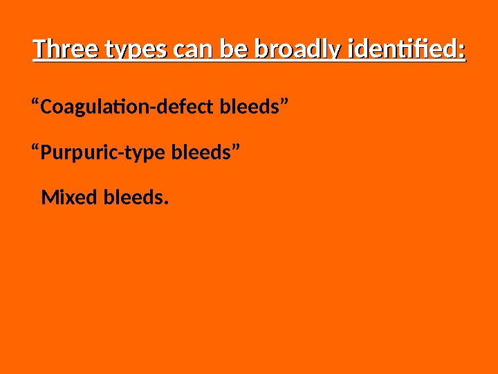
Three types can be broadly identified: “ Coagulation-defect bleeds” “ Purpuric-type bleeds” Mixed bleeds.
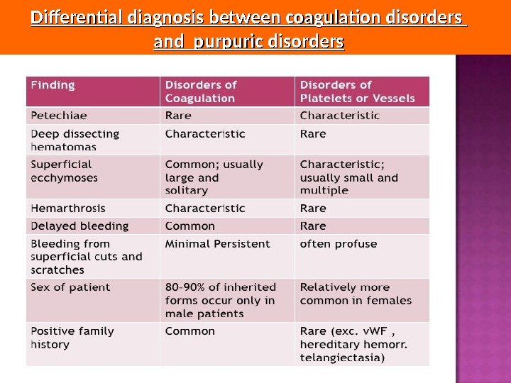
Differential diagnosis between coagulation disorders and purpuric disorders
Bleeding history The bleeding history forms the basis of the laboratory tests and therapy. Asking patients if they are bleeders is not always helpful, as patients with mild to moderate bleeding abnormalities may not admit that their bleeding episodes are significant. Valuable information may be obtained if a patient has undergone major surgery; however, if this is not the case, inquiries as to minor surgical procedures, such as dental extractions and tonsillectomy, should be made. It is important to get information on the duration of bleeding, the type of bleeding and what procedure was necessary to stop the bleeding (blood transfusion? ).
Physical Examination Several clinical as well as laboratory features help differentiate clinical disorders associated with qualitative and quantitative platelet defects and abnormalities of the blood vessel wall (diseases of primary hemostasis) from those associated with disorders of the coagulation factors. Diseases of primary hemostasis have also been referred to as “purpuric syndromes. ” Purpuric syndromes are characterized by capillary hemorrhages occurring chiefly in the skin and mucous membranes.
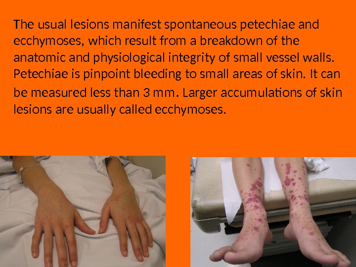
The usual lesions manifest spontaneous petechiae and ecchymoses, which result from a breakdown of the anatomic and physiological integrity of small vessel walls. Petechiae is pinpoint bleeding to small areas of skin. It can be measured less than 3 mm. Larger accumulations of skin lesions are usually called ecchymoses.
Gastrointestinal and genitourinary bleeding may occur spontaneously with abnormalities of platelets and/or coagulation factors. Deep hematomas, areas of palpable skin or soft tissue bleeding, and hemarthroses are most often associated with coagulation factor deficiencies or abnormalities. It is important to note that antecedent trauma or surgery, such as tooth extraction, tonsillectomy, or circumcision can be referred to excessive bleeding. Recurrent bleeding for several days usually indicates as an underlying bleeding disorder. In women, a careful history of their menstrual bleeding pattern gives valuable information about the nature of their hemostatic mechanism.
LABORATORY EVALUATION OF HEMOSTATIC DISORDERS Understanding of the physiology of primary and secondary hemostasis is important for the interpretation of diagnostic laboratory tests and for the subsequent management of patients with hemostatic disorders. The type of bleeding may be of significant help in designing the program of hemostatic laboratory tests. This is accomplished by performing a platelet count, a bleeding time, an activated partial thromboplastin time (a. PTT) and a prothrombin time (PT), and a thorough review of the patient’s peripheral blood smear.
The platelet count is performed to detect thrombocytopenia, which is defined as a platelet count of less than 150, 000/μL. The test can be considered as reliable down to a platelet count of 30, 000/μL. The finding of an unexpected thrombocytopenia should be confirmed by a review of the peripheral blood smear. The possibility of the existence of red blood cell fragments or of a pseudothrombocytopenia may provide clues for further evaluation of the patient.
The bleeding time is defined as the time between the infliction of a small standard cut and the moment the bleeding stops. Bleeding time measures the interactions of the platelets with the vessel wall and the subsequent formation of the primary hemostatic plug. A long bleeding time will be recorded either when the number of platelets is decreased, their function is abnormal, or there is defect of the vessel wall. The bleeding time may also be prolonged when there is a decrease of plasmatic factors, especially the VWF (von Willebrand factor) or fibrinogen.
The various methods for performing the bleeding time are basically modifications of two techniques: the bleeding time according to Duke; the bleeding time according to Mielke.
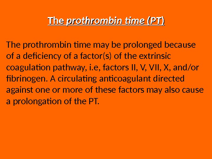
The prothrombin time (PT )) The prothrombin time may be prolonged because of a deficiency of a factor(s) of the extrinsic coagulation pathway, i. e, factors II, V, VII, X, and/or fibrinogen. A circulating anticoagulant directed against one or more of these factors may also cause a prolongation of the PT.
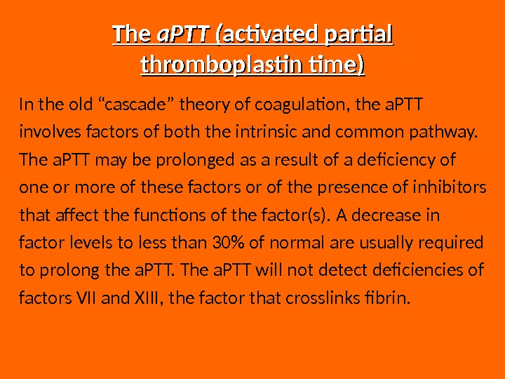
The a. PTT ( activated partial thromboplastin time) In the old “cascade” theory of coagulation, the a. PTT involves factors of both the intrinsic and common pathway. The a. PTT may be prolonged as a result of a deficiency of one or more of these factors or of the presence of inhibitors that affect the functions of the factor(s). A decrease in factor levels to less than 30% of normal are usually required to prolong the a. PTT. The a. PTT will not detect deficiencies of factors VII and XIII, the factor that crosslinks fibrin.
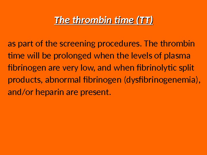
The thrombin time (TT) as part of the screening procedures. The thrombin time will be prolonged when the levels of plasma fibrinogen are very low, and when fibrinolytic split products, abnormal fibrinogen (dysfibrinogenemia), and/or heparin are present.
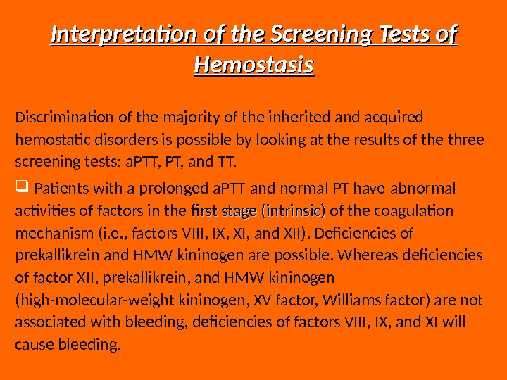
Interpretation of the Screening Tests of Hemostasis Discrimination of the majority of the inherited and acquired hemostatic disorders is possible by looking at the results of the three screening tests: a. PTT, PT, and TT. Patients with a prolonged a. PTT and normal PT have abnormal activities of factors in the first stage (intrinsic) of the coagulation mechanism (i. e. , factors VIII, IX, XI, and XII). Deficiencies of prekallikrein and HMW kininogen are possible. Whereas deficiencies of factor XII, prekallikrein, and HMW kininogen (high-molecular-weight kininogen, XV factor, Williams factor) are not associated with bleeding, deficiencies of factors VIII, IX, and XI will cause bleeding.
A prolonged PT and a normal a. PTT and TT may indicate a factor VII deficiency. Inherited or acquired deficiencies of factors II, V, VII, and X have to be considered if a prolonged PT and a. PTT and a normal thrombin time are found. A prolongation of a. PTT, PT, and thrombin time may reflect fibrinogen deficiency or dysfibrinogenemia.
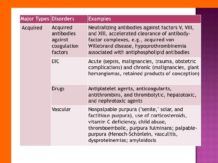
Most congenital deficiencies are single, whereas acquired abnormalities caused by vitamin K deficiency, liver disease, disseminated intravascular coagulation (DIC), or anticoagulant therapy which causes multiple coagulation defects. A prolonged bleeding time in the presence of a normal platelet amount is usually a sign of an abnormality of platelet–blood vessel interaction (e. g. , von Willebrand disease).
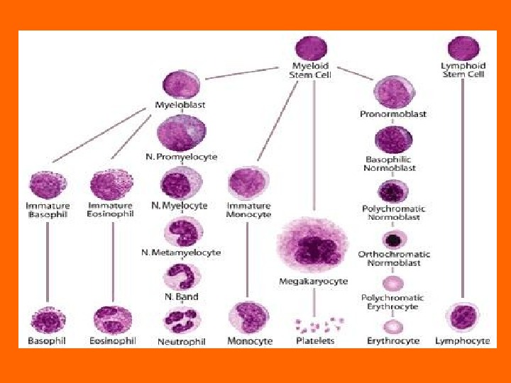
PLATELET DISORDERS Though platelets are classified as cells, they are actually cytoplasmatic fragments derived from megakaryocytes in the bone marrow. Platelet formation and release probably occur through the sinus endothelial cells. Platelets remain in the circulation for approximately 8– 10 days. The normal platelet amount ranges from 150, 000/μL to 400, 000/μL. The diameter of normal platelets is 1– 4 μm. At a given point, 70% of the platelets are in the circulation and 30% are in the spleen (splenic pool). The daily platelet production is 40, 000/μL and can be increased eightfold.
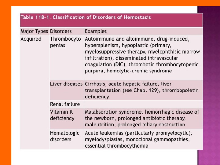
Thrombocytopenia thrombocytopenia occurs when the platelet amount drops below 150, 000/μL. The bleeding usually occurs when the platelet amount is less than 100, 000/μL. Spontaneous bleeding usually occurs with platelet amount less than 20, 000/μL. Thrombocytopenia is due to either decreased bone marrow production of platelets or increased destruction and sequestration of the platelets from the circulation, or both.
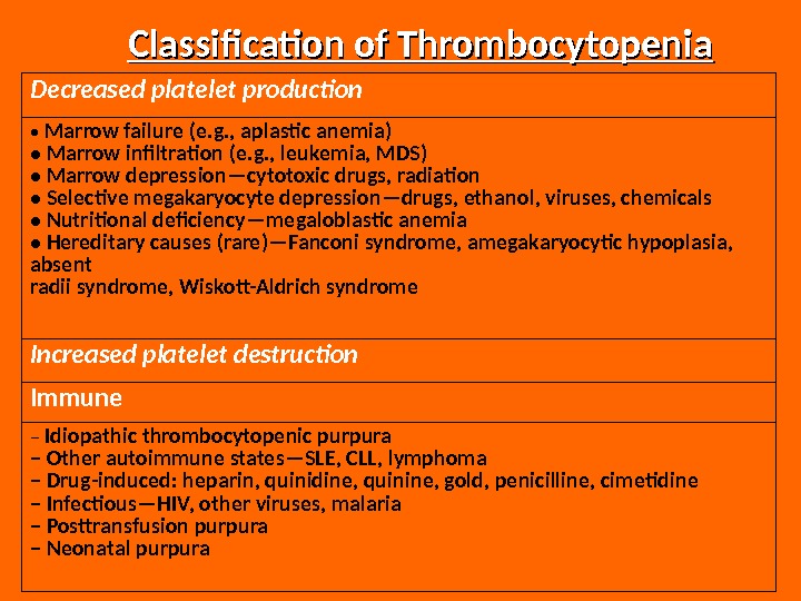
Classification of Thrombocytopenia Decreased platelet production • Marrow failure (e. g. , aplastic anemia) • Marrow infiltration (e. g. , leukemia, MDS) • Marrow depression—cytotoxic drugs, radiation • Selective megakaryocyte depression—drugs, ethanol, viruses, chemicals • Nutritional deficiency—megaloblastic anemia • Hereditary causes (rare)—Fanconi syndrome, amegakaryocytic hypoplasia, absent radii syndrome, Wiskott-Aldrich syndrome Increased platelet destruction Immune – Idiopathic thrombocytopenic purpura – Other autoimmune states—SLE, CLL, lymphoma – Drug-induced: heparin, quinidine, quinine, gold, penicilline, cimetidine – Infectious—HIV, other viruses, malaria – Posttransfusion purpura – Neonatal purpura
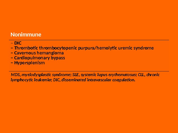
Nonimmune – DIC – Thrombotic thrombocytopenic purpura/hemolytic uremic syndrome – Cavernous hemangioma – Cardiopulmonary bypass – Hypersplenism MDS, myelodysplastic syndrome; SLE, systemic lupus erythematosus; CLL, chronic lymphocytic leukemia; DIC, disseminated intravascular coagulation.
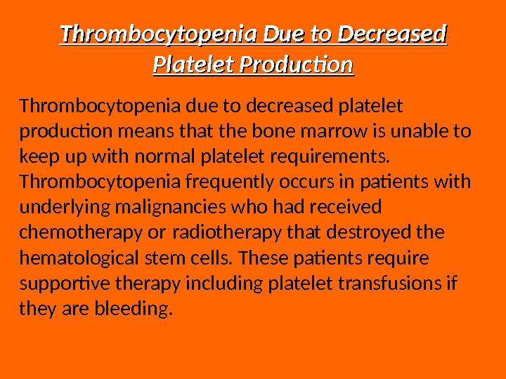
Thrombocytopenia Due to Decreased Platelet Production Thrombocytopenia due to decreased platelet production means that the bone marrow is unable to keep up with normal platelet requirements. Thrombocytopenia frequently occurs in patients with underlying malignancies who had received chemotherapy or radiotherapy that destroyed the hematological stem cells. These patients require supportive therapy including platelet transfusions if they are bleeding.
Thrombocytopenia Due to Increased Platelet Destruction Isolated thrombocytopenia is caused by increased platelet destruction. In these patients, the rate of platelet destruction cannot be compensated by the bone marrow with increased platelet production. These patients have isolated thrombocytopenia, and the number of megakaryocytes in the bone marrow is normal or increased. The platelet life span is shortened.
Immune Thrombocytopenia Immune thrombocytopenia is an increase of platelet destruction caused by immunological mechanisms. The sensitization of platelets by immunoglobulin G (Ig. G) and Ig. M antibodies reacting with antigenic sites (usually glycoprotein [GP]IIb/IIIa in idiopathic thrombocytopenic purpura [ITP], platelet alloantigens in post-transfusion purpura, and neonatal isoimmune purpura) on the platelet membrane is the main cause of platelet destruction.
Idiopathic Thrombocytopenic Purpura Acute ITP in children is equally common in boys and girls, has its peak incidence at age 2 — 4 yr, and frequently follows a viral infection or vaccination. More than 80% of these children have a spontaneous remission of their illness in 2 — 4 wk. Serious morbidity or mortality is rare (approximately 1%), but the recovery of the platelets can be forced by the administration of high doses of intravenous Ig. G. Incidence Children: 5 per 100, 000.
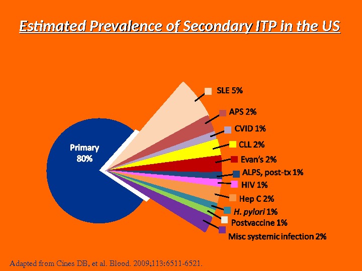
Estimated Prevalence of Secondary ITP in the US Adaptedfrom. Cines. DB, etal. Blood. 2009; 113: 65116521.
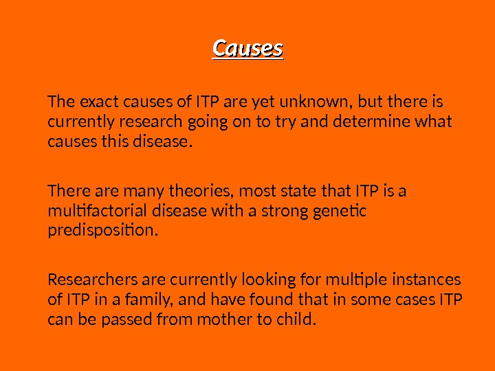
Causes The exact causes of ITP are yet unknown, but there is currently research going on to try and determine what causes this disease. There are many theories, most state that ITP is a multifactorial disease with a strong genetic predisposition. Researchers are currently looking for multiple instances of ITP in a family, and have found that in some cases ITP can be passed from mother to child.
Theories Three most common theories for ITP are: • The Microbial Trigger Theory • The Molecular Mimicry Theory • The Free Radical Damage Theory
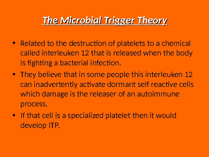
The Microbial Trigger Theory • Related to the destruction of platelets to a chemical called interleuken 12 that is released when the body is fighting a bacterial infection. • They believe that in some people this interleuken 12 can inadvertently activate dormant self reactive cells which damage is the releaser of an autoimmune process. • If that cell is a specialized platelet then it would develop ITP.
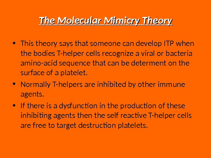
The Molecular Mimicry Theory • This theory says that someone can develop ITP when the bodies T-helper cells recognize a viral or bacteria amino-acid sequence that can be determent on the surface of a platelet. • Normally T-helpers are inhibited by other immune agents. • If there is a dysfunction in the production of these inhibiting agents then the self reactive T-helper cells are free to target destruction platelets.
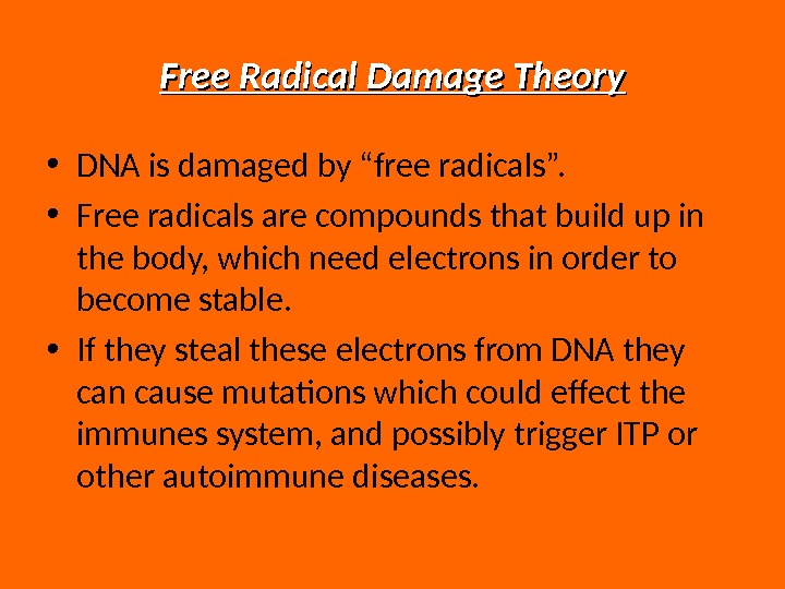
Free Radical Damage Theory • DNA is damaged by “free radicals”. • Free radicals are compounds that build up in the body, which need electrons in order to become stable. • If they steal these electrons from DNA they can cause mutations which could effect the immunes system, and possibly trigger ITP or other autoimmune diseases.
Pathophysiology ITP is caused by an autoantibody — in generally Ig. G — that binds to specific platelet glycoproteins, especially GP IIb, IIIa. These antibodies attached to the platelet membranes may bind complement and cause accelerated platelet destruction through phagocytosis by the reticuloendothelial cells in the spleen and the liver. A compensatory increase in bone-marrow megakaryopoiesis usually occurs, which may initially prevent or delay the development of more severe thrombocytopenia. In a few cases, the measurement of the platelet life-span may be helpful. On rare occasions, platelets of patients with apparent ITP have a nearly normal life-span. The cause of the thrombocytopenia in these patients is unclear, an early myelodysplastic syndrome should be ruled out.
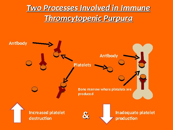
Increased platelet destruction && Inadequate platelet production. Two Processes Involved in Immune Thromcytopenic Purpura Platelets. Antibody Bone marrow where platelets are produced
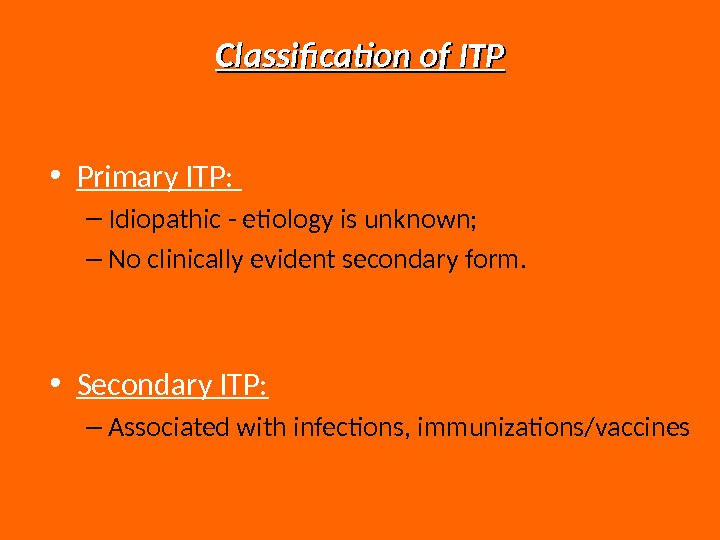
Classification of ITP • Primary ITP: – Idiopathic — etiology is unknown; – No clinically evident secondary form. • Secondary ITP: – Associated with infections, immunizations/vaccines
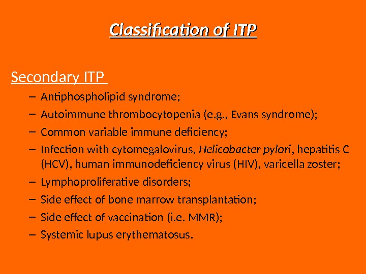
Classification of ITP Secondary ITP – Antiphospholipid syndrome; – Autoimmune thrombocytopenia (e. g. , Evans syndrome); – Common variable immune deficiency; – Infection with cytomegalovirus, Helicobacter pylori , hepatitis C (HCV), human immunodeficiency virus (HIV), varicella zoster; – Lymphoproliferative disorders; – Side effect of bone marrow transplantation; – Side effect of vaccination (i. e. MMR); – Systemic lupus erythematosus.
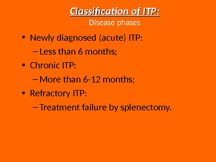
• Newly diagnosed (acute) ITP: – Less than 6 months; • Chronic ITP: – More than 6 -12 months; • Refractory ITP: – Treatment failure by splenectomy. Classification of ITP: Disease phases
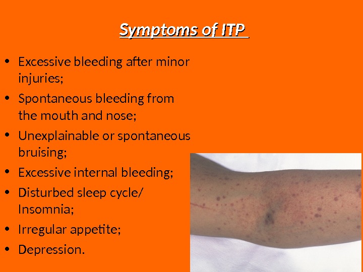
Symptoms of ITP • Excessive bleeding after minor injuries; • Spontaneous bleeding from the mouth and nose; • Unexplainable or spontaneous bruising; • Excessive internal bleeding; • Disturbed sleep cycle/ Insomnia; • Irregular appetite; • Depression.
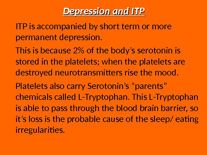
Depression and ITP ITP is accompanied by short term or more permanent depression. This is because 2% of the body’s serotonin is stored in the platelets; when the platelets are destroyed neurotransmitters rise the mood. Platelets also carry Serotonin’s “parents” chemicals called L-Tryptophan. This L-Tryptophan is able to pass through the blood brain barrier, so it’s loss is the probable cause of the sleep/ eating irregularities.
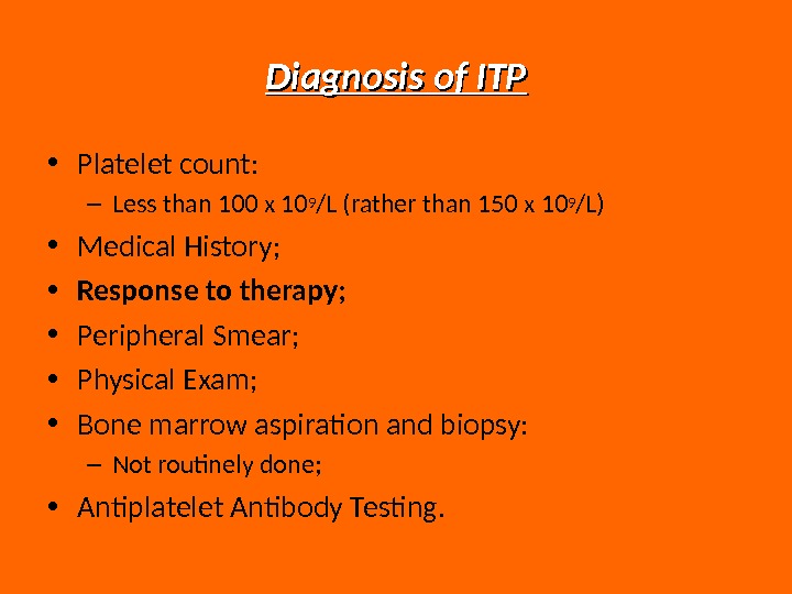
Diagnosis of ITP • Platelet count: – Less than 100 x 10 9 /L (rather than 150 x 10 9 /L) • Medical History; • Response to therapy; • Peripheral Smear; • Physical Exam; • Bone marrow aspiration and biopsy: – Not routinely done; • Antiplatelet Antibody Testing.
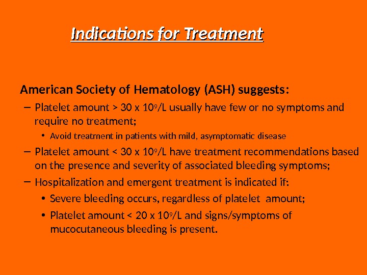
Indications for Treatment American Society of Hematology (ASH) suggests : – Platelet amount > 30 x 10 9 /L usually have few or no symptoms and require no treatment; • Avoid treatment in patients with mild, asymptomatic disease – Platelet amount < 30 x 10 9 /L have treatment recommendations based on the presence and severity of associated bleeding symptoms; – Hospitalization and emergent treatment is indicated if: • Severe bleeding occurs, regardless of platelet amount; • Platelet amount < 20 x 10 9 /L and signs/symptoms of mucocutaneous bleeding is present.
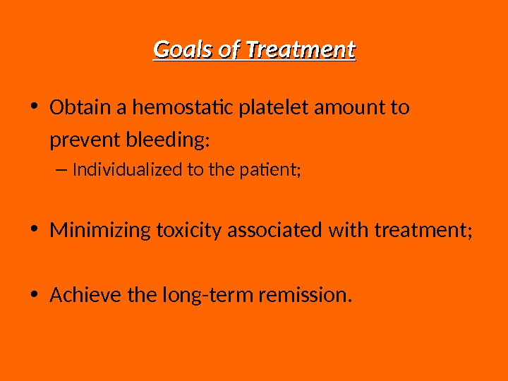
Goals of Treatment • Obtain a hemostatic platelet amount to prevent bleeding: – Individualized to the patient; • Minimizing toxicity associated with treatment; • Achieve the long-term remission.
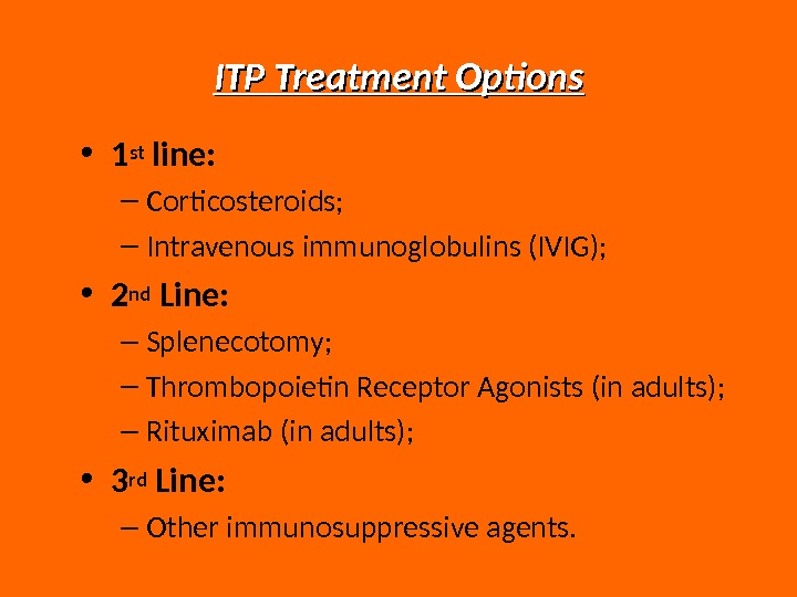
ITP Treatment Options • 1 st line: – Corticosteroids; – Intravenous immunoglobulins (IVIG); • 2 nd Line: – Splenecotomy; – Thrombopoietin Receptor Agonists (in adults); – Rituximab (in adults); • 3 rd Line: – Other immunosuppressive agents.
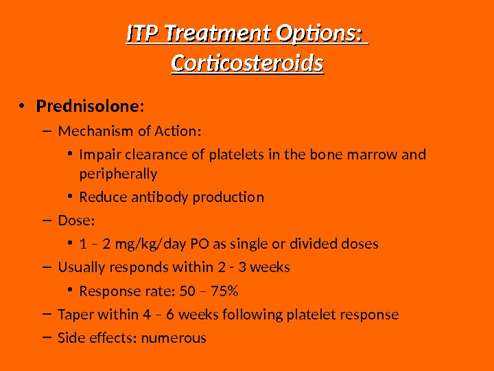
ITP Treatment Options: Corticosteroids • Prednisolone: – Mechanism of Action: • Impair clearance of platelets in the bone marrow and peripherally • Reduce antibody production – Dose: • 1 – 2 mg/kg/day PO as single or divided doses – Usually responds within 2 — 3 weeks • Response rate: 50 – 75% – Taper within 4 – 6 weeks following platelet response – Side effects: numerous
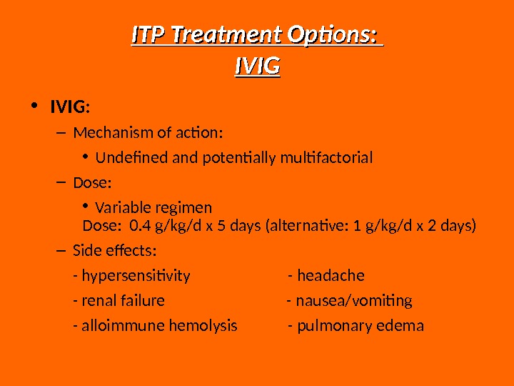
ITP Treatment Options: IVIG • IVIG: – Mechanism of action: • Undefined and potentially multifactorial – Dose: • Variable regimen Dose: 0. 4 g/kg/d x 5 days (alternative: 1 g/kg/d x 2 days) – Side effects: — hypersensitivity — headache — renal failure — nausea/vomiting — alloimmune hemolysis — pulmonary edema
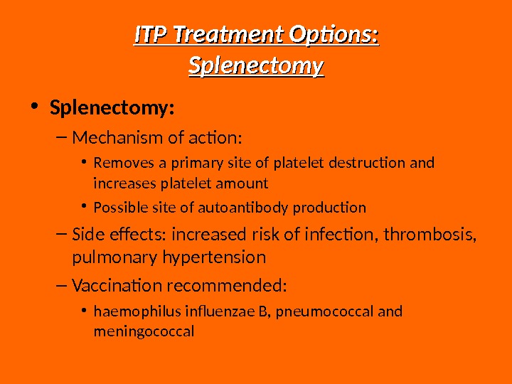
ITP Treatment Options: Splenectomy • Splenectomy: – Mechanism of action: • Removes a primary site of platelet destruction and increases platelet amount • Possible site of autoantibody production – Side effects: increased risk of infection, thrombosis, pulmonary hypertension – Vaccination recommended: • haemophilus influenzae B, pneumococcal and meningococcal
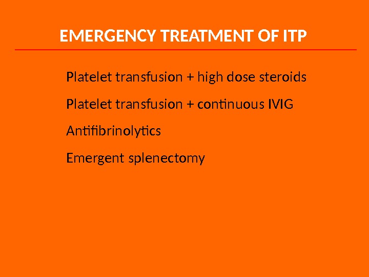
EMERGENCY TREATMENT OF ITP Platelet transfusion + high dose steroids Platelet transfusion + continuous IVIG Antifibrinolytics Emergent splenectomy
Neonatal Purpura Neonatal thrombocytopenia may develop due to isoimmunization of the mother against fetal platelets with subsequent transplacental transfer. The incidence of alloimmune neonatal purpura is 1 in 5000 deliveries. The most commonly involved antigen is PIA 1, an antigen that is present on platelets in 98% of the population. The infant shows generalized purpura at delivery, and platelet amount are usually below 30, 000/μL. Intracranial hemorrhage is a severe complication that occurs in about 10% of affected infants.
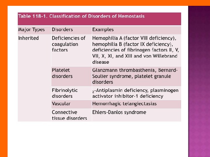
INHERITED DISORDERS OF COAGULATION There is a large number of inherited disorders of coagulation; however, only three are relatively common: I. Von Willebrand’s Disease (v. WD) , II. Factor VIII deficiency ( hemophilia A ), III. Factor IX deficiency ( hemophilia B ; Christmas disease ). All of the others are rare.
von Willebrand’s Disease (v. WD) von Willebrand disease is the most common inherited disorder of primary hemostasis. The prevalence of v. WD mutations may be as high as 1 to 2% of the population, although most of cases are never diagnosed; the prevalence of clinically evident of v. WD is much lower. The clinical and laboratory manifestations of v. WD are extremely heterogeneous, and diagnosis can sometimes be difficult.
Three main subtypes of v. WD have been defined: Type 1 : the most common (≥ 70% of cases of clinical v. WD). There is a decrease in the concentration of v. WF in the plasma (i. e. , a quantitative defect ), but all sizes of multimers, including the very high-molecular-weight multimers, are present. The inheritance pattern is autosomal dominant.
Type 2 : In Type 2 v. WD, there is a qualitative defect in v. WF. Several different subtypes of v. WD Type 2 are described. Inheritance is autosomal dominant in most cases, although a few types display autosomal recessive inheritance. Type 2 A : In Type 2 A, there is a deficiency of the high-molecular-weight multimers of v. WD, but the absolute level of total v. WF is normal. This is the most common variant of v. WD Type 2 B : In Type 2 B, the v. WF appears with an abnormally increased affinity for the v. WF receptor (GP Ib-IX/V) on the platelet surface. Too much v. WF is tied up on the platelet surface, rather than being in the plasma, and is therefore not able to bind to subendothelial collagen. The diagnosis of Type 2 B v. WD is made by demonstrating increased platelet agglutination with ristocetin rather than decreased ristocetin agglutination, as seen in the other types of v. WD. Mild thrombocytopenia is common. Type 2 M : In Type 2 M v. WD, there is a mutation that affects some important functional domains of the protein (think “ M ” for M utation). A variety of mutations in different regions of the protein have been described.
Type 3 : : In Type 3 v. WD, there is a total or near-total absence of v. WF in the plasma. Type 3 v. WD is rare. The patients have markedly decreased factor VIII level and thus they may have bleeding manifestations resembling hemophilia. The inheritance pattern is autosomal recessive; some cases appear to represent homozygous Type 1 v. WD.
Clinical Manifestations of von Willebrand’s Disease Most cases of v. WD present with the typical picture of a primary hemostatic defect : mucocutaneous bleeding (epistaxis, bleeding gums), easy bruising, and immediate bleeding from cuts, incisions, and dental extractions. Most patients have a mild to moderate bleeding tendency. The severity of illness in different patients is highly variable , and it can also vary over time in individual patients. The clinical phenotype can vary between different members of the same family.
The laboratory manifestations of v. WD can also be highly variable; sometimes laboratory tests must be repeated several times to make a firm diagnosis. As noted, the inheritance pattern of most cases of v. WD is autosomal dominant. Type 3 v. WD presents with a mixed picture. Since the factor VIII levels are low, the patients may have hemarthroses, muscle hematomas, and other manifestations of defects of secondary hemostasis in addition to mucocutaneous bleeding.
Laboratory Diagnosis of von Willebrand’s Disease The most important diagnostic tests for v. WD are the bleeding time , ristocetin cofactor assay , a quantitative assay of v. WF concentration (ELISA or Laurell rocket immunoelectrophoresis), ristocetin-induced platelet aggregation, and agarose gel electrophoresis to determine whether the high-molecular-weight multimers are present or absent.
In v. WD Type 1, the BT will usually be prolonged. The PTT may also be slightly prolonged due to decreased factor VIII concentration. The absolute level of v. WF is decreased; the v. WF multimer pattern is normal (the highmolecular-weight multimers are present, but the concentration of all sizes of multimers is decreased). Platelet aggregation with ristocetin and the ristocetin cofactor activity are decreased; platelet aggregation with other agents is usually normal.
Type 2 A is diagnosed by demonstrating an absence of the high-molecular-weight multimers by agarose gel electrophoresis. The absolute concentration of v. WF is usually normal. Type 2 B is diagnosed by demonstrating increased platelet agglutination with ristocetin. Plasma from patients with The absolute level of v. WF is decreased; agarose gel electrophoresis demonstrates a decrease in high-molecular-weight multimers (because they are all stuck on the platelets). Mild thrombocytopenia is common.
Diagnosis of Type 2 M requires sophisticated techniques, which are not widely available; therefore, specimens must usually be sent to reference laboratories that specialize in coagulation. Type 3 is diagnosed by demonstrating a total or near-total absence of v. WF in the plasma.
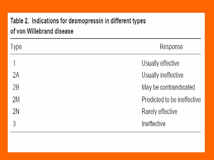
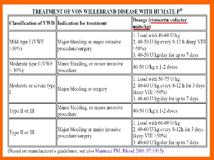
Treatment of von Willebrand’s Disease Most cases of v. WD Type 1 can be very successfully treated with desmopressin acetate ( DDAVP ), which causes release of preformed v. WD from endothelial cells. Desmopressin acetate is cheap, is safe, and has no infectious risk. It can be administered either intravenously or intranasally (0. 3 mcg/kg IV, or 150 mcg per nostril). Other types of v. WD, or a patient with Type 1 requiring major surgery and may need replacement therapy. There are no commercially available v. WF concentrate preparations, but some factor VIII concentrate preparations contain enough v. WF to be effective. Factor VIII concentrates have therefore replaced cryoprecipitate as the treatment of choice for v. WD requiring factor replacement.
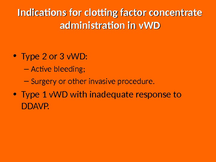
Indications for clotting factor concentrate administration in v. WD • Type 2 or 3 v. WD: – Active bleeding; – Surgery or other invasive procedure. • Type 1 v. WD with inadequate response to DDAVP.
The Hemophilias The hemophilias are inherited disorders of the coagulation cascade. Deficiency of factor VIII ( hemophilia A ) is the most common (~85% of cases). Factor IX deficiency ( hemophilia B ) is second (~15%), and all others are rare. The incidence of hemophilia A is estimated at 1 per 5, 000 to 10, 000 male births in the United States; the incidence of hemophilia B is approximately 1 in 30, 000 male births.
Hemophilia A and B are both inherited as X-linked recessive : women are carriers, men develop the disease. All of the other factor deficiencies are inherited as autosomal recessive. Factor XI deficiency (sometimes called hemophilia C ) is the third most common inherited disorder of coagulation factors but is much less common than deficiency of factors VIII or IX.
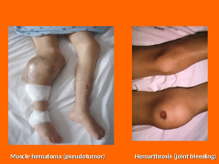
Clinical Features The clinical features of hemophilia A and B are identical. The manifestations are those of deficiencies of secondary hemostasis: hemarthroses , muscle hematomas , soft tissue bleeding , and delayed but prolonged bleeding from cuts or incisions. Recurrent hemarthroses result in joint destruction and can be disabling. Bleeding into muscle or soft tissue can result in a compartment syndrome, with compression of nerves and blood vessels; this may require fasciotomy for relief.
Muscle hematoma (pseudotumor) Hemarthrosis (joint bleeding)
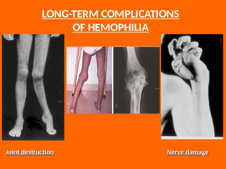
LONG-TERM COMPLICATIONS OF HEMOPHILIA Joint destruction Nerve damage
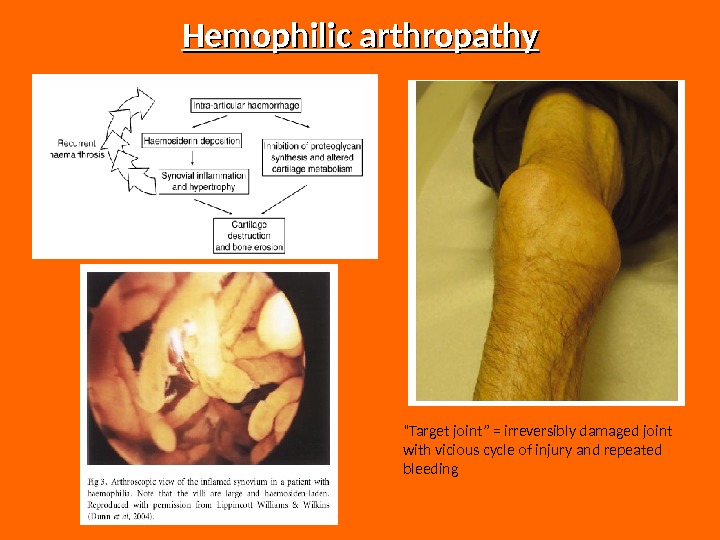
Hemophilic arthropathy “ Target joint” = irreversibly damaged joint with vicious cycle of injury and repeated bleeding
Intracranial bleeding is anespecially serious complication, and even minor head trauma in a severe hemophiliac should be treated with factor replacement. There is no such thing as minor head trauma in a severe hemophiliac. Another potentially lethal complication is bleeding into the soft tissues of the oropharynx; dissection of the hematoma into the trachea can result in airway occlusion and asphyxia.
Children with severe hemophilia usually begin to have problems at about 9 to 12 months of age, at about the time they begin to walk. Some will come to medical attention earlier due to prolonged bleeding after circumcision or other surgery. Mild or moderate cases may not have problems until they undergo a severe challenge to hemostasis, such as a dental extraction, major surgery, or severe injury.
Most patients will have a family history of pathologic bleeding on the maternal side, including maternal brothers, uncles, and other male relatives. A history of bleeding on the paternal side, or a history of bleeding in female relatives, suggests v. WD or another clotting factor deficiency. The severity of bleeding depends on the level of the deficient factor. Approximately half of hemophilia A cases have severe disease, with a total absence of detectable factor activity.
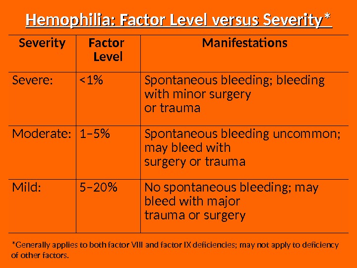
Hemophilia: Factor Level versus Severity* Severity Factor Level Manifestations Severe: <1% Spontaneous bleeding; bleeding with minor surgery or trauma Moderate: 1– 5% Spontaneous bleeding uncommon; may bleed with surgery or trauma Mild: 5– 20% No spontaneous bleeding; may bleed with major trauma or surgery *Generally applies to both factor VIII and factor IX deficiencies; may not apply to deficiency of other factors.
Laboratory Diagnosis The PTT is prolonged; the PT is normal. Mixing studies show correction of the PTT with normal plasma. Specific diagnosis and distinction of factor VIII deficiency from factor IX deficiency require assay of the factor levels and demonstration of a deficiency. It is critical to distinguish factor VIII deficiency from factor IX deficiency because the treatment is completely different. Remember, it is impossible to distinguish hemophilia A from hemophilia B based on clinical or family history. Clotting factor assays also allow you to predict clinical severity if it is not obvious from the history.
Treatment depends on which factor is deficient, the severity of the deficiency, and the nature of the bleeding, injury, or planned surgery. Various highly purified preparations of factors VIII and IX are available. These have been intensively treated to prevent infection, and are generally very safe. Recombinant forms of both are also available.
Always be sure to determine which factor the patient is deficient in before you start replacement therapy. For severe bleeding or major surgery, it is desirable to achieve peak factor levels of 100% and maintain minimum levels >50%. For minor bleeding or surgery, aim for peak levels of ~50% and minimum levels ≥ 25%. A variety of different formulas are available for dosing factor concentrates.
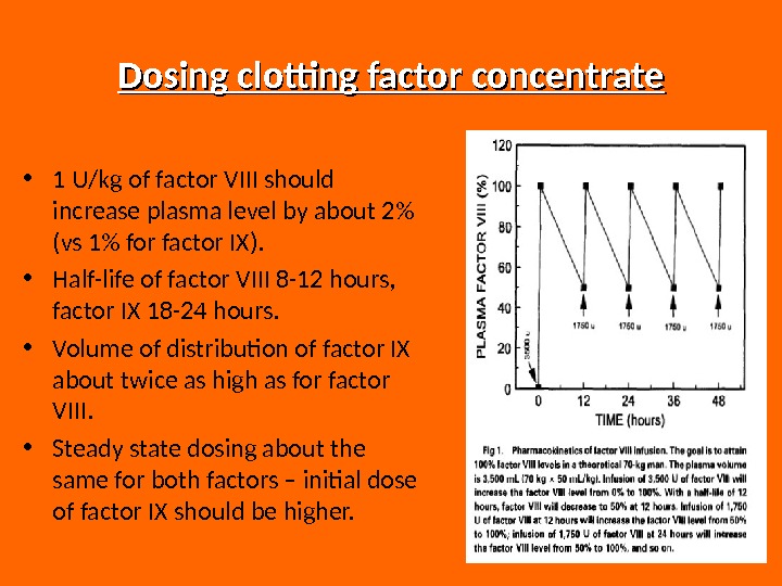
Dosing clotting factor concentrate • 1 U/kg of factor VIII should increase plasma level by about 2% (vs 1% for factor IX). • Half-life of factor VIII 8 -12 hours, factor IX 18 -24 hours. • Volume of distribution of factor IX about twice as high as for factor VIII. • Steady state dosing about the same for both factors – initial dose of factor IX should be higher.
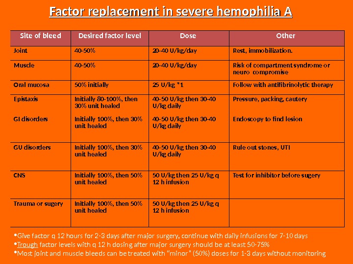
• Give factor q 12 hours for 2 -3 days after major surgery, continue with daily infusions for 7 -10 days • Trough factor levels with q 12 h dosing after major surgery should be at least 50 -75% • Most joint and muscle bleeds can be treated with “minor” (50%) doses for 1 -3 days without monitoring Factor replacement in severe hemophilia A Site of bleed Desired factor level Dose Other Joint 40 -50% 20 -40 U/kg/day Rest, immobilization. Muscle 40 -50% 20 -40 U/kg/day Risk of compartment syndrome or neuro compromise Oral mucosa 50% initially 25 U/kg *1 Follow with antifibrinolytic therapy Epistaxis Initially 80 -100%, then 30% unit healed 40 -50 U/kg then 30 -40 U/kg daily Pressure, packing, cautery GI disorders Initially 100%, then 30% unit healed 40 -50 U/kg then 30 -40 U/kg daily Endoscopy to find lesion GU disorders Initially 100%, then 30% unit healed 40 -50 U/kg then 30 -40 U/kg daily Rule out stones, UTI CNS Initially 100%, then 50% unit healed 50 U/kg then 25 U/kg q 12 h infusion Test for inhibitor before sugery Trauma or sugery Initially 100%, then 50% unit healed 50 U/kg then 25 U/kg q 12 h infusion
Liver disease in hemophilia • Hepatitis C is still a problem, though incidence is falling with safer factor concentrates • Treatment for hepatitis C with interferon often causes thrombocytopenia • Liver transplantation is made occasionally (cures hemophilia) • All newly diagnosed hemophiliacs should be vaccinated against hepatitis A and
ACQUIRED FACTOR VIII DEFICIENCY • Due to antibody to factor VIII (most common autoimmune factor deficiency) • Most patients elderly • Often presents with severe soft tissue or mucosal bleeding (different bleeding pattern than inherited hemophilia) • Laboratory: prolonged a. PTT not corrected by mixing, very low factor VIII activity – Normal thrombin time and platelet amount • Treatment: r. VIIa, FEIBA, immunosuppression