
Презентация auscultation heart


- Размер: 4.9 Mегабайта
- Количество слайдов: 65
Описание презентации Презентация auscultation heart по слайдам
• “ Symptoms and syndromes based on the data of auscultation of a heart «
Auscultation was inculcated by French physitian Rene Laennec Рис. 10. Стетоскопи тверд!.
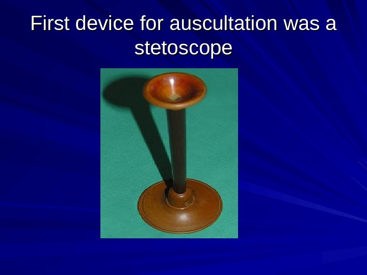
First device for auscultation was a stetoscope
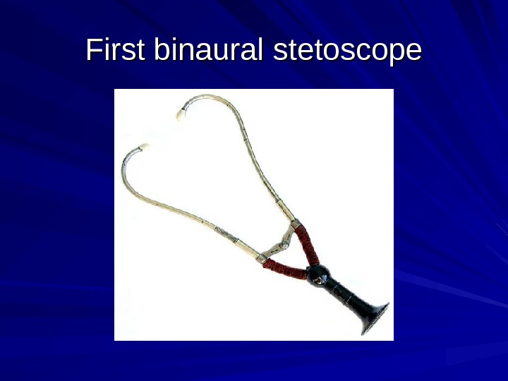
First binaural stetoscope
First phonendoscope
Modern stetophonendoscope
The heart is usually auscultated by a stethoscope or a phonendoscope, but direct (immediate) auscultation is also used. The condition of the patient permitting, the heart sounds should be heard in various postures of the patient: erect, recumbent, after exer sice (e. g. after repeated squatting). Sounds associated with the mitral valve ’s’s pathology are well heard when the patient lies on his left side, since the heart apex is at its nearest position to the chest wall; aortic valve defects ar ee best heard when the patient is in the upright posture or when he lies on his right side. The heart sounds are better heard if the patient is asked to inhale deeply and then exhale deeply and keep breath for short periods of time so that the respiratory sounds should not interfere with auscultation of the heart. The valve sounds should be heard in the order of decreasing frequency of their affection.
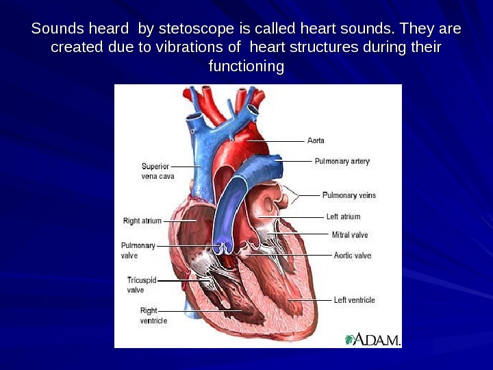
Sounds heard by stetoscope is called heart sounds. They are created due to vibrations of heart structures during their functioning
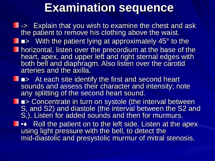
Examination sequence -> Explain that you wish to examine the chest and ask the patient to remove his clothing above the waist. ■■ > With the patient lying at approximately 45° to the horizontal, listen over the precordium at the base of the heart, apex, and upper left and right sternal edges with both bell and diaphragm. Also listen over the carotid arteries and the axilla. ■■ > At each site identify the first and second heart sounds and assess their character and intensity; note any splitting of the second heart sound. ■■ > Concentrate in turn on systole (the interval between S, and S 2) and diastole (the interval between the S 2 and S, ). Listen for added sounds and then for murmurs. • ♦ Roll the patient on to the left side. Listen at the apex using light pressure with the bell, to detect the mid-diastolic and presystolic murmur of mitral stenosis.
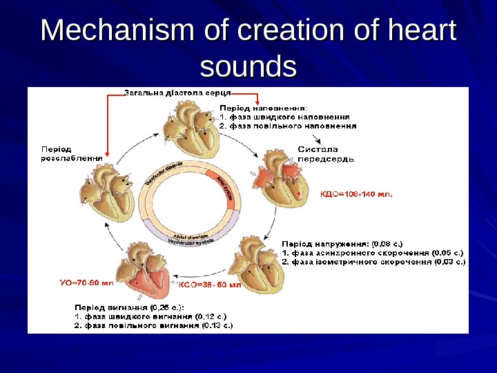
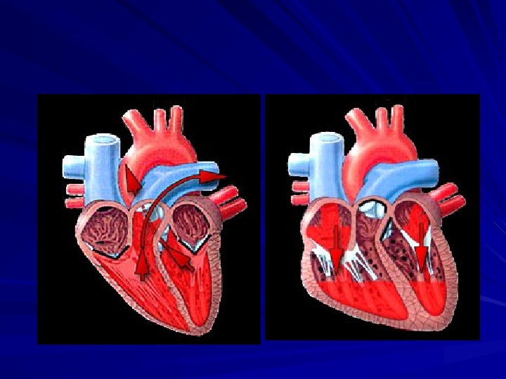
Mechanism of creation of heart sounds
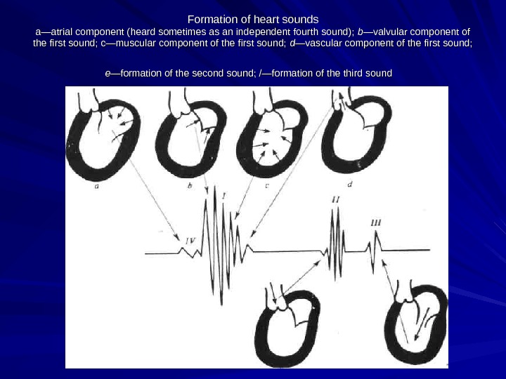
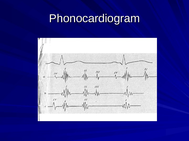
Formation of heart sounds a—atrial component (heard sometimes as an independent fourth sound); b—b— valvular component of the first sound; c—muscular component of the first sound; dd —vascular component of the first sound; ee —formation of the second sound; /—formation of the third sound
Auscultation involves listening for heart sounds with the stethoscope, similar to the procedure used in assessing breath sounds The sounds produced by a working heart are called heart sounds. Two sounds can be well heard in a healthy subject; the first sound, which is produced during systole and the second sound, which occurs during diastole.
СС omponents of heart sounds I heart sound: – the valve component, i. e. vibrations of the cusps of the atrioventricular valves during the isometric contraction phase – the muscular one due to the myocardial isometric contraction – the vascular one. This is due to vibrations of the nearest portions of the aorta and the pulmonary trunk caused by their distention with the blood during the ejection phase – Atrial one is generated by vibrations caused by atrial contractions II heart sound: The second sound is generated by vibrations arising at the early diastole when the semilunar cusps of the aortic valve and the pulmonary trunk are shut (the valve component) and by vibration of the walls at the point of origination of these vessels (the vascular component). The intensity of myocardial and valvular vibrations depends on the rate of ventricular contractions: the higher the rate of their contractions and the faster the intraventricular pressure grows, the greater is the intensity of these vibrations.
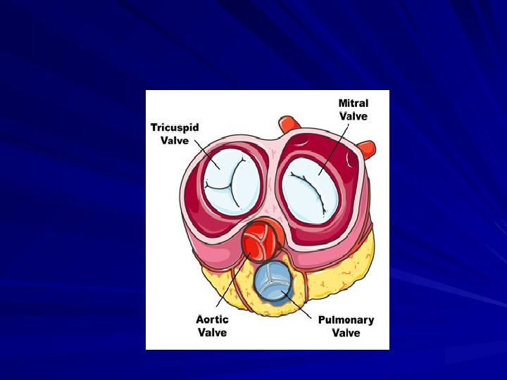
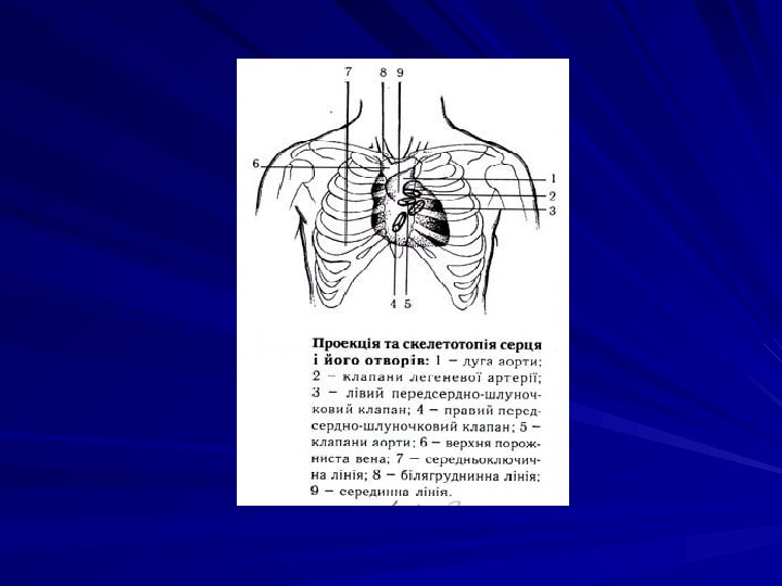
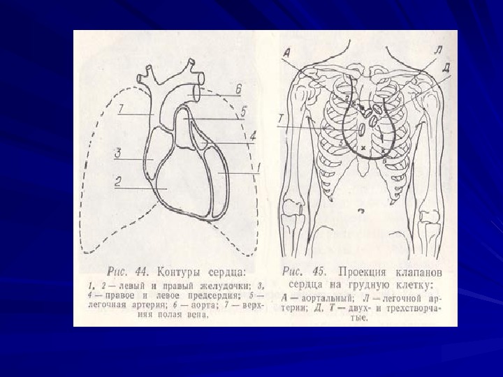
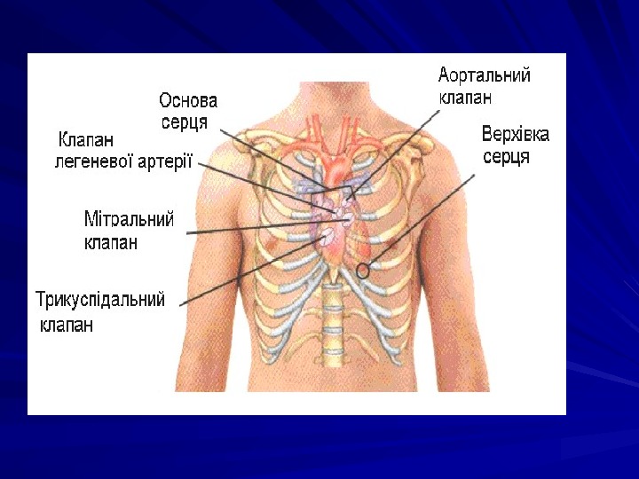
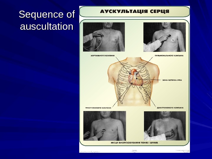
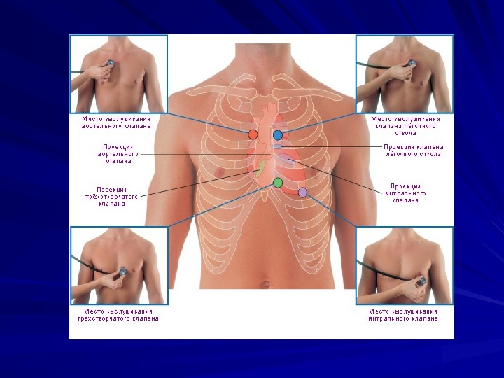
Sequence of auscultation The mitral valve — at the heart apex; the aortic valve — in the second intercostal space to the right of the sternum), the pulmonary valve — in the second intercostal space, to the left of the sternum, tricuspid valve — at the base of the xyphoid process, the aortic valve again at the Botkin-Erb point.
Points of auscultation
Rules for auscultation of the heart. The heart is usually auscultated by a stethoscope or a phonendoscope, but direct (immediate) auscultation is also used. The condition of the patient permitting, the heart sounds should be heard in various postures of the patient: erect, recumbent, after exercise (e. g. after repeated squatting). Sounds associated with the mitral valve pathology are well heard when the patient lies on his left side, since the heart apex is at its nearest position to the chest wall; aortic valve defects are best heard when the patient is in the upright posture or when he lies on his right side. The heart sounds are better heard if the patient is asked to inhale deeply and then exhale deeply and keep breath for short periods of time so that the respiratory sounds should not interfere with auscultation of the heart. The valve sounds should be heard in the order of decreasing frequency of their affection. The mitral valve should be heard first (at the heart apex); next follows the aortic valve (in the second intercostal space to the right of the sternum), the pulmonary valve (in the second intercostal space, to the left of the sternum), tricuspid valve (at the base of the xiphoid pro cess), and finally the aortic valve again at the Botkin-Erb point. If any deviations from normal sounds have been revealed at these points, the en tire heart area should be auscultated thoroughly.
Sequence of auscultation
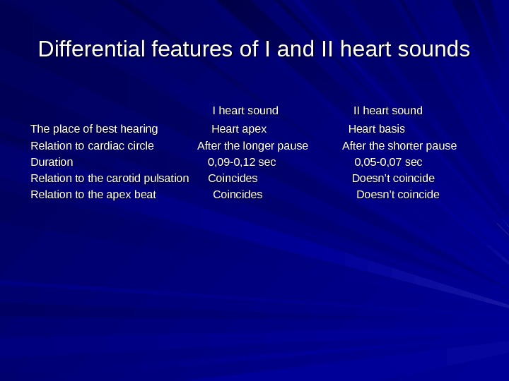
Differential features of I and II heart sounds I heart sound II heart sound The place of best hearing Heart apex Heart basis Relation to cardiac circle After the longer pause After the shorter pause Duration 0, 09 -0, 12 sec 0, 05 -0, 07 sec Relation to the carotid pulsation Coincides Doesn’t coincide Relation to the apex beat Coincides Doesn’t coincide
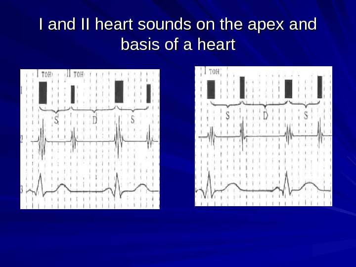
I and II heart sounds on the apex and basis of a heart
For differentiation of I and II heart sounds in tachycardia it is necessary to check which of them is synchronous with carotic artery pulsation
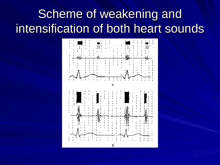
Intensity of the heart sounds may depend on conditions of the sound wave transmission TT he intensity of both heart sounds decreases if if their transmission to the chest becomes difficult : : – subcutaneous fat oror muscles of the chest are overdeveloped, – lung emphysema, – liquid in the left pleural cavity, – other affections that separate the heart from the anterior chest wall. – If conditions for sound transmission are improved in decreased myocardial contractility: – in myocarditis, – myocardial dystrophy, – cardiosclerosis, – collapse, – accumulation of fluid in the pericardial cavity.
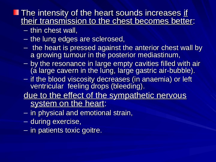
The intensity of the heart sounds increases if if their transmission to the chest becomes better : : – thin chest wall, – the lung edges are sclerosed, – the heart is pressed against the anterior chest wall by a growing tumour in the posterior mediastinum, – by the resonance in large empty cavities filled with air (a large cavern in the lung, large gastric air-bubble). – if the blood viscosity decreases (in anaemia) or left ventricular feeling drops (bleeding). due to the effect of the sympathetic nervous system on the heart : : – in physical and emotional strain, – during exercise, – in patients toxic goitre.
Scheme of weakening and intensification of both heart sounds
Separate changes of one heart sound (I or II): FF irst heart sound diminishes: – in the mitral and aortic valve insufficiency (at the apex). In tricuspid and pulmonary valve failure, the diminution of the first heart sound will be better heard at the base of the xiphoid process, – at the heart apex in stenotic aortal orifice, – In diffuse affections of the myocardium (due to dystrophy, cardiosclerosis or myocarditis), the first heart sound only may be diminished because its muscular component also diminishes in these cases. The first sound increases at the heart apex if the left ventricle is not adequately filled with blood during diastole: – in stenosis of the left atrioventricular orifice, – In extrasystole. The second sound can be inaudible over the aorta if: – the aortic valve is much destroyed, – diminishes over the aorta in cases with marked hypo tt ee nn sion; – diminishes over the pulmonary trunk in cases with aortic valve incompetence (in very rare cases), – in decreased pressure in the lesser circulation. The second sound may increase either over the aorta or over the pulmonary trunk indicating hypertension in the proper circle of circulation.
Splitting or reduplication of the sounds occurs in asynchronous work and right chambers of the heart Asynchronous closure of the right- and left ventricular valves splits the first sound while asynchronous closure of the semilunar valves causes reduplication of the second heart sound. Reduplication or or splitting of the first sound is due to asynchronous closure of the atrioventricular valves, e. g. during very deep expiration, when the blood is ejected into the left atrium with a greater force to prevent the closure of the mitral valve; Pathological reduplication of the first sound can occur in impaired intraventricular conduction (through the His bundle) as a result of delays systole of one of the ventricles.
The second sound is reduplicated more frequently Reduplication occurs due to asynchronous closure of the valve of the aorta and pulmonary trunk because of the different length of contractions of the left and the right ventricles. The second heart sound can be duplicated in cases with, diminished or increased filling of one of the ventricles or when pressure in the aorta or the pulmonary artery changes. Physiological reduplication of the second sound is mostly connected with various respiratory phases: the filling of the right and left ventricles differs during inspiration and expiration and the length of their systole changes, as well as the tinted of closure of the valve of the aorta and pulmonary trunk. The amount oil blood flowing to the left ventricle decreases during inspiration because part of blood is retained in the distended vessels of the lungs. The left ventricular systolic blood volume decreases during inspiration, its systole ends earlier, and the aortic valve therefore closes earlier as well. At the same time, the stroke volume of the right ventricle increases, its systole prolongs, the pulmonary valve closure is delayed and the second sound is thus doubled. Pathological reduplication of the second sound can be due to delayed closure of the aortic valve in persons suffering from essential hypertension, or if the closure of the pulmonary valve is delayed at increased pressure in the lesser circulation (e. g. in mitral stenosis or emphysema of the lungs).
Scheme of reduplication of I and II heart sounds
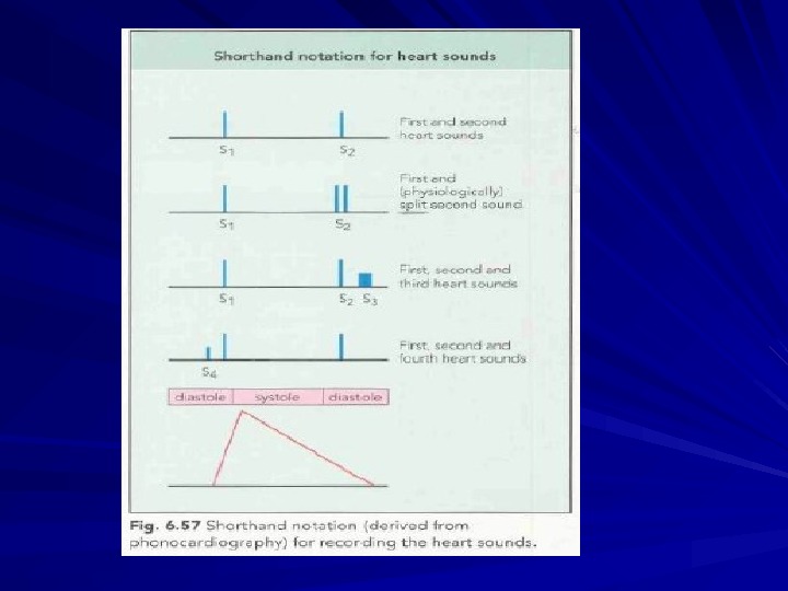
Adventitious heart sounds The third heart sound (S 3) is the result of vibrations produced during ventricular filling. It is normally heard only in some children and young adults, but it is considered abnormal in older individuals. It arises in 0. 15— 1. 12 s from the beginning of the second soun d. d. The forth heart sound (S 4) is caused by the recoil of vibrations between the atria and ventricles following atrial contraction, at the end of diastole. It is rarely heard as a normal heart sound; usually it is considered indicative of further cardiac evaluation. Both S 3 and S 4 may be recorded in heart failure indicating poor muscular tone of the left ventricle. The mitral valve opening sound (opening snup) is heard at the heart apex of patients with mitral stenosis 0. 07 -0. 13 s following the second sound, during diastole. Extra-pericardial-sound can occur in pericardial adhesion. It originates during diastole, 0. 08 -0. 14 s after the second sound, and is generated by the vibrating pericardium during the rapid dilatation of the ventricles at the beginning of diastole.
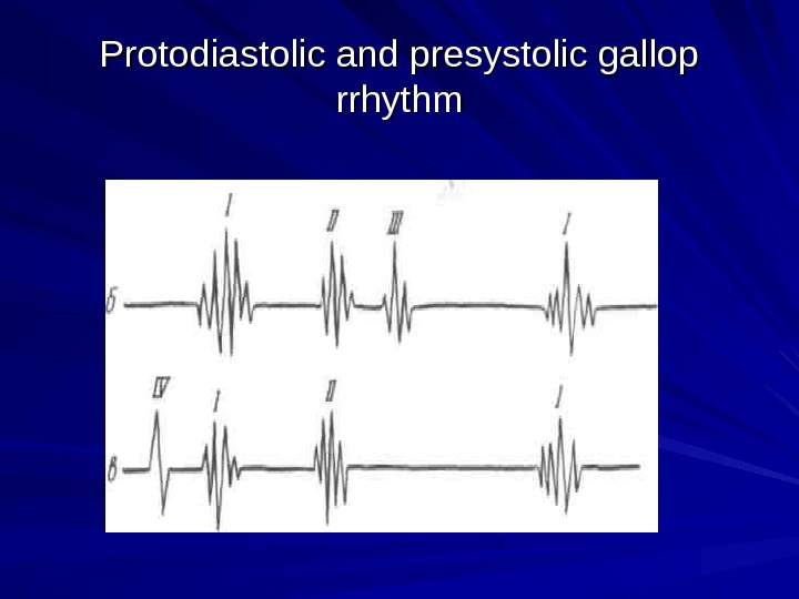
Heart melodies Intensification of S 3 or S 4 sounds gives a three-sound or even four- three-sound rhythm, known as the gallop rhythm (because it resembles the galloping of a horse). The rrhythm indicates heavy lesions of cardiac muscle (inflammatory, degenerative, toxic), it is called as » cry of a heart for help». The gallop rrhythm is conditionally divides into protodiastolic (intensified III sound arises up though 0, 12 -0, 2 sec. after second sound), mesodiastolic(at tachicardia descend coalescence of III and IV sounds and it is accepted at auscultation as a single sound) and presystolic (( is conditioned by pathological IV cardiac sound). A gallop rhythm is better auscultated directly by ear (together with a note is accepted mild impetus transmitted from heart on thoracal cage in diastole phase ) ) in the apical region at left lateral recumbent position of the patient, in III- IV intercostal spaes to the left.
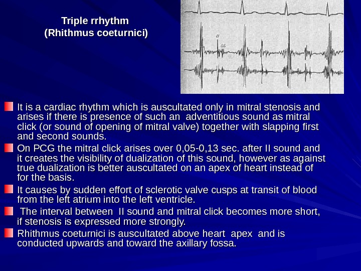
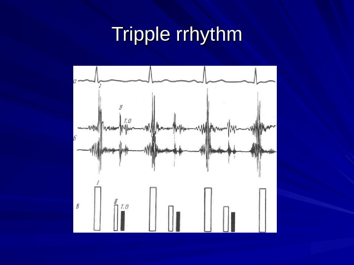
Triple rrhythm (( Rhithmus coeturnici) It is a cardiac rhythm which is auscultated only in mitral stenosis and arises if there is presence of such an adventitious sound as mitral click (or sound of opening of mitral valve) together with slapping first and second sounds. On PCG the mitral click arises over 0, 05 -0, 13 sec. after II sound and it creates the visibility of dualization of this sound, however as against true dualization is better auscultated on an apex of heart instead of for the basis. It causes by sudden effort of sclerotic valve cusps at transit of blood from the left atrium into the left ventricle. The interval between II sound and mitral click becomes more short, if stenosis is expressed more strongly. Rhithmus coeturnici is auscultated above heart apex and is conducted upwards and toward the axillary fossa.
Tripple rrhythm
Pendulum rhythm In the case of pendulum rhythm the large (diastolic) heart pause is so shortened, that becomes an equal to small (systolic) pause. The sound phenomenon, which one arises thus, reminds of even pendulum swinging. Such rhythm disturbance meets usually at heavy lesions of heart muscle . . If pendulum rhythm is accompaning by sharp heart acceleration, this phenomenon is called as embriocardia.
Protodiastolic and presystolic gallop rrhythm
Cardiac murmurs- phenpmena which arise due to pathological blood flow in the heart Intracardial murmurs: – Organic and functional (relative), – Systolic and diastolic, – Ejection and regurgitation murmurs, – They are also different in character, intensity, duration. Extracardial (pericarial friction murmur and pleuropericardial murmur)
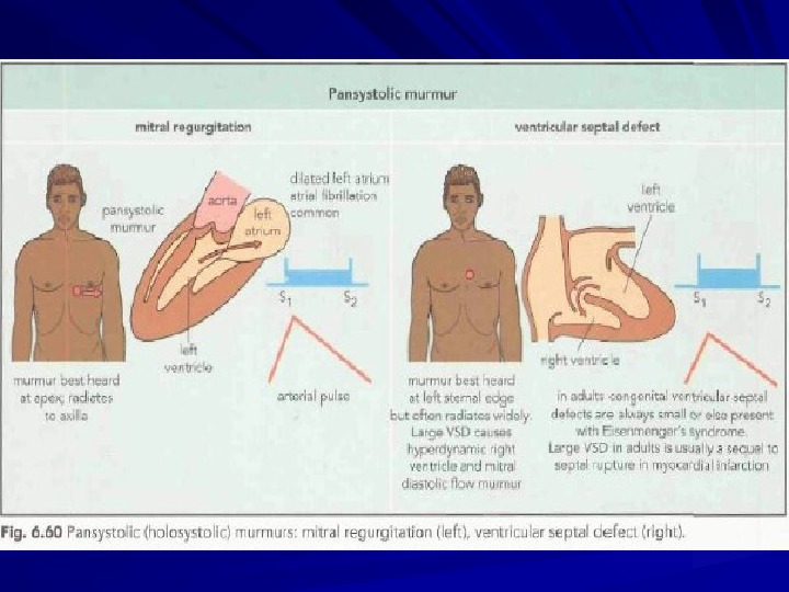
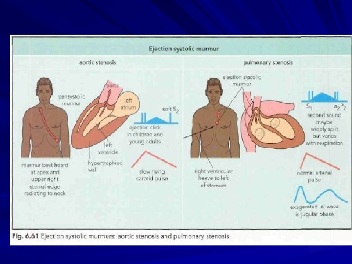
Properties of murmurs Duration The murmurs of mitral (and tricuspid) regurgitation start simultaneously with the first heart sound and continue throughout systole (pansystolic). The murmur produced by mitral valve prolapse does not begin until the mitral valve leaflet has prolapsed during systole, producing a late systolic murmur (Fig. 3. 25). The ejection systolic murmur of aortic or pulmonary stenosis begins after the first heart sound, reaches maximal intensity in midsystole, then fades, stopping before the second heart sound. Character and pitch The quality of murmurs is hard to define. Terms such as harsh, blowing, musical, rumbling, high or low pitched arc used. High-pitched murmurs often correspond with high-pressure gradients, so the diastolic murmur of aortic incompetence is higher pitched than that of mitral stenosis. Location Record the sitc(s) where you hear the murmur best. This helps to differentiate diastolic murmurs (mitral stenosis al the apex, aortic regurgitation at the left sternal edge), but is less helpful with systolic murmurs, which arc often loud and audible all over the precordium. Radiation Murmurs radiate in the direction of the blood flow causing the murmur to specific sites out with the precordium. Do nol j
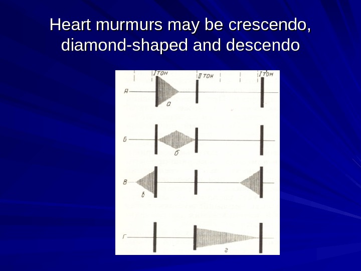
Heart murmurs may be crescendo, diamond-shaped and descendo
Intensity There are six grades of intensity used to describe murmurs. Diastolic murmurs are rarely louder than grade 4. The severity of valve dysfunction cannot be determined from the intensity of the murmur. For instance the murmur of critical aortic stenosis can be quiet and occasionally inaudible. Changes in intensity arc important as they often denote progression of a valve lesion. Rapidly changing murmurs arc sometimes heard with infective endocarditis because of valve destruction. Grades of intensity of murmur – Heard by an expert in optimum conditions – Heard by a non-expert in optimum conditions – Easily heard; no thrill – A loud murmur, with a thrill – Very loud, often heard over wide area, with thrill – Extremely loud, heard without stethoscope
Causes of systolic murmurs Ejection systolic murmur Increased flow through normal valves • • ‘Innocent systolic murmur’: fever athletes (bradycardia -> large stroke volume) pregnancy (cardiac output maximum at 15 weeks) Atrial septal defect (pulmonary flow murmur) Severe anaemia Normal or reduced flow though stenotic valve Aortic stenosis Pulmonary stenosis Other causes of flow murmurs Hypertrophic obstructive cardiomyopathy (obstruction at subvalvular level) Aortic regurgitation (aortic flow murmur) Pansystolic murmurs I caused by a systolic leak from a high to a lower pressure chamber Mitral regurgitation Tricuspid regurgitation Ventricular septal defect Leaking mitral or tricuspid prosthesis or bradycardia. Atrial septal defect is characterized
At an auscultation it is necessary to determine: 1) 1) relation of murmur to the phase of cardiac cycle (systole or diastole); 2) 2) properties of murmur, its character, intensity, duration; 3) 3) localization of murmur, i. e. place of the best auscultation; 4) 4) condution of murmur (irradiation).
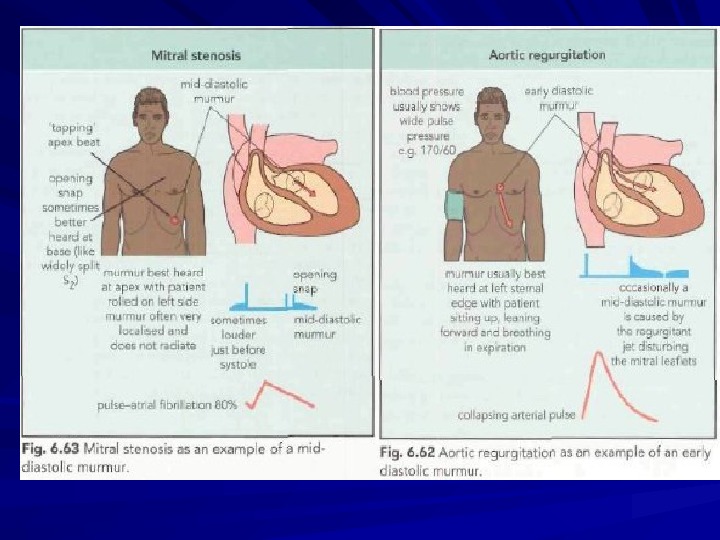
MM urmurs are auscultated better at points of auscultation of those valves, in which they were formed. Only in some cases murmurs are better heard in a distance from a place of originating beouse of their good conduction. The murmurs are well spent on a direction of a blood flow; they are better auscultated in that range, where heart to a chest and where it is not covered mild. The systolic murmur in mitral valve incompetene is best auscultated at heart apex; it can be conduted to axillary region or with blood bukflow from a left ventricle in the left atrium — — to the second and third intercostal space to the left of a breast bone. The diastolic murmur in narrowing of the left atrioventricular aperture is usually auscultated on a circumscribed field in apex area. The systolic murmur in stenosis of aortic rout is audible in the second intercostal space to the right of a breast bone. As a rule, he is well onduted with blood flow towards caroti arteries. As for this defeect rasping and loud (sawing, scratching) murmur is characteristic it can be determined by auscultation above all heart region and can be onduted to interscapular space. The diastolic murmur aortic valve inompetence is often better auscultated not above the aorti valve, but at Botkin-Erb’s point, where it is onduted with blood bukflow from the aorta to the left ventricle.
Differentiation of functional and organic murmurs in the most cases functional murmurs are systolic; the murmurs are changeable, can arise and decrease in intensity or even disappear at various positions of a body, after an exercise, stress, in different phases of respiration; most often they are auscultated above a pulmonary trunk, less often — — above heart apex, the murmurs are short, seldom occupy all systole; mild and blowing in character; the murmurs are usually auscultated on a circumscribed field and are not conducted far from the place of occurence; The functional murmurs are not accompanied by other attributes of valvular lesions (enlargement of heart chambers, change of sounds etc. ).
The pericardial friction It is develops in change of visceral and parietal pericardiac layers, when the fibrin (is postponed at a pericarditis), or cancerous nodules are deposied on them. The mechanism of its development is similar to the mechanism of creation of a pleural friction, only instead of respiratory movements the cause of its appearance is the movements of a heart during systole and diastole. Differential features: – It is heart equally over the whole heart area, – It intensifies if to press motightly to the heart area with a phonendoscope and at inclination of a trunk forward , , – It is sinchronous with heart contractions (is heart in systole and diastole), – it i s changeable, disappear and appear again.
The pleuropericardial friction murmur It arises in inflammation of pleura, immediately accumbent to heart, owing to friction of pleural layers, synchronic with activity of a heart. As opposite to pericardial friction: – it is auscultated on the left edge of relative cardiac dullness; – is usually combined with pleural friction, – changes the intensity in different phases of respiration strengthens at a penetrating inspiration, when the edge mild adjoins to more closely to the heart, and weakens at expiration, at fall of edge mild sharply.
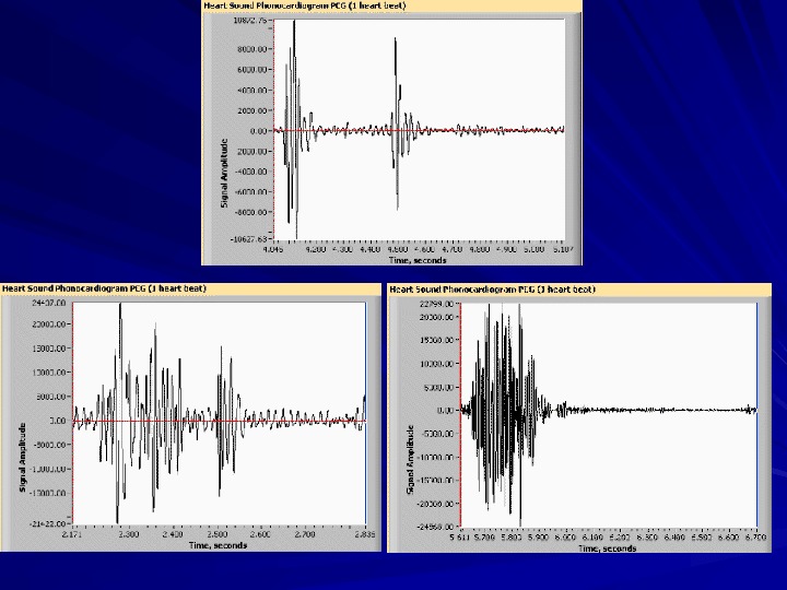
Phonocardiogram
AUSCULTATION OF VESSELS Auscultation of arteries. Arteries of medium calibre, such as the carotid, subclavian, or femoral artery, are usually auscultated. The artery is first palpated, then heard by a phonendoscope without applying pressure, since stenotic murmurs may otherwise appear. Sounds and mur murs can be heard over arteries. These can be generated either in the arteries themselves or be transmitted from the heart and aortic valves. The transmitted sounds and murmurs can only be heard on the arteries that are located close to the heart, such as the carotid and the subclavian arteries.
In norm: – Two sounds can be heard on the carotid and subclavian arteries in healthy persons. – The first sound is due to the tension of the arterial wall distended by the running pulse wave, and the second sound is transmitted onto these arteries from the aortic semilunar valve. – One systolic sound can sometimes be heard on the femoral artery. . In aortic incompetence: – the first sound over the arteries becomes louder because of the higher pulse wave, and it can be heard at greater distances from the heart, e. g. on the brachial and radial arteries. – Two sounds can sometimes be heard on the femoral artery in aortic incompetence. This doubled tone (Traube’s doubled tone) is generated by intense vibration of the vascular wall during both systole and diastole. – The Vinogradov-Duroziez doubled tone can be heard in aortic incompetence over the femoral artery when it is compressed by a stethoscope bell. The first of these tones is stenotic murmur, which is due to the blood flow through a narrowed (by the pressure of the stethoscope) vessel, while the second sound is explained by the accelerated backflow to the heart during diastole.
Systolic sound produced by the stenosed aortal orifice is usually well transmitted onto the carotid and subclavian arteries. Systolic sound associated with decreased viscosity of blood and increased flow rate (e. g. in anaemia, fever, exophthalmic goitre) can also be heard on these vessels. Systolic sound sometimes appears in stenosis or aneurysmal dilation of large vessels. .
Auscultation of veins Neither sounds nor murmurs are normally heard over veins. Auscultation of the jugular veins, over which the so-called nun’s murmur may be heard, is diagnostically important. This is a permanent blowing or humming sound, which is produced by accelerated flow of blood with decreased viscosity in anaemic patients. It is better heard on the right jugular vein and becomes more intense when the patient turns the head in the opposite side.
Thank you!