
Презентация Абдоминальная травма En


- Размер: 1.7 Mегабайта
- Количество слайдов: 62
Описание презентации Презентация Абдоминальная травма En по слайдам
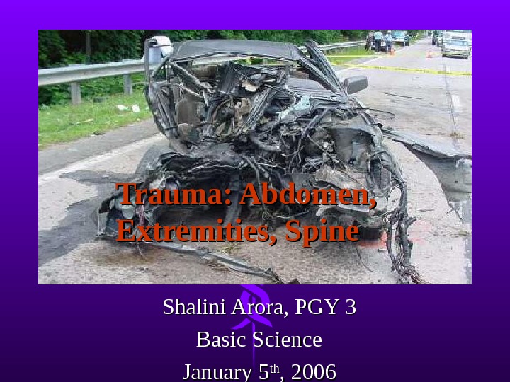
Shalini Arora, PGY 3 Basic Science January 5 thth , 2006Trauma: Abdomen, Extremities, Spine
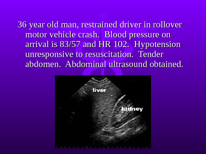
36 year old man, restrained driver in rollover motor vehicle crash. Blood pressure on arrival is 83/57 and HR 102. Hypotension unresponsive to resuscitation. Tender abdomen. Abdominal ultrasound obtained.
Next step? a. a. Exploratory laparotomy b. b. Diagnostic peritoneal lavage c. c. Abdominal CT scan d. d. Serial observation
28 y/o woman, unrestrained driver in a motor vehicle crash. Stable vital signs and LUQ tenderness, but no signs of peritonitis. Next step? a. a. Exploratory laparotomy b. b. Diagnostic peritoneal lavage c. c. Serial observation d. d. Abdominal CT scan e. e. Abdominal ultrasonography
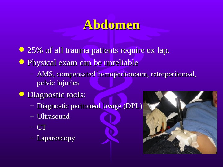
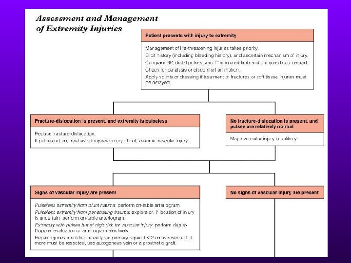
Abdomen 25% of all trauma patients require ex lap. Physical exam can be unreliable – AMS, compensated hemoperitoneum, retroperitoneal, pelvic injuries Diagnostic tools: – Diagnostic peritoneal lavage (DPL) – Ultrasound – CTCT – Laparoscopy
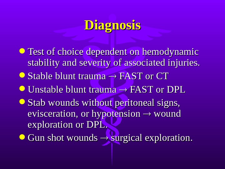
Diagnosis Test of choice dependent on hemodynamic stability and severity of associated injuries. Stable blunt trauma FAST or CT Unstable blunt trauma FAST or DPL Stab wounds without peritoneal signs, evisceration, or hypotension wound exploration or DPL. Gun shot wounds surgical exploration.
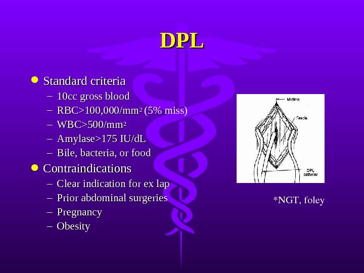
DPLDPL Standard criteria – 10cc gross blood – RBC>100, 000/mm 2 2 (5% miss) – WBC>500/mm 22 – Amylase>175 IU/d. L – Bile, bacteria, or food Contraindications – Clear indication for ex lap – Prior abdominal surgeries – Pregnancy – Obesity *NGT, foley
DPLDPL Highly sensitive to intraperitoneal blood, but low specificity nontherapeutic explorations. Supraumbilical if pelvic fracture present Significant injuries may be missed – Diaphragm – Retroperitoneal hematomas – Renal, pancreatic, duodenal – Minor intestinal – Extraperitoneal bladder injuries
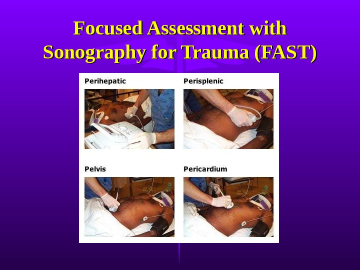
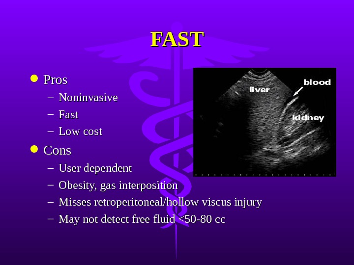
Focused Assessment with Sonography for Trauma (FAST)
FAST Pros – Noninvasive – Fast – Low cost Cons – User dependent – Obesity, gas interposition – Misses retroperitoneal/hollow viscus injury – May not detect free fluid <50-80 cc
CT Scan Hemodynamically stable patient Pros – Retroperitoneal assessment – Nonoperative management of solid organ injury – High specificity Cons – Hardware, cost, radiation – Hollow viscus injuries, diaphragm injury
Laparoscopy Role still being defined Good for diaphragm injury evaluation Cons – Invasive – Expensive – Missed small bowel, splenic, retroperitoneal injuries
Gastric Injury Mostly penetrating trauma. <1% from blunt trauma – Including iatrogenic injury from CPR NGT + aspirate for blood Intraop evaluation includes complete visualization of posterior wall Most penetrating wounds treated by debridement and primary closure in layers. Evacuation of hematomas. Major tissue loss may necessitate resection.
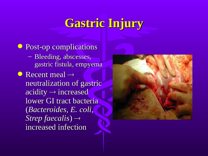
Gastric Injury Post-op complications – Bleeding, abscesses, gastric fistula, empyema Recent meal neutralization of gastric acidity increased lower GI tract bacteria (( Bacteroides, E. coli, Strep faecalis ) ) increased infection
Duodenal Injury Majority due to penetrating trauma. Blunt injury usually secondary to steering wheel blow to the epigastrium Retroperitoneal location is protective, but also prevents early diagnosis. Isolated injury to the duodenum is rare Hyperamylasemia in 50% with blunt injury.
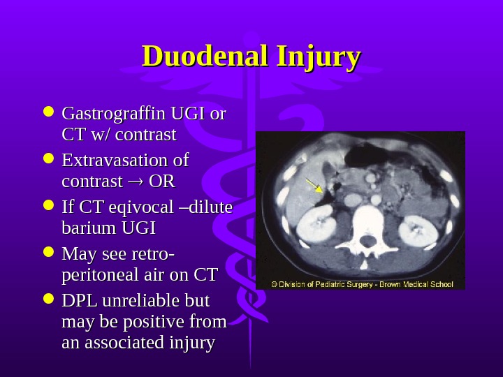
Duodenal Injury Gastrograffin UGI or CT w/ contrast Extravasation of contrast OR OR If CT eqivocal –dilute barium UGI May see retro- peritoneal air on CT DPL unreliable but may be positive from an associated injury
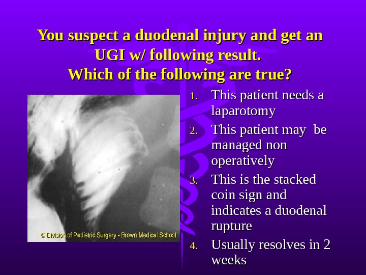
You suspect a duodenal injury and get an UGI w/ following result. Which of the following are true? 1. 1. This patient needs a laparotomy 2. 2. This patient may be managed non operatively 3. 3. This is the stacked coin sign and indicates a duodenal rupture 4. 4. Usually resolves in 2 weeks
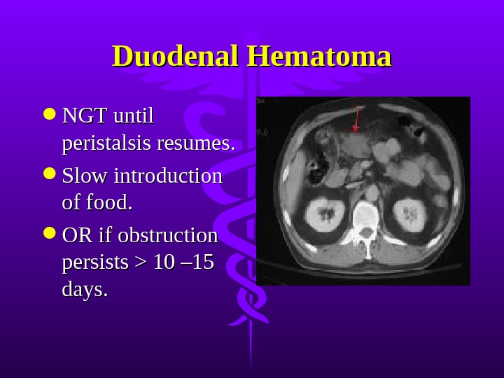
Duodenal Hematoma NGT until peristalsis resumes. Slow introduction of food. OR if obstruction persists > 10 – 15 days.
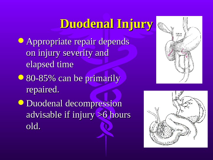
Duodenal Injury Appropriate repair depends on injury severity and elapsed time 80-85% can be primarily repaired. Duodenal decompression advisable if injury >6 hours old.
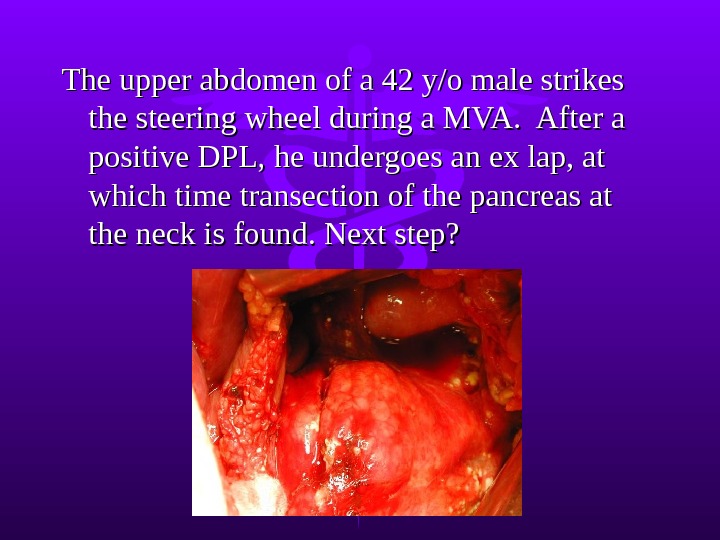
The upper abdomen of a 42 y/o male strikes the steering wheel during a MVA. After a positive DPL, he undergoes an ex lap, at which time transection of the pancreas at the neck is found. Next step?
Next step? a. a. Distal pancreatectomy with oversewing and drainage of proximal stump. b. b. Primary repair and drainage of the pancreatic duct. c. c. Roux-en-Y pancreaticojejunostomy to the distal pancreas with oversewing and drainge of the proximal stump. d. d. Total pancreatectomy
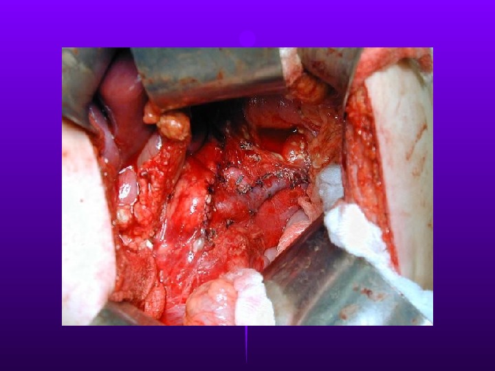
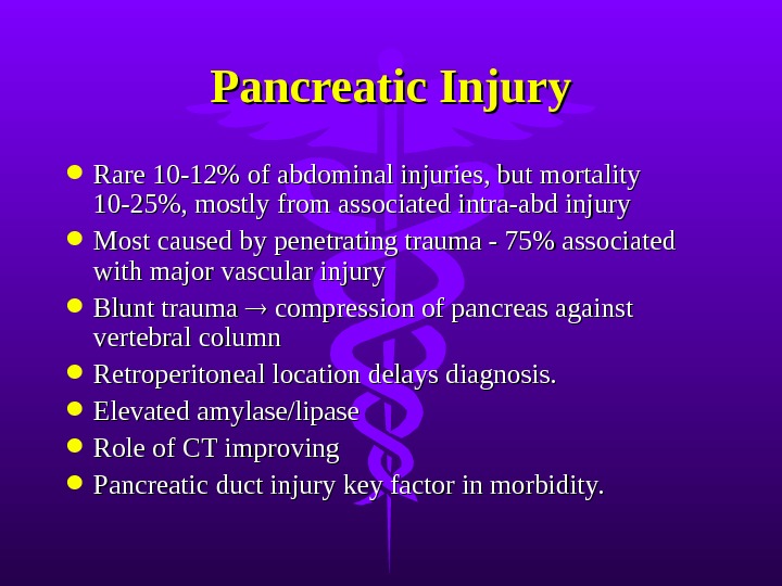
Pancreatic Injury Rare 10-12% of abdominal injuries, but mortality 10-25%, mostly from associated intra-abd injury Most caused by penetrating trauma — 75% associated with major vascular injury Blunt trauma compression of pancreas against vertebral column Retroperitoneal location delays diagnosis. Elevated amylase/lipase Role of CT improving Pancreatic duct injury key factor in morbidity.
Pancreatic Injury
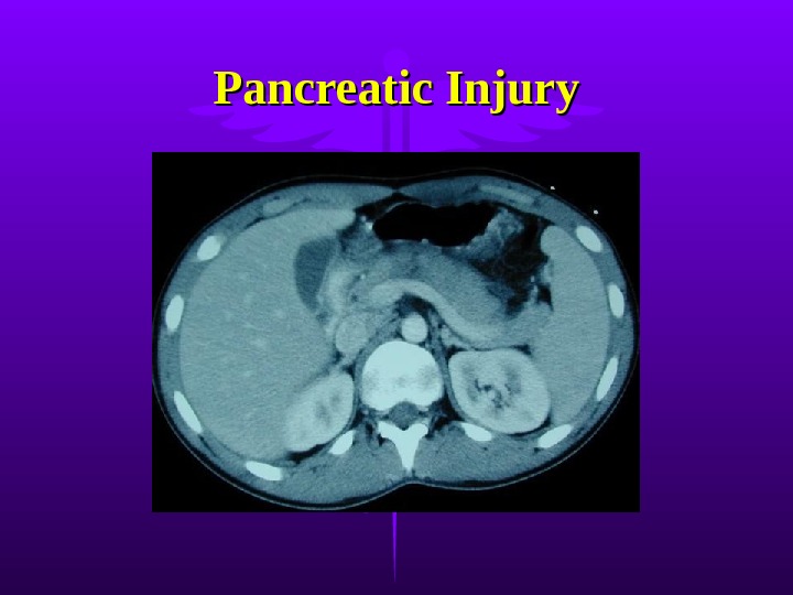
GSW to Pancreatic Head
Pancreatic Injury Divided into proximal or distal according to location on the R or L of SMV Contusions (Grade I-II) should be drained. Distal duct injury (Grade III) distal resection with splenic preservation Proximal injury (Grade IV) – Oversewing and distal resection or pancreaticojejunostomy in diabetic patients. Extensive pancreatic head injuries (Grade V) – 40% pancreatic fistula development – Simple external wide drainage
Complications after Pancreatic Trauma High complication rate 35-40% Most common are pancreatic fistulas & abscesses Most fistulas close spontaneously if well drained Somatostatin to expedite healing Abscesses — surgical debridement & drainage Incidence of pancreatitis 8-18% Pseudocysts are infrequent
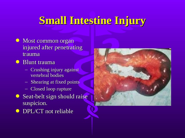
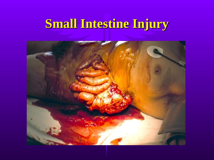
Small Intestine Injury Most common organ injured after penetrating trauma Blunt trauma – Crushing injury against vertebral bodies – Shearing at fixed points – Closed loop rupture Seat-belt sign should raise suspicion. DPL/CT not reliable
Small Intestine Injury
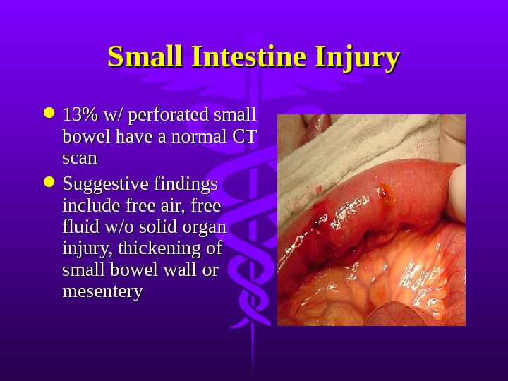
Small Intestine Injury 13% w/ perforated small bowel have a normal CT scan Suggestive findings include free air, free fluid w/o solid organ injury, thickening of small bowel wall or mesentery
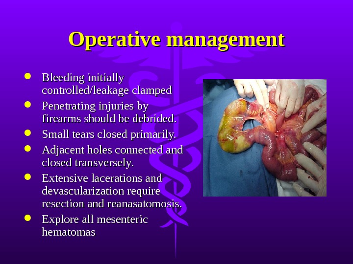
Operative management Bleeding initially controlled/leakage clamped Penetrating injuries by firearms should be debrided. Small tears closed primarily. Adjacent holes connected and closed transversely. Extensive lacerations and devascularization require resection and reanasatomosis. Explore all mesenteric hematomas
Colon Injury Second most frequent injured organ, usually from penetrating trauma Repair within 2 hours dramatically reduces infectious complications. Pre-operative antibiotics important adjunct. PE blood per rectum, stab to flanks or back CT w/rectal contrast, XR- pneumoperitoneum WWI primary repair led to 60% mortality. WWII colostomy led to 35% mortality.
Colon Injury Primary repair criteria – Early diagnosis (within 4-6 hours) – Absence of prolonged shock/hypotension – Absence of gross contamination – Absence of associated colonic vascular injury – Less than 6 units blood transfusion – No requirement for use of mesh for closure Extensive wounds – Right colon hemicolectomy +/- ileostomy – Left colon resection + colostomy
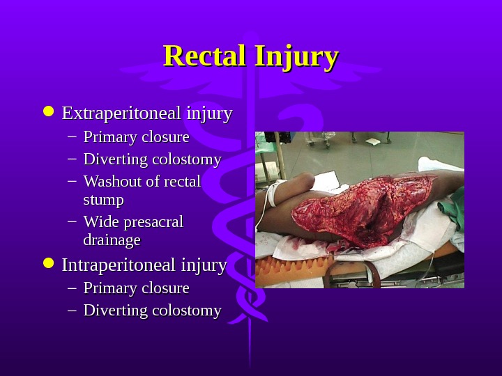
Rectal Injury Most from GSW Other causes — foreign body, impalement, pelvic fractures, and iatrogenic Lower abdomen/buttock penetrating injury should raise suspicion. May be intra- or extraperitoneal Rectal exam may reveal blood or laceration Work-up includes anoscopy and rigid sigmoidoscopy.
Rectal Injury Extraperitoneal injury – Primary closure – Diverting colostomy – Washout of rectal stump – Wide presacral drainage Intraperitoneal injury – Primary closure – Diverting colostomy
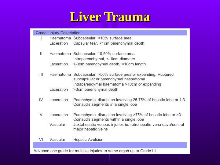
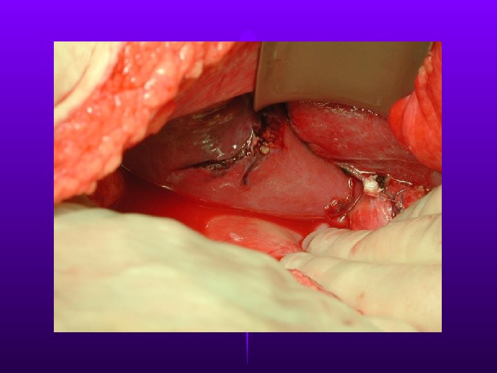
Liver Trauma Frequently injured in both blunt & penetrating trauma. Control of profuse bleeding from deep lacerations a formidable challenge. – Simple suture, mattress sutures, packing, debridement, resection, mesh hepatorrhaphy Nonoperative treatment (blunt trauma) – Stable without peritoneal signs U/S CT CT – Low-grade liver lesions (1-3, 95% success) – ICU monitoring
Liver Trauma
Liver Trauma
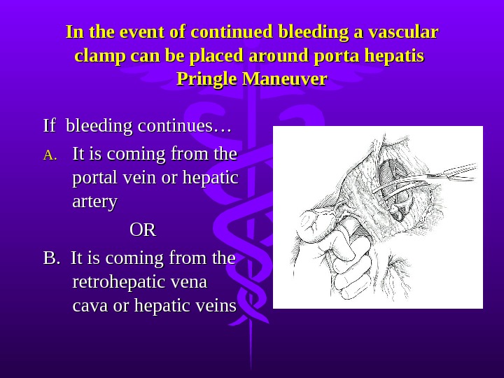
In the event of continued bleeding a vascular clamp can be placed around porta hepatis Pringle Maneuver If bleeding continues… A. A. It is coming from the portal vein or hepatic artery OROR B. It is coming from the retrohepatic vena cava or hepatic veins
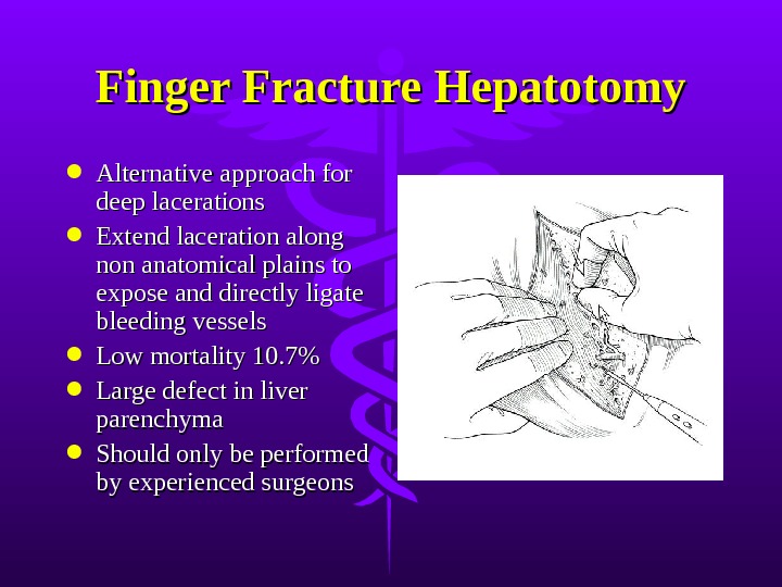
Finger Fracture Hepatotomy Alternative approach for deep lacerations Extend laceration along non anatomical plains to expose and directly ligate bleeding vessels Low mortality 10. 7% Large defect in liver parenchyma Should only be performed by experienced surgeons
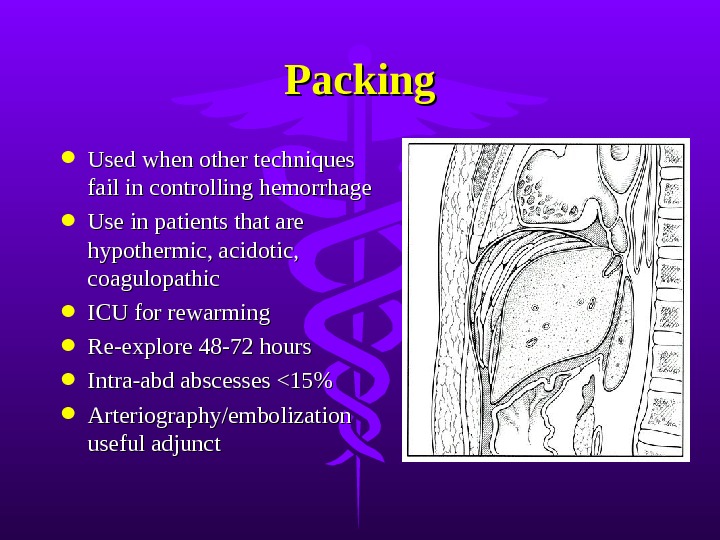
Packing Used when other techniques fail in controlling hemorrhage Use in patients that are hypothermic, acidotic, coagulopathic ICU for rewarming Re-explore 48-72 hours Intra-abd abscesses <15% Arteriography/embolization useful adjunct
Of the following hemodynamically stable patients, who is most likely to fail non-operative management. A. A. 8 y/o girl s/p left lateral abdominal blow playing soccer. CT with 3cm laceration with blood around spleen and liver. B. B. 22 y/o male restrained low speed MVA with left lower rib fractures. CT with 3cm laceration with blood around spleen and liver. C. C. 15 y/o boy tackled playing football. CT with 3 splenic lacerations, blood around spleen, liver, and in pelvis. D. D. 21 y/o intoxicated restrained high speed MVA. CT with deep splenic laceration, upper pole contusion, and perisplenic blood. E. E. 25 y/o male pinned under car when it feel from its lumberjack and landed on his upper chest. CT with deep splenic laceration, blush of intravenous contrast by laceration, and perisplenic blood.
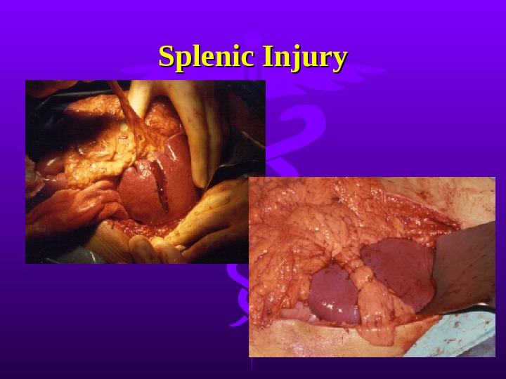
Splenic Injury Most frequently injured intra-abdominal organ in blunt trauma. Splenic preservation when possible OPSI (0. 6% in children, 0. 3% in adults) More than 70% can be treated non-operatively
Splenic Injury Nonoperative criteria – Hemodynamic stability – Negative abdominal examination – Absence of contrast extravasation Angiography/embolization an option – No other clear indications for ex lap – No coagulopathy – Low grade injuries (1-3)
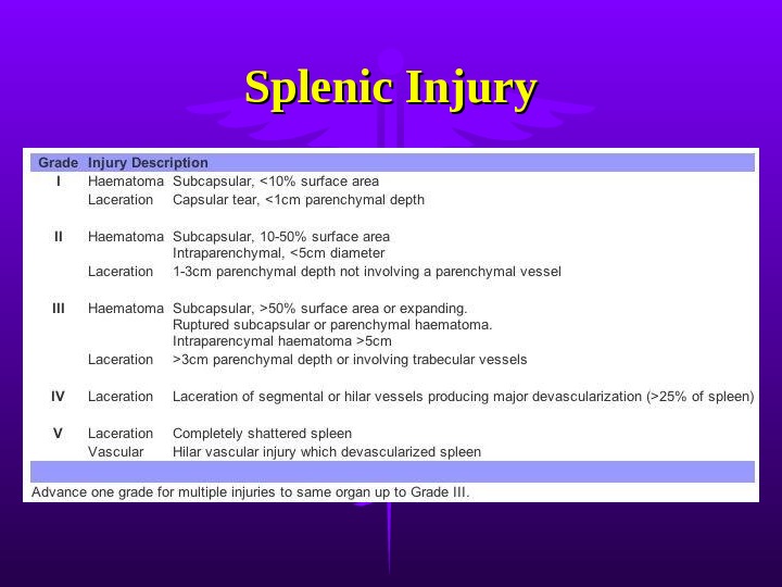
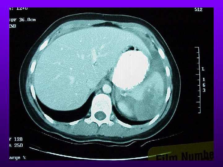
Splenic Injury
Splenic Injury
Splenic Injury
30 year-old man ejected from automobile after head-on collision at high speed. Sustained pelvic fracture. Grossly positive supra-umbilical DPL. On exploration, a pelvic hematoma and an expanding central hematoma are noted. Next step? a. a. Observation of both hematomas. b. b. Exploration of both hematomas. c. c. Exploration of central hematoma after obtaining proximal and distal vascular control; observation of the pelvic hematoma. d. d. Observation of central hematoma, and exploration of the pelvic hematoma after application of external fixators.
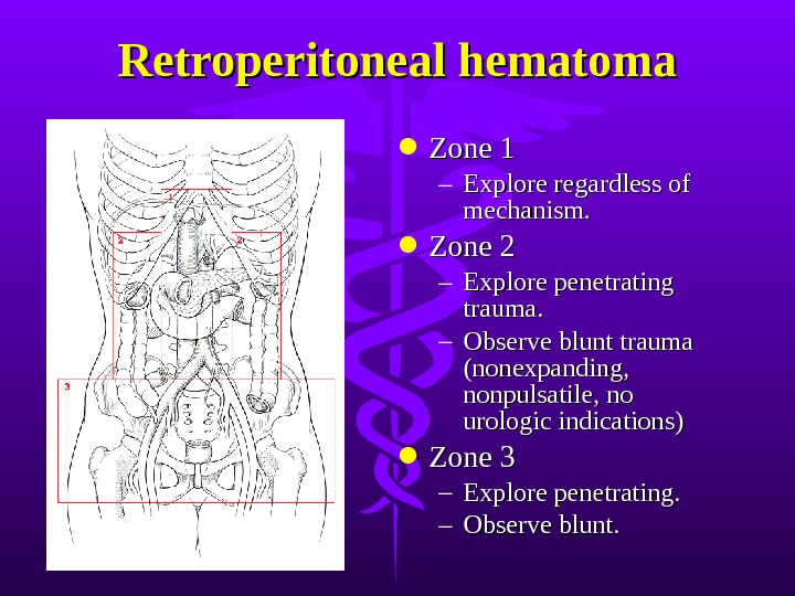
Retroperitoneal hematoma Zone 1 – Explore regardless of mechanism. Zone 2 – Explore penetrating trauma. – Observe blunt trauma (nonexpanding, nonpulsatile, no urologic indications) Zone 3 – Explore penetrating. – Observe blunt.
Damage Control Abbreviated laparotomy and temporary packing Effort to blunt physiologic response to shock and hemorrhage – Severe metabolic acidosis, coagulopathy, and hypothermia ICU resuscitation Return to OR in 48-72 hours
Damage Control
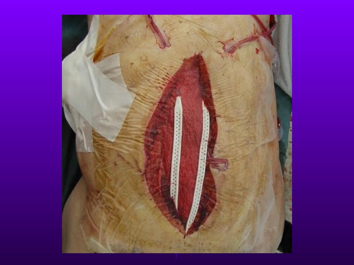
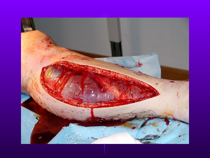
30 y/o woman sustained crushing injury to right lower leg. Arrived at hospital 12 hours later. PE reveals tense calf and closed tibia-fibula fracture. Unable to dorsiflex foot, absent pedal pulses. Next step? a. a. Angiography b. b. Below knee amputation c. c. Four compartment fasciotomy d. d. Surgical exploration of popliteal artery e. e. Internal fixation of tibial fracture
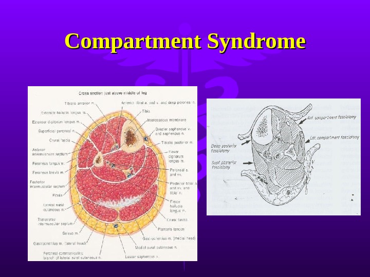
Compartment Syndrome Common in forearm and lower leg secondary to defined fascial boundaries. Four Ps: pressure, pain, paresthesia, and intact pulses Compartment pressure measurement – Critical pressure? (20-30mm Hg) – MAP — compartment pressure < 40mm Hg
Compartment Syndrome
Fasciotomy
Extremity Injuries
With regard to cervical spine injury, which of the following is/are true? a. a. Jefferson fractures (C 1) are usually caused by axial load and involve blowout of the ring. b. b. Hangman’s fractures are unstable and are best treated by operative spinal fusion. c. c. Type II odontoid fractures are considered stable.
Spine Trauma C 1 burst fractures (Jefferson’s) – Axial loading force – Considered stable – Treat with rigid cervical collar Hangman’s fracture – Extension and distraction force – Posterior C 2 elements – Unstable fracture – Traction halo vest
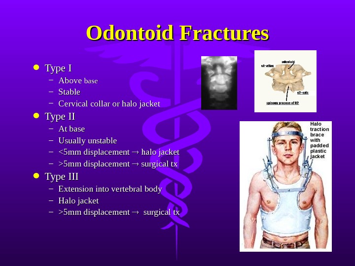
Odontoid Fractures Type I – Above base – Stable – Cervical collar or halo jacket Type II – At base – Usually unstable – 5mm displacement surgical tx Type III – Extension into vertebral body – Halo jacket – >5mm displacement surgical tx
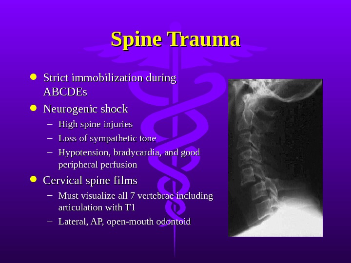
Spine Trauma Strict immobilization during ABCDEs Neurogenic shock – High spine injuries – Loss of sympathetic tone – Hypotension, bradycardia, and good peripheral perfusion Cervical spine films – Must visualize all 7 vertebrae including articulation with T 1 – Lateral, AP, open-mouth odontoid
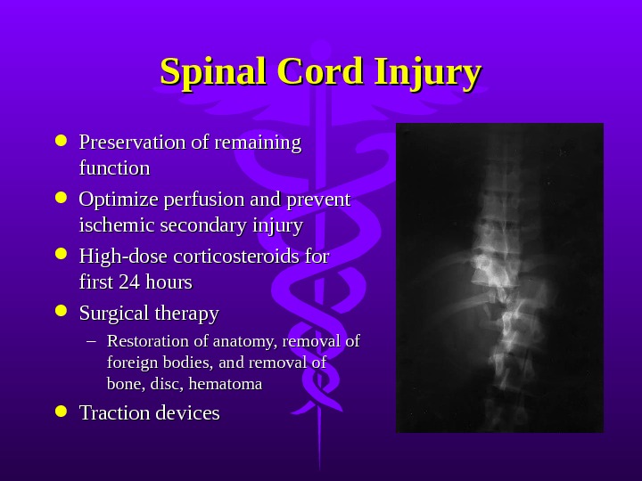
Spinal Cord Injury Preservation of remaining function Optimize perfusion and prevent ischemic secondary injury High-dose corticosteroids for first 24 hours Surgical therapy – Restoration of anatomy, removal of foreign bodies, and removal of bone, disc, hematoma Traction devices
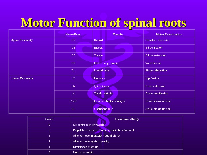
Nerve Root Muscle Motor Examination Upper Extremity C 5 Deltoid Shoulder abduction C 6 Biceps Elbow flexion C 7 Triceps Elbow extension C 8 Flexor carpi ulnaris Wrist flexion T 1 Lumbricales Finger abduction Lower Extremity L 2 Iliopsoas Hip flexion L 3 Quadriceps Knee extension L 4 Tibialis anterior Ankle dorsiflexion L 5-S 1 Extensor hallucis longus Great toe extension S 1 Gastrocnemius Ankle plantarflexion Score Functional Ability 0 No contraction of muscle 1 Palpable muscle contraction, no limb movement 2 Able to move in gravity-neutral plane 3 Able to move against gravity 4 Diminished strength 5 Normal strength. Motor Function of spinal roots