
Pneumonia in children V. N. Karazin Kharkiv National


acute_pneumonia_in_childr.ppt
- Размер: 3.5 Mегабайта
- Количество слайдов: 104
Описание презентации Pneumonia in children V. N. Karazin Kharkiv National по слайдам
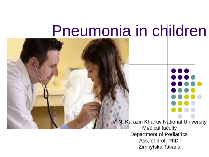
Pneumonia in children V. N. Karazin Kharkiv National University Medical faculty Department of Pediatrics Ass. of prof. Ph. D Zimnytska Tatiana
Pneumonia – – polyetiological infectious disease of respiratory system lower parts with alveolar exudation which is confirmed by radiological method (( European pulmonological society ))
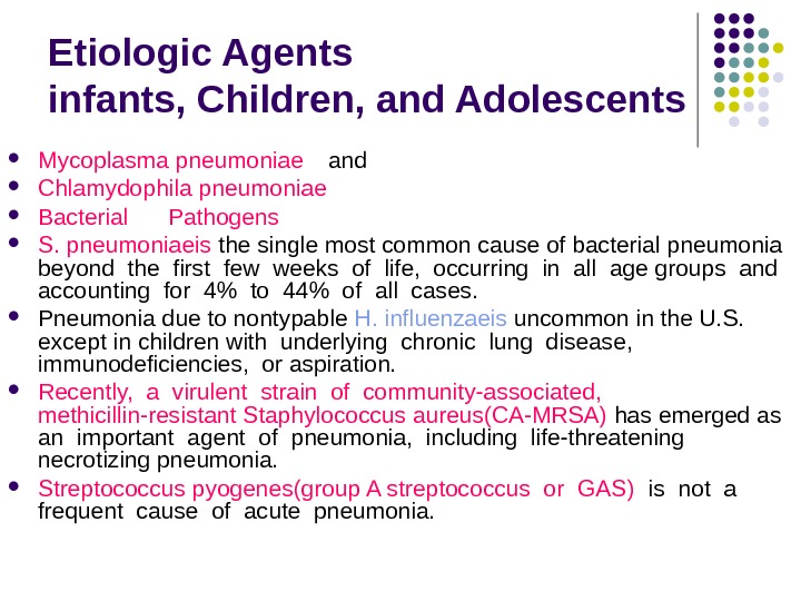
Etiologic Agents
Etiologic Agents Neonates and Young Infants Pneumonia in neonates can manifest as early-onset disease (within the first week of life) or late-onset disease (≥ 7 days of life). Aspiration of either infected amniotic fluid or genital secretions at delivery is the cause of most early-onset infections. Group B streptococcus is the most frequent cause of early-onset pneumonia, but Listeria monocytogenes, Escherichia coli , and other gramnegative bacilli can cause severe respiratory distress resembling hyaline membrane disease, usually as a part of a widespread systemic infection. Prenatal and perinatal risk factors, including preterm delivery, maternal chorioamnionitis and prolonged rupture of membranes, increase the risk for development of neonatal pneumonia. Hematogenous dissemination also can occur from an infected mother Chlamydia trachomatis pneumonia can occur 2 to 3 weeks after birth in 10% of neonates born to mothers colonized with the organism in their genital tract. Bordetella pertussis infection cause secondary bacterial pneumonia or pulmonary hypertension (simulating pneumonia). Viruses are a less common cause compared with older infants. Congenital or perinatal infection with cytomegalovirus (CMV), herpes simplex virus (HSV), or Treponema pallidum can cause severe pneumonia. Genital Mycoplasmaspecies and Ureaplasma urealyticum can cause LRTI in very-low-birthweight infants
Etiologic Agents i nfants, Children, and Adolescents Viruses account for approximately 14% to 35% of childhood CAPbut for 80% of CAP in children <2 years. RSV is the predominant respiratory tract viral pathogen. Other viruses include , human metapneumovirus (HMPV), parainfluenza viruses (PIV) types 1, 2, and 3, influenza viruses (A and B), adenoviruses, rhinoviruses, and enteroviruses. Rhinoviruses have been recovered in 2% to 24% cases of childhood pneumonia. Varicella-zoster virus (VZV), CMV , and HSV can cause LRTI in immunocompromised children.
Etiologic Agents i nfants, Children, and Adolescents Mycoplasma pneumoniae and Chlamydophila pneumoniae Bacterial Pathogens S. pneumoniaeis the single most common cause of bacterial pneumonia beyond the first few weeks of life, occurring in all age groups and accounting for 4% to 44% of all cases. Pneumonia due to nontypable H. influenzaeis uncommon in the U. S. except in children with underlying chronic lung disease, immunodeficiencies, or aspiration. R ecently, a virulent strain of community-associated, methicillin-resistant Staphylococcus aureus(CA-MRSA) has emerged as an important agent of pneumonia, including life-threatening necrotizing pneumonia. Streptococcus pyogenes(group A streptococcus or GAS) is not a frequent cause of acute pneumonia.
Pathogenesis and Pathology
The pulmonary defense mechanisms Physical barriers of the respiratory tract include the presence of hairs in the anterior nares that can trap particles >10 µm in size, configuration of the nasal turbinates, and acute branching of the respiratory tract Physiologic protection includes filtration and humidification in the upper airways, mucus production, and protection of the airway by the epiglottis and cough reflex.
The pulmonary defense mechanisms Mucociliary transport moves normally aspirated oropharyngeal flora and particulate matter up the tracheobronchial tree, minimizing the presence of bacteria below the carina. However, particles less than 1 µm can escape into the lower airways. Immunoglobulin A (Ig. A), is the major protective antibody secreted by the upper airways; Ig. G and Ig. M primarily protect the lower airways. Substances found in alveolar fluid – including surfactant, fibronectin, complement, lysozyme, and iron-binding proteins – have antimicrobial activity. The LRT has distinct populations of macrophages. Alveolar macrophages are the pre-eminent phagocytic cells that ingest and kill bacteria.
Pneumonia is inflammatory process developed after entry of infectious agent in respiratory portions of airway tract. Entry routes of foreign agents in respiratory system (lungs) may be via inhalation or hematogenous. Inflammatory cascade is disease trigger and causes plasma exudation and loss of surfactant, causing difficult air exchange and consolidation.
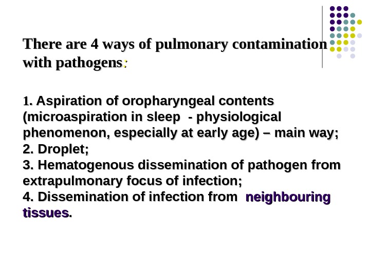
There are 4 4 ways of pulmonary contamination with pathogens : : 1. 1. Aspiration of oropharyngeal contents (( microaspiration in sleep — — physiological phenomenon, especially at early age ) – main way ; ; 2. 2. Droplet ; ; 3. 3. Hematogenous dissemination of pathogen from extrapulmonary focus of infection ; ; 4. 4. Dissemination of infection from neighbouring tissues. .
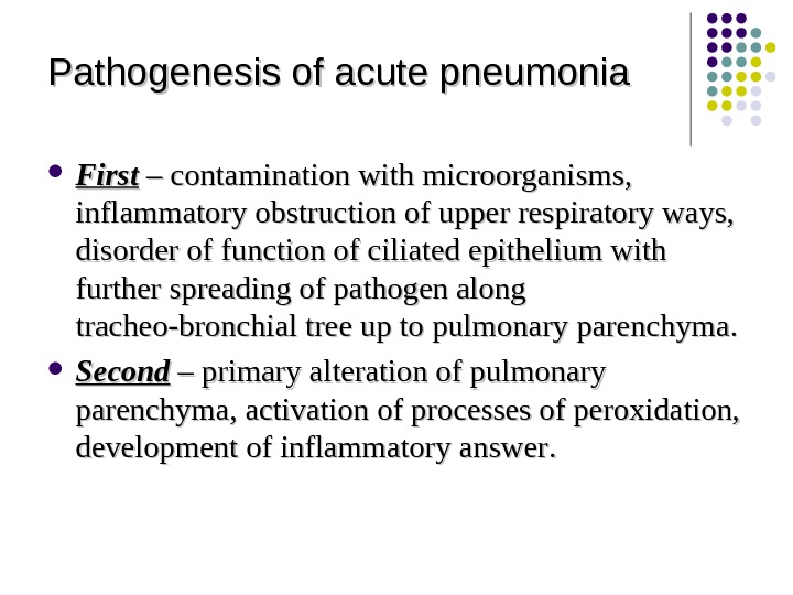
Pathogenesis of acute pneumonia First – – contamination with microorganisms, inflammatory obstruction of upper respiratory ways, disorder of function of ciliated epithelium with further spreading of pathogen along tracheo-bronchial tree up to pulmonary parenchyma. Second – – primary alteration of pulmonary parenchyma, activation of processes of peroxidation, development of inflammatory answer. .
Pathogenesis of acute pneumonia Third – – alteration of not only pathogen but of own organism including surfactant, destabilization of biological membranes of subcellular structures – phase of secondary toxic autoagression. Forth – – disorders of tissue respiration, central regulation of respiration, ventilation, gas exchange and pulmonary perfusion. .
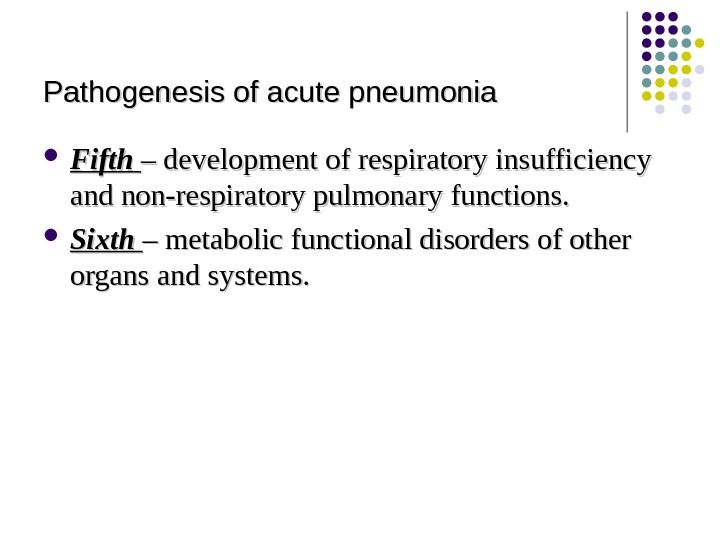
Pathogenesis of acute pneumonia Fifth – – development of respiratory insufficiency and non-respiratory pulmonary functions. Sixth – – metabolic functional disorders of other organs and systems.
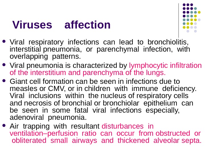
Viruses affection Viral respiratory infections can lead to bronchiolitis, interstitial pneumonia, or parenchymal infection, with overlapping patterns. Viral pneumonia is characterized by lymphocytic infiltration of the interstitium and parenchyma of the lungs. Giant cell formation can be seen in infections due to measles or CMV, or in children with immune deficiency. Viral inclusions within the nucleus of respiratory cells and necrosis of bronchial or bronchiolar epithelium can be seen in some fatal viral infections especially, adenoviral pneumonia. Air trapping with resultant disturbances in ventilation–perfusion ratio can occur from obstructed or obliterated small airways and thickened alveolar septa.
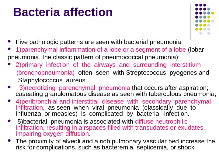
Bacteria affection Five pathologic patterns are seen with bacterial pneumonia: 1) parenchymal inflammation of a lobe or a segment of a lobe (lobar pneumonia, the classic pattern of pneumococcal pneumonia); 2) primary infection of the airways and surrounding interstitium (bronchopneumonia) often seen with Streptococcus pyogenes and Staphylococcus aureus; 3) necrotizing parenchymal pneumonia that occurs after aspiration; caseating granulomatous disease as seen with tuberculous pneumonia; 4) peribronchial and interstitial disease with secondary parenchymal infiltration , as seen when viral pneumonia (classically due to influenza or measles) is complicated by bacterial infection. 5)b acterial pneumonia is associated with diffuse neutrophilic infiltration, resulting in airspaces filled with transudates or exudates, impairing oxygen diffusion. The proximity of alveoli and a rich pulmonary vascular bed increase the risk for complications, such as bacteremia, septicemia, or shock.
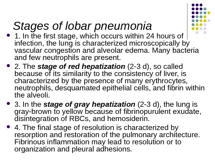
Stages of lobar pneumonia 1. In the first stage, which occurs within 24 hours of infection, the lung is characterized microscopically by vascular congestion and alveolar edema. Many bacteria and few neutrophils are present. 2. The stage of red hepatization (2 -3 d), so called because of its similarity to the consistency of liver, is characterized by the presence of many erythrocytes, neutrophils, desquamated epithelial cells, and fibrin within the alveoli. 3. In the stage of gray hepatization (2 -3 d), the lung is gray-brown to yellow because of fibrinopurulent exudate, disintegration of RBCs, and hemosiderin. 4. The final stage of resolution is characterized by resorption and restoration of the pulmonary architecture. Fibrinous inflammation may lead to resolution or to organization and pleural adhesions.
Classification
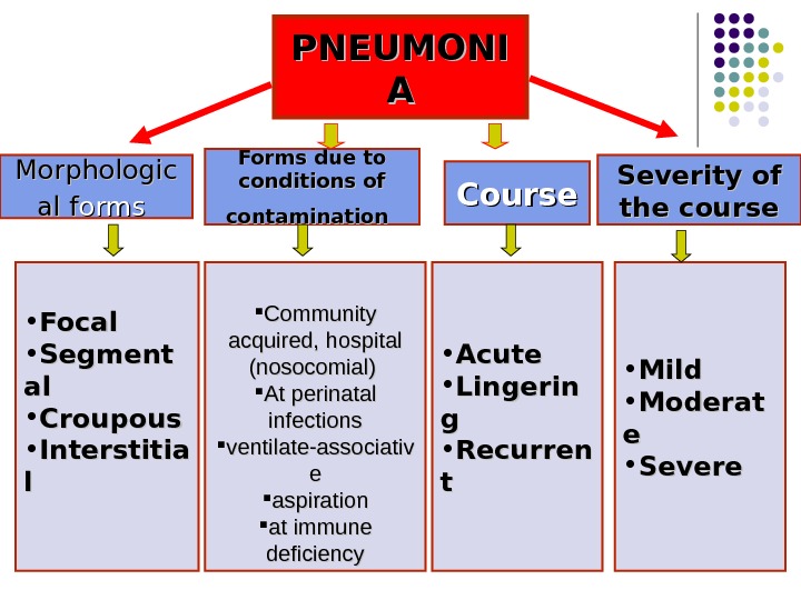
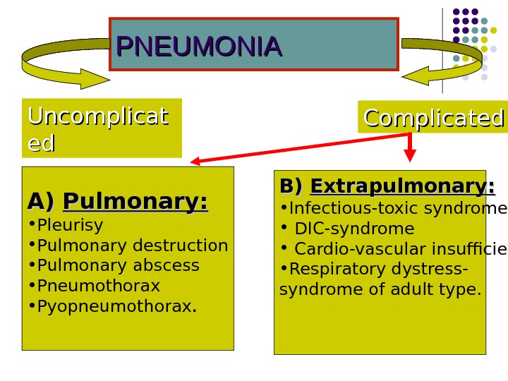
PNEUMONI AA Morphologic alal ff orms Forms due to conditions of contamination Course Severity of the course • Focal • Segment alal • Croupous • Interstitia ll Community acquired, hospital (nosocomial) At perinatal infections ventilate-associativ ee aspiration at immune deficiency • Acute • Lingerin gg • Recurren tt • Mild • Moderat ee • Severe
PNEUMONIA Complicated. Uncomplicat eded А)А) Pulmonary: • Pleurisy • Pulmonary destruction • Pulmonary abscess • Pneumothorax • Pyopneumothorax. BB ) ) Extrapulmonary : : • Infectious-toxic syndrome • DIC-syndrome • Cardio-vascular insufficiency • Respiratory dystress- syndrome of adult type.
Clinical symptoms
Main signs of pneumonia Symptoms of intoxication, fever Cough (recently started) Tachypnoea Dyspnoea Chest wall retractions Nasal flaring Percustory change s–decrease of resonance or dull sound over lung consolidation Auscultatory – bronchial breathing, diminished breathing over consolidation, 3 -5 day of illness – fine crackles, crepitaion
Pneumonia indications in children younger 5 years of age: Nasal flaring (before 12 months) Oxygen saturation <94% Tachypnoea Chest wall retractions
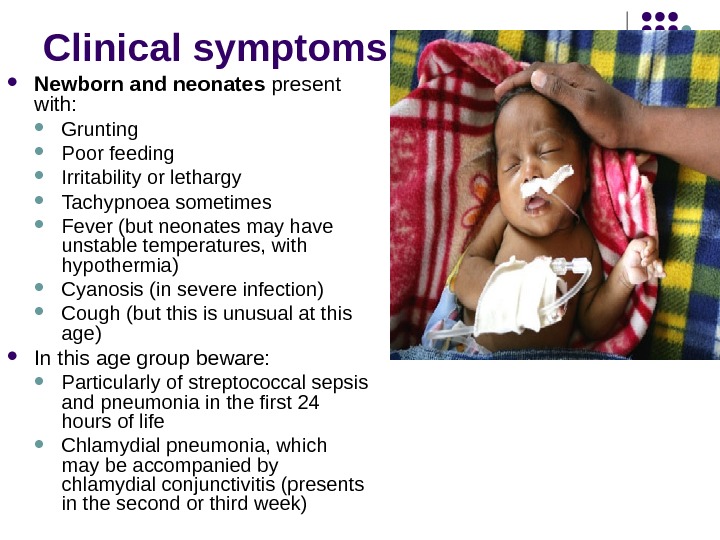
Clinical symptoms Newborn and neonates present with: Grunting Poor feeding Irritability or lethargy Tachypnoea sometimes Fever (but neonates may have unstable temperatures, with hypothermia) Cyanosis (in severe infection) Cough (but this is unusual at this age) In this age group beware: Particularly of streptococcal sepsis and pneumonia in the first 24 hours of life Chlamydial pneumonia, which may be accompanied by chlamydial conjunctivitis (presents in the second or third week)
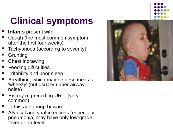
Clinical symptoms I nfants present with: Cough (the most common symptom after the first four weeks) Tachypnoea (according to severity) Grunting Chest indrawing Feeding difficulties Irritability and poor sleep Breathing, which may be described as ‘wheezy’ (but usually upper airway noise) History of preceding URTI (very common) In this age group beware: Atypical and viral infections (especially pneumonia) may have only low-grade fever or no fever
Clinical symptoms Toddlers/pre-school children : Again, preceding URTI is common Cough is the most common symptom Fever occurs most noticeably with bacterial organisms Pain occurs more often in this age group (chest and abdominal) Vomiting with coughing is common (post-tussive vomiting) Be aware that: Lower lobe pneumonias can cause abdominal pain Severe infections will compromise breathing more
Clinical symptoms Older children : There will be additional symptoms to those above More expressive and articulate children will report a wider range of symptoms Constitutional symptoms may be more vividly described Be aware that: Atypical organisms are more likely in older children
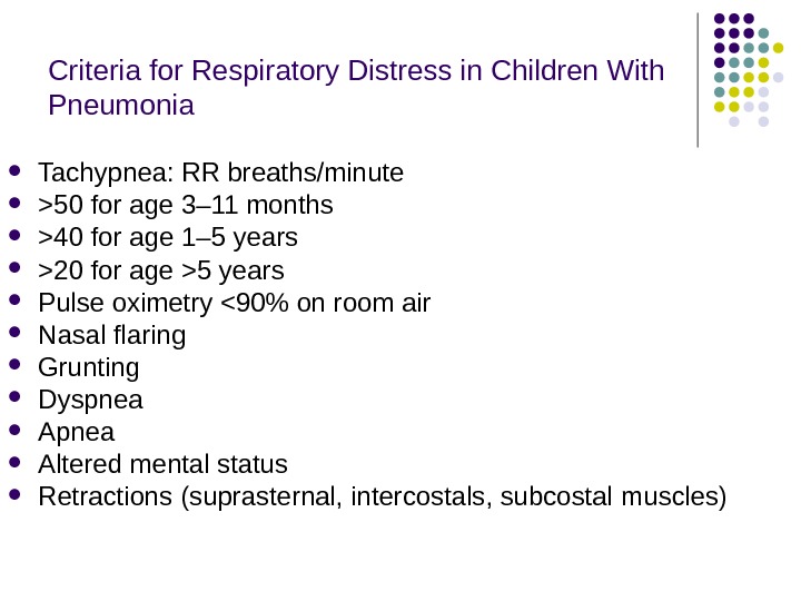
Criteria for Respiratory Distress in Children With Pneumonia Tachypnea: RR breaths/minute >50 for age 3– 11 months >40 for age 1– 5 years >20 for age >5 years Pulse oximetry <90% on room air Nasal flaring Grunting Dyspnea Altered mental status Retractions (suprasternal, intercostals, subcostal muscles)
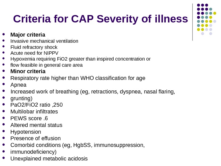
Criteria for CAP Severity of i llness Major criteria Invasive mechanical ventilation Fluid refractory shock Acute need for NIPPV Hypoxemia requiring Fi. O 2 greater than inspired concentration or flow feasible in general care area Minor criteria Respiratory rate higher than WHO classification for age Apnea Increased work of breathing (eg, retractions, dyspnea, nasal flaring, grunting) Pa. O 2/Fi. O 2 ratio , 250 Multilobar infiltrates PEWS score. 6 Altered mental status Hypotension Presence of effusion Comorbid conditions (eg, Hgb. SS, immunosuppression, immunodeficiency) Unexplained metabolic acidosis
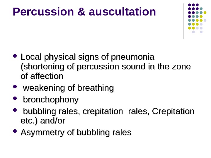
Percussion & auscultation Local physical signs of pneumonia (( shortening of percussion sound in the zone of affection weakening of breathing bronchophony bubbling rales, crepitation rales, Crepitation etc. ) and/or Asymmetry of bubbling rales
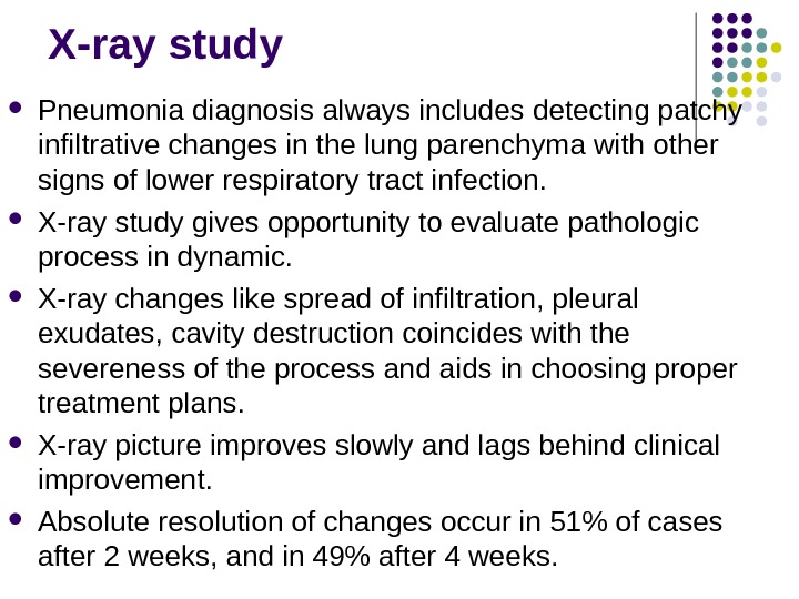
X-ray study Pneumonia diagnosis always includes detecting patchy infiltrative changes in the lung parenchyma with other signs of lower respiratory tract infection. X-ray study gives opportunity to evaluate pathologic process in dynamic. X-ray changes like spread of infiltration, pleural exudates, cavity destruction coincides with the severeness of the process and aids in choosing proper treatment plans. X-ray picture improves slowly and lags behind clinical improvement. Absolute resolution of changes occur in 51% of cases after 2 weeks, and in 49% after 4 weeks.
X-ray study used If the diagnosis is questionable This is repeated episode The patient is ill enough to be admitted The child is younger than 3 y. o. has Fever > 39 without a sourse Leucocytosis >15. 000 mm*3 A complicated pneumonia is suspected
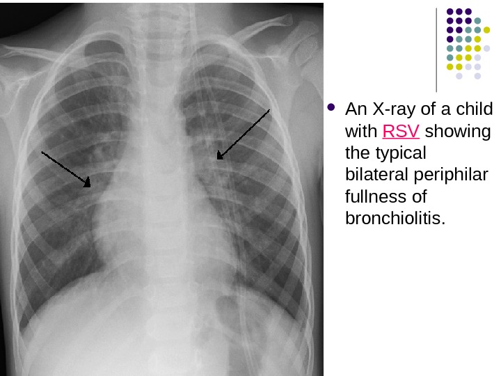
An X-ray of a child with RSV showing the typical bilateral periphilar fullness of bronchiolitis.
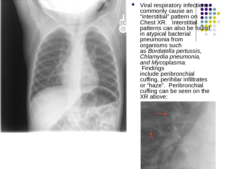
V iral respiratory infections commonly cause an “interstitial” pattern on Chest XR. Interstitial patterns can also be found in atypical bacterial pneumonia from organisms such as Bordatella pertussis, Chlamydia pneumonia, and Mycoplasma. Findings include peribronchial cuffing, perihilar infiltrates or “haze”. Peribronchial cuffing can be seen on the XR above:
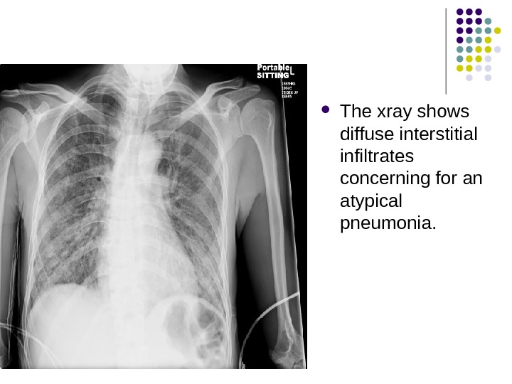
The xray shows diffuse interstitial infiltrates concerning for an atypical pneumonia.
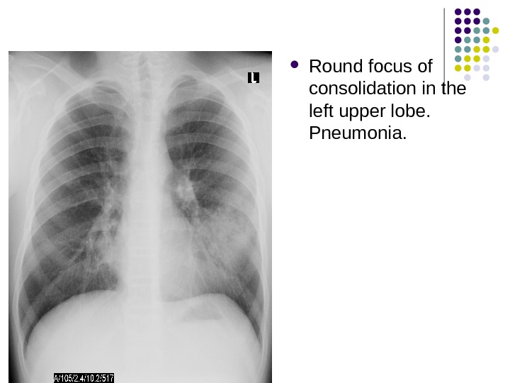
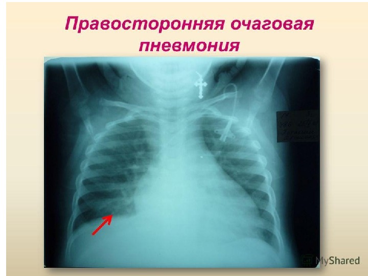
Round focus of consolidation in the left upper lobe. Pneumonia.
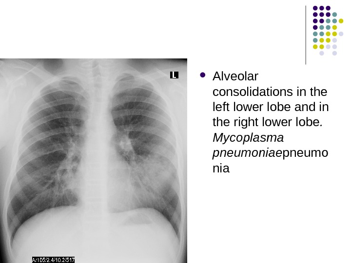
Alveolar consolidations in the left lower lobe and in the right lower lobe. Mycoplasma pneumoniae pneumo nia
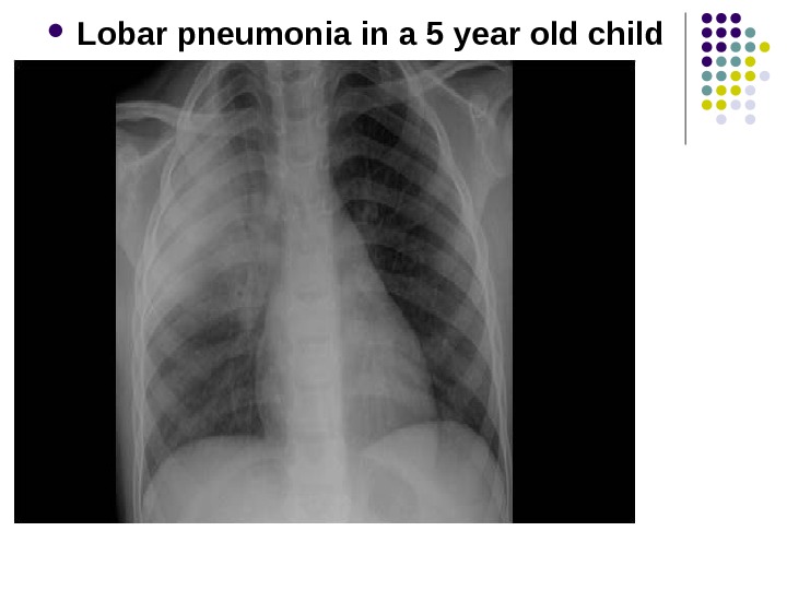
Lobar pneumonia in a 5 year old child
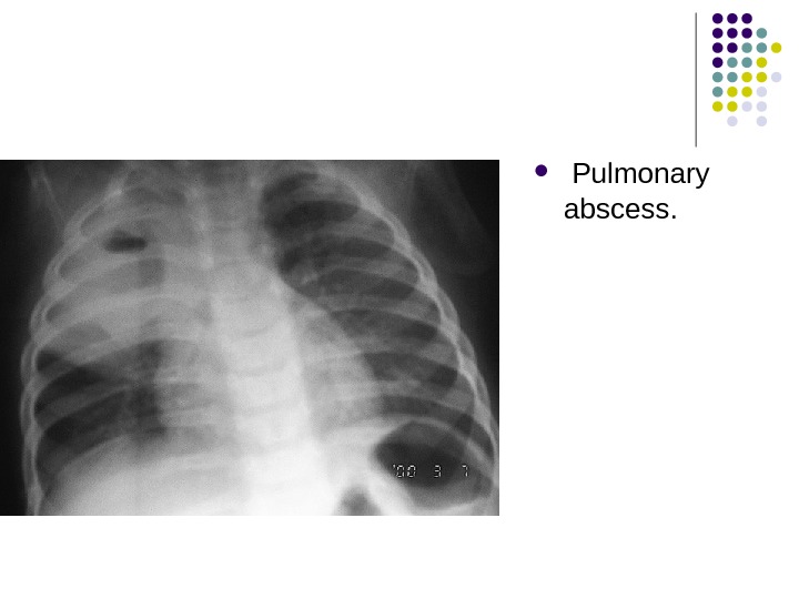
Pulmonary abscess.
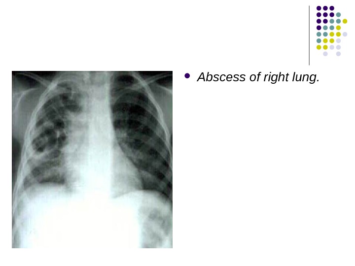
Abscess of right lung.
Sputum Gram Stain and Culture Sputum is rarely produced in children younger than 10 years, and samples are always contaminated by oral flora. In situations in which a microbiologic diagnosis is essential, endotracheal cultures and/or bronchoalveolar lavage culture can be sent for the isolation of offending pathogens. This is most important in patients with enigmatic and/or severe pneumonia, and it should be considered a priority in patients with compromised immune systems.
Rapid antigen tests are available for RSV, parainfluenza 1, 2, and 3, influenza A and B, and adenovirus. These assays, which are performed on specimens collected from the nasopharynx, can help determine the etiology of viral pneumonia Nasopharyngeal specimens for bacterial culture or antigen assays are less useful, because bacteria commonly colonize on the nasopharynx. Antigen and antibody assays for pneumococcal infection are not sensitive enough to be helpful in diagnosing S. pneumoniae infection. In the future, detection of pneumococcal immune complexes may offer a rapid etiologic diagnosis in children older than two years.
Serologic testing for Ig. M or an increase in Ig. G titers may be performed for Mycoplasma and Chlamydia species. However, serologic tests often provide only a retrospective diagnosis and are more useful in establishing the causative agent during an outbreak than in treating individual children. Cultures for Mycoplasma and Chlamydia are not routinely recommended. Polymerase chain reaction testing is not readily accessible, and positive results do not necessarily imply causality.
The complete blood count Complete Blood Cell Count may help in determining if an infection is bacterial (leukocytosis) or viral (leukopenia). In cases of pneumococcal pneumonia, the WBC count and ESR is often elevated
Acute-phase reactants erythrocytesedimentation rate (ESR) C-reactive protein (CRP)concentration serum procalcitonin concentration
Oxygen saturation should be assessed by pulse oximetry in children with respiratory distress, significant tachypnea, or pallor. Hypoxaemia is defined as the arterial oxygen saturation of less than 90% in room air at sea level as recorded by the pulse oximetry, which is the most serious Invasive diagnostic methods (bronchoscopy, trans-tracheal aspiration, transthoracic biopsy, etc) are carried out in hospital when Tuberculosis or bronchogenic cancer is suspecte
Classification of hypoxaemia There are two ways of classifying hypoxaemia in children: (i) WHO classification and (ii) British Thoracic Society (BTS) classification as defined below: (i) WHO classification of hypoxaemia Experts from WHO often classifies hypoxaemia as mild, moderate and severe as defined below: Mild hypoxaemia : when the arterial oxygen saturation lies between 85 to 90%, the patient is known to have mild hypoxaemia. Moderate hypoxaemia : when the arterial oxygen saturation lies between 80 to 85%, the patient is known to have moderate hypoxaemia. Severe hypoxaemia : when the arterial oxygen saturation is less than 80%, the patient is known to have severe hypoxaemia.
Clinical picture of focal pneumonia In children of pre-school and school age: Respiratory complaints, symptoms of intoxication, signs of respiratory insufficiency and local physical changes In infants: Signs of respiratory insufficiency and intoxication are dominant, and local physical changes in lungs appear later, process is more often bilateral Onset may be abrupt or gradual Course is favorable Duration depends upon etiology and reactivity o f the organism
Clinical picture of segmental pneumonia : : First variant: -course is favourable, sometimes they aren’t diagnosed because local changes are present only several days, respiratory insufficiency, intoxication and sometimes even cough are absent, and diagnosis is possible only with the help of X-ray. Probably it is segmental edema on the background of viral infections.
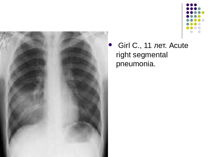
Girl С. , 11 лет. Acute right segmental pneumonia.
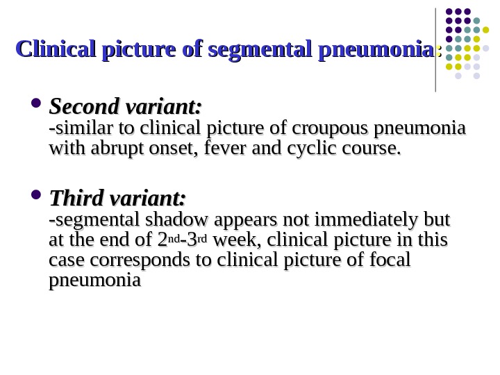
Clinical picture of segmental pneumonia : : Second variant: -similar to clinical picture of croupous pneumonia with abrupt onset, fever and cyclic course. Third variant: -segmental shadow appears not immediately but at the end of 2 ndnd -3 -3 rdrd week, clinical picture in this case corresponds to clinical picture of focal pneumonia
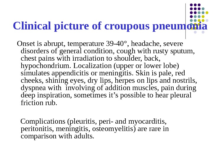
Clinical picture of croupous pneumonia Onset is abrupt, temperature 39 -40 ° , headache, severe disorders of general condition, cough with rusty sputum, chest pains with irradiation to shoulder, back, hypochondrium. Localization (upper or lower lobe) simulates appendicitis or meningitis. Skin is pale, red cheeks, shining eyes, dry lips, herpes on lips and nostrils, dyspnea with involving of addition muscles, pain during deep inspiration, sometimes it’s possible to hear pleural friction rub. Complications (pleuritis, peri- and myocarditis, peritonitis, meningitis, osteomyelitis) are rare in comparison with adults.
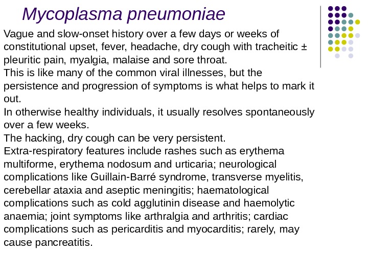
Mycoplasma pneumoniae Vague and slow-onset history over a few days or weeks of constitutional upset, fever, headache, dry cough with tracheitic ± pleuritic pain, myalgia, malaise and sore throat. This is like many of the common viral illnesses, but the persistence and progression of symptoms is what helps to mark it out. In otherwise healthy individuals, it usually resolves spontaneously over a few weeks. The hacking, dry cough can be very persistent. Extra-respiratory features include rashes such as erythema multiforme, erythema nodosum and urticaria; neurological complications like Guillain-Barré syndrome, transverse myelitis, cerebellar ataxia and aseptic meningitis; haematological complications such as cold agglutinin disease and haemolytic anaemia; joint symptoms like arthralgia and arthritis; cardiac complications such as pericarditis and myocarditis; rarely, may cause pancreatitis.
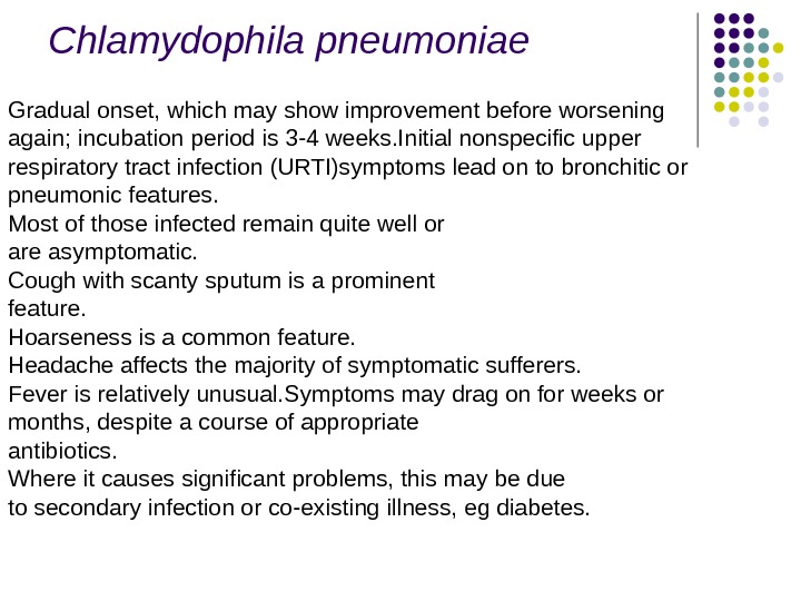
Chlamydophila pneumoniae Gradual onset, which may show improvement before worsening again; incubation period is 3 -4 weeks. Initial nonspecific upper respiratory tract infection (URTI)symptoms lead on to bronchitic or pneumonic features. Most of those infected remain quite well or are asymptomatic. Cough with scanty sputum is a prominent feature. Hoarseness is a common feature. Headache affects the majority of symptomatic sufferers. Fever is relatively unusual. Symptoms may drag on for weeks or months, despite a course of appropriate antibiotics. Where it causes significant problems, this may be due to secondary infection or co-existing illness, eg diabetes.
Legionella pneumophila This tends to be the most severe of the pneumonias due to atypical pathogens. Focal outbreaks centred around poorly maintained air-conditioning or humidification systems (although this is often noted retrospectively by public health physicians). 2 -10 days’ incubation period. Initial mild headache and myalgia leading to high fever, chills and repeated rigors; non-chest symptoms often predominate early on Cough is nearly always present, initially unproductive, but may lead to expectoration later. Dyspnoea, pleuritic pain and haemoptysis are not uncommon. Gastrointestinal upset, such as diarrhoea, nausea and vomiting or loss of appetite/anorexia, may occur. There may be neurological complications such as confusion, disorientation and focal neurological deficit. Arthralgia and myalgia are often reported. Severe complications include pancreatitis, peritonitis, pericarditis, myocarditis, endocarditis and glomerulonephritis.
Hospital-acquired pneumonia This is defined as a new infection of lung parenchyma appearing more than 48 hours after admission to the hospital. It occurs mostly in patients who are severely debilitated, immunocompromised or mechanically ventilated. Infection occurring during the first four days of the hospital stay is usually caused by S. pneumoniae , H. influenzae and Moraxella catarrhalis. Onset more than four days after admission is more often caused by Gram-negative enterobacteria, S. aureus or L. pneumophila. Hospital-acquired pneumonia is often caused by multiple organisms.
DIFFERENTAIL DIAGNOSIS OF THE PNEUMONIA Asthma Inhaled foreign body Pneumothorax Cardiac dyspnoea Pneumonitis from other causes: Extrinsic allergic alveolitis Smoke inhalation Gastro-oesophageal reflux Cronical broncho-pulmonary diseases , acute stage
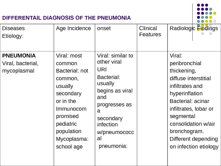
Diseases Etiology: Age Incidence onset Clinical Features Radiologic Findings PNEUMONIA Viral, bacterial, mycoplasmal Viral: most common Bacterial: not common, usually secondary or in the Immunocom promised pediatric population Mycoplasma: school age Viral: similar to other viral URI Bacterial: usually begins as viral and progresses as a secondary infection w/pneumococc al pneumonia: Viral: peribronchial thickening, diffuse interstitial infiltrates and hyperinflation Bacterial: acinar infiltrates, lobar or segmental consolidation w/air bronchogram. Different depending on infection etiology DIFFERENTAIL DIAGNOSIS OF THE PNEUMONI
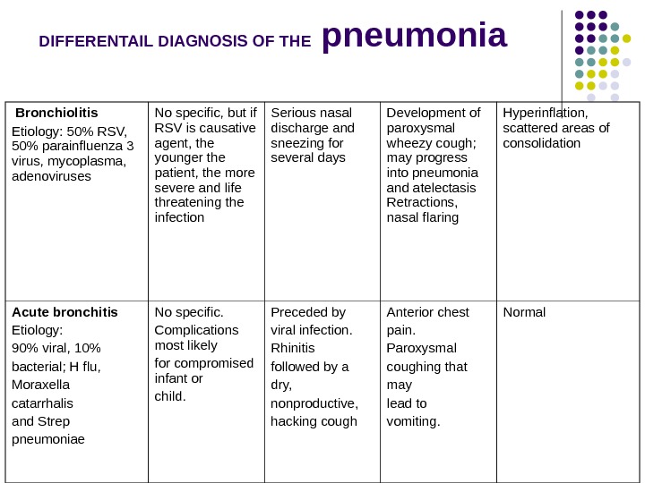
DIFFERENTAIL DIAGNOSIS OF THE pneumonia Bronchiolitis Etiology: 50% RSV, 50% parainfluenza 3 virus, mycoplasma, adenoviruses No specific, but if RSV is causative agent, the younger the patient, the more severe and life threatening the infection Serious nasal discharge and sneezing for several days Development of paroxysmal wheezy cough; may progress into pneumonia and atelectasis Retractions, nasal flaring Hyperinflation, scattered areas of consolidation Acute bronchitis Etiology: 90% viral, 10% bacterial; H flu, Moraxella catarrhalis and Strep pneumoniae No specific. Complications most likely for compromised infant or child. Preceded by viral infection. Rhinitis followed by a dry, nonproductive, hacking cough Anterior chest pain. Paroxysmal coughing that may lead to vomiting. Normal
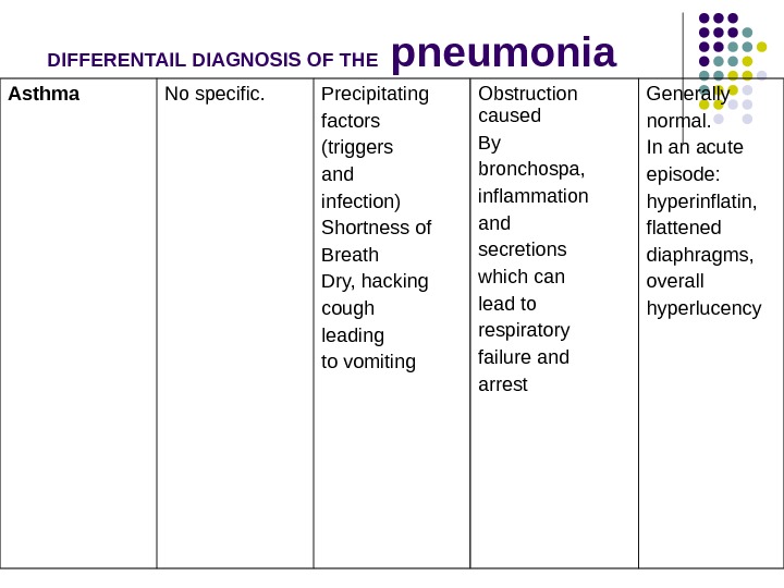
DIFFERENTAIL DIAGNOSIS OF THE pneumonia Asthma No specific. Precipitating factors (triggers and infection) Shortness of Breath Dry, hacking cough leading to vomiting Obstruction caused By bronchospa, inflammation and secretions which can lead to respiratory failure and arrest Generally normal. In an acute episode: hyperinflatin, flattened diaphragms, overall hyperlucency
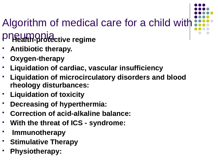
Algorithm of medical care for a child with pneumonia Health-protective regime Antibiotic therapy. Oxygen-therapy Liquidation of cardiac, vascular insufficiency Liquidation of microcirculatory disorders and blood rheology disturbances: Liquidation of toxicity Decreasing of hyperthermia: Correction of acid-alkaline balance: With the threat of ICS — syndrome: Immunotherapy Stimulative Therapy Physiotherapy:
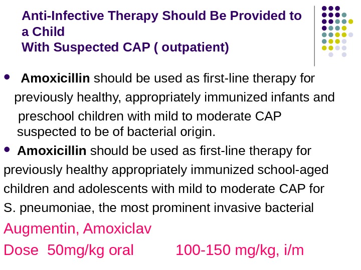
Anti-Infective Therapy Should Be Provided to a Child With Suspected CAP ( outpatient) Amoxicillin should be used as first-line therapy for previously healthy, appropriately immunized infants and preschool children with mild to moderate CAP suspected to be of bacterial origin. Amoxicillin should be used as first-line therapy for previously healthy appropriately immunized school-aged children and adolescents with mild to moderate CAP for S. pneumoniae, the most prominent invasive bacterial Augmentin, Amoxiclav Dose 50 mg/kg oral 100 -150 mg/kg, i/m
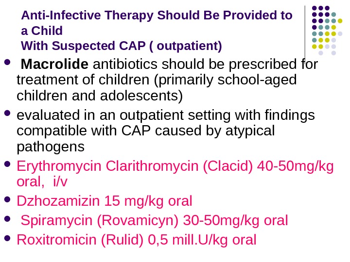
Anti-Infective Therapy Should Be Provided to a Child With Suspected CAP ( outpatient) Macrolide antibiotics should be prescribed for treatment of children (primarily school-aged children and adolescents) evaluated in an outpatient setting with findings compatible with CAP caused by atypical pathogens Erythromycin Clarithromycin (Clacid) 40 -50 mg/kg oral, i/v Dzhozamizin 15 mg/kg oral Spiramycin (Rovamicyn) 30 -50 mg/kg oral Roxitromicin (Rulid) 0, 5 mill. U/kg oral
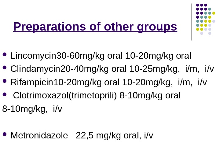
Preparations of other groups Lincomycin 30 -60 mg/kg oral 10 -20 mg/kg oral Clindamycin 20 -40 mg/kg oral 10 — 25 mg/kg, i/m, i/v Rifampicin 10 -20 mg/kg oral 10 -20 mg/kg, i/m, i/v Clotrimoxazol (trimetoprili) 8 -10 mg/kg oral 8 -10 mg/kg, i/v Metronidazole 22, 5 mg/kg oral, i/v
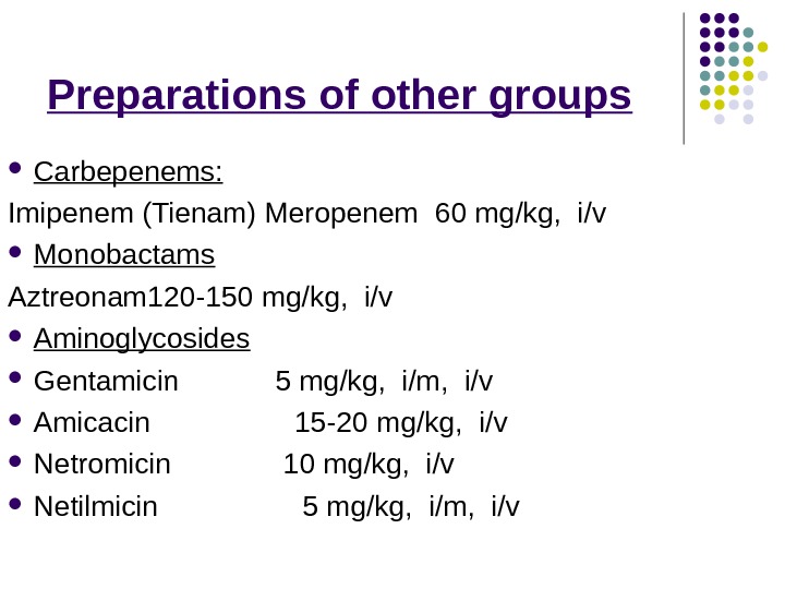
Preparations of other groups Carbepenems: Imipenem (Tienam) Meropenem 60 mg/kg, i/v Monobactams Aztreonam 120 -150 mg/kg, i/v Aminoglycosides Gentamicin 5 mg/kg, i/m, i/v Amicacin 15 -20 mg/kg, i/v Netromicin 10 mg/kg, i/v Netilmicin 5 mg/kg, i/m, i/v
Anti-Infective Therapy Should Be Provided to a Child With Suspected CAP ( outpatient) Influenza antiviral therapy should be administered as soon as possible to children with moderate to severe CAP consistent with influenza virus Infection during widespread local circulation of influenza viruses, particularly for those with clinically worsening disease documented at the time of an outpatient visit. Because early antiviral treatment has been shown to provide maximal benefit, treatment should not be delayed until confirmation of positive influenza test results
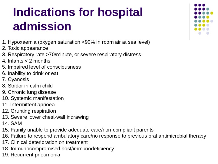
Indications for hospital admission 1. Hypoxaemia (oxygen saturation 70/minute, or severe respiratory distress 4. Infants < 2 months 5. Impaired level of consciousness 6. Inability to drink or eat 7. Cyanosis 8. Stridor in calm child 9. Chronic lung disease 10. Systemic manifestation 11. Intermittent apnoea 12. Grunting respiration 13. Severe lower chest-wall indrawing 14. SAM 15. Family unable to provide adequate care/non-compliant parents 16. Failure to respond ambulatory care/no response to previous oral antimicrobial therapy 17. Clinical deterioration on treatment 18. Immunocompromised host/immunodeficiency 19. Recurrent pneumonia
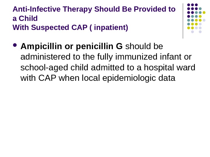
Anti-Infective Therapy Should Be Provided to a Child With Suspected CAP ( inpatient) Ampicillin or penicillin G should be administered to the fully immunized infant or school-aged child admitted to a hospital ward with CAP when local epidemiologic data
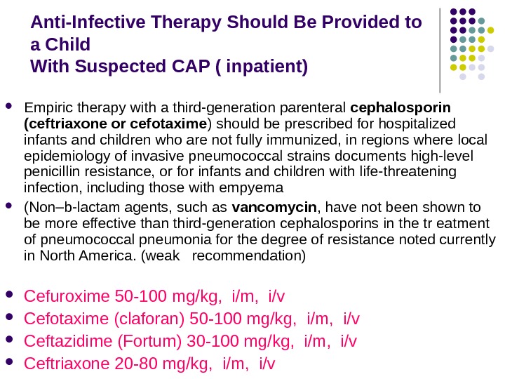
Anti-Infective Therapy Should Be Provided to a Child With Suspected CAP ( inpatient) Empiric therapy with a third-generation parenteral cephalosporin (ceftriaxone or cefotaxime ) should be prescribed for hospitalized infants and children who are not fully immunized, in regions where local epidemiology of invasive pneumococcal strains documents high-level penicillin resistance, or for infants and children with life-threatening infection, including those with empyema (Non–b-lactam agents, such as vancomycin , have not been shown to be more effective than third-generation cephalosporins in the tr eatment of pneumococcal pneumonia for the degree of resistance noted currently in North America. (weak recommendation) Cefuroxime 50 -100 mg/kg, i/m, i/v Cefotaxime (claforan) 50 -100 mg/kg, i/m, i/v Ceftazidime (Fortum) 30 -100 mg/kg, i/m, i/v Ceftriaxone 20 -80 mg/kg, i/m, i/v
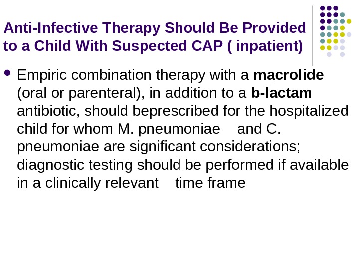
Anti-Infective Therapy Should Be Provided to a Child With Suspected CAP ( inpatient) Empiric combination therapy with a macrolide (oral or parenteral), in addition to a b-lactam antibiotic, should beprescribed for the hospitalized child for whom M. pneumoniae and C. pneumoniae are significant considerations; diagnostic testing should be performed if available in a clinically relevant time frame
Anti-Infective Therapy Should Be Provided to a Child With Suspected CAP ( inpatient) Vancomycin or clindamycin (based on local susceptibility data) should be provided in addition to b-lactam therapy iclinical, laboratory, or imaging characteristics are consistent with infection caused by S. aureus
Management of atypical pneumonia Macrolides , such as erythromycin, clarithromycin and azithromycin , have been shown to be effective in the treatment of all three most common infective organisms. Resistance to macrolides is a growing concern. Severe legionella infections may require rifampicin as well as a macrolide. Tetracycline, doxycycline and fluoroquinolones are also effective against all three of the common infective organisms. Fluoroquinolones Ciprofloxacin (Ciprobay, Ciprinol) 15 mg/kg oral 10 mg/kg, i/v Oflaxacin (Tarivid) 7, 5 mg/kg oral 5 mg/kg, i/v
Indications for oxygen therapy 1. Hypoxaemia (oxygen saturation 70 breaths/min 8. Head Nodding
oxygen therapy a) To ensure free airway, optimization of ventilation (throwing head back, the output of the lower jaw forward — to prevent the retraction of the tongue) b) the removal of mucus from the nasopharynx, large bronchi – the stimulation of cough, aspiration of mucus, the appointment of stimulants, for thinning the phlegm (Bromhexine, acetylcysteine, mixtures based on the herbas), vibrating massage with postur al drainage Euphyllin i/v 2, 4% 0, 1 ml/ kg for child till 1 y. o. 1 ml / 1 yr. of life after 1 y. o. Inhalation warm humidity.
Methods of oxygen administration Nasal prongs : are recommended for most children. Nasal prongs give a maximum fractional concentration of inspired oxygen (F 1 O 2) of 28 -35% except in small infants when higher concentrations may be obtained. This method does not require humidification of oxygen and ensures that the child receives oxygen during feeding. Oxygen flow rates of 0. 5 -1 l/minute are required in children less than 2 months old and 2 -3 l/minute in infants and children aged 2 months to 5 years. Nasal catheters : are usually well tolerated and humidification is not required, but they can be blocked by mucous. Oxygen via nasal catheters gives a maximum F 1 O 2 of 35 -40%. Nasopharyngeal catheters : have the advantage of requiring the lowest flow rate to achieve a given oxygen concentration in the airways. Infants under the age of 2 months can usually be treated with 0. 5 minute and infants up to 1 year with 1 minute. However, humidification of oxygen is required and the catheter may be easily blocked. Further, potentiallylethal complications including gastric distension, airway obstruction, apnoea, pneumo-orbitus and pneumocephalus may occur. Continuous skilled nursing is therefore necessary to prevent these complications. Consequently, oxygen administration via nasopharyngeal catheter is not recommended.
Methods of oxygen administration Headbox: oxygen is well tolerated by young infants. Headbox oxygen requires no humidification but requires a high flow and a mixing device to ensure the correct F 1 O 2 is delivered. This is the least preferred method as there is wastage of oxygen and delivered F 1 O 2 is unpredictable. Facemask : oxygen is designed to deliver 28%-65% oxygen at a flow rate of 6 -10 minutes. Polymask : In severely hypoxaemic infants who are not ventilated, oxygen should be administered using a polymask whereby F 1 O 2 concentrations of 60 -80% may be achieved. The flow rate should be regulated to keep the bag of the mask inflated during inspiration and expiration. Using the prone position for infants may improve hypoxaemia and the respiratory system compliance (Chaisupamongkollarp et al. , 1999) and should be attempted if hypoxaemia is difficult to treat. Oxygen should be discontinued when the child is improving and the transcutanous saturation is above 90% in room air, as recorded by the pulse oximetry.
Antipyretics and analgesics drugs Children with CAP are generally pyrexial and may also have some pain, including headache, chest pain, arthralgia (in cases of Mycoplasma pneumoniae), referred abdominal pain, and possibly earache from associated otitis media. Pleural pain may interfere with the depth of breathing and may impair the ability of the child to cough. Antipyretics and analgesics can be used to keep the child comfortable and to help coughing. Minimal handling helps to reduce metabolic and oxygen requirements and this should be considered when planning and carrying out procedures, investigations, and treatments. Pain associated with pneumonia may be due to pleurisy or to pathology involving the upper airways. Pain or discomfort should be treated as it may severely compromise respiratory function and adequate clearance of secretions
Indications for the use of antipyretics and analgesics in CAP Rectal temperature >39 Celsius There is a known risk of febrile convulsions There is central nervous system pathology that may be aggravated by high fever
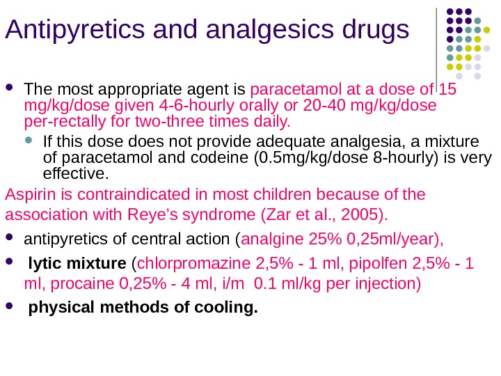
Antipyretics and analgesics drugs The most appropriate agent is paracetamol at a dose of 15 mg/kg/dose given 4 -6 -hourly orally or 20 -40 mg/kg/dose per-rectally for two-three times daily. If this dose does not provide adequate analgesia, a mixture of paracetamol and codeine (0. 5 mg/kg/dose 8 -hourly) is very effective. Aspirin is contraindicated in most children because of the association with Reye’s syndrome (Zar et al. , 2005). antipyretics of central action ( analgine 25% 0, 25 ml/year), lytic mixture ( chlorpromazine 2, 5% — 1 ml, pipolfen 2, 5% — 1 ml, procaine 0, 25% — 4 ml, i/m 0. 1 ml/kg per injection) physical methods of cooling.
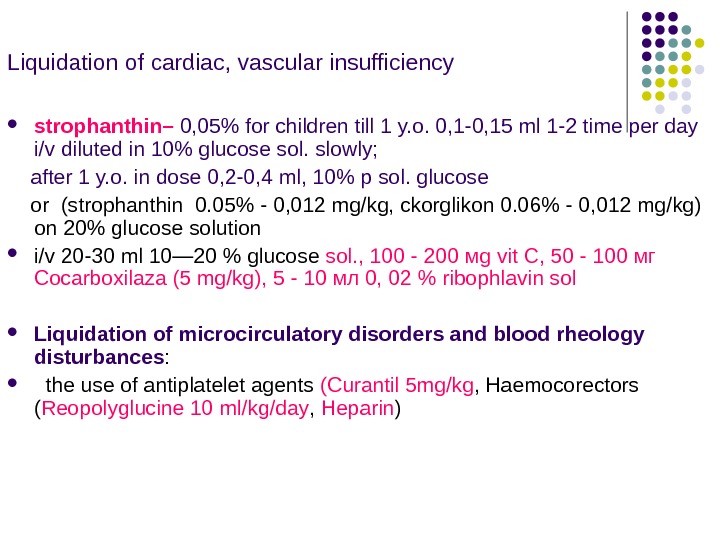
Liquidation of cardiac, vascular insufficiency strophanthin – 0, 05% for children till 1 y. o. 0, 1 -0, 15 ml 1 -2 time per day i/v diluted in 10% glucose sol. slowly ; after 1 y. o. in dose 0, 2 -0, 4 ml , 10% р sol. glucose or (strophanthin 0. 05% — 0, 012 mg/kg, ckorglikon 0. 06% — 0, 012 mg/kg) on 20% glucose solution i/v 20 -30 ml 10— 20 % glucose sol. , 100 — 200 м g vit C , 50 — 100 мг Cocarboxilaza (5 mg/kg) , 5 — 10 мл 0, 02 % ribophlavin sol Liquidation of microcirculatory disorders and blood rheology disturbances : the use of antiplatelet agents (Curantil 5 mg/kg , Haemocorectors ( Reopolyglucine 10 ml/kg/day , Heparin )
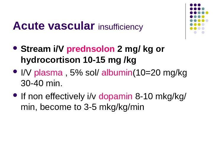
Acute vascular insufficiency Stream i/V prednsolon 2 mg/ kg or hydrocortison 10 -15 mg /kg I/V plasma , 5% sol/ albumin (10=20 mg/kg 30 -40 min. If non effectively i/v dopamin 8 -10 mkg/kg/ min, become to 3 -5 mkg/kg/min
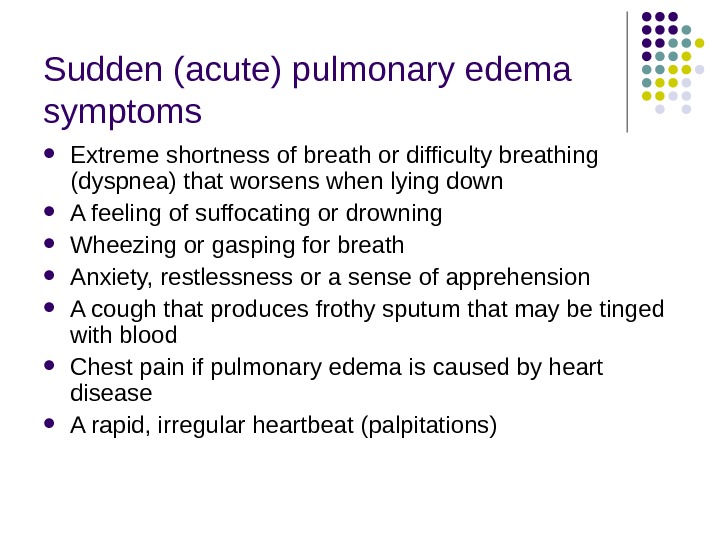
Sudden (acute) pulmonary edema symptoms Extreme shortness of breath or difficulty breathing (dyspnea) that worsens when lying down A feeling of suffocating or drowning Wheezing or gasping for breath Anxiety, restlessness or a sense of apprehension A cough that produces frothy sputum that may be tinged with blood Chest pain if pulmonary edema is caused by heart disease A rapid, irregular heartbeat (palpitations)
Prevention of lung edema oxygen therapy use antifoam drugs ( inhalation 30 % C 2 H 5 OH 30 — 40 min , Antifomsilan keep free airway diuretic drugs Furosemid i/v 2 -3 mg/kg
Anticonvulsion therapy Deacresing hypoxia and Deacresing edema of brain Furosemid i/v 2 -3 mg/kg Deacresing excitability – 0, 5 % sol seduxen 0, 5 -0. 1 ml/kg Fenobarbital i/v or i/m initial dose 20 mg/kg, following dose 3 -4 mg/kg daily 25% sol. Mg. SO 4 0, 2 ml/kg on 1 injection For increasing effect 0, 25% sol. droperidol o, 1 ml/kg Lumbal punction
Liquidation of toxicity: albumin , plasma, Haemodesum 5 -10 ml/kg/day. Correction of acid-alkaline balance : 4% solution of sodium carbonate (3. 5 ml/kg in 2 -3 reception With the threat of ICS — syndrome : heparin 200 -250 U/kg/day in the stage of hypercoagulation, 50 -100 U/kg/day in stage of hypocoagulation. Immunotherapy of directed action (at Staphilicoccal, Proteus, Pseudomonaspneumonia): hyperimmune plasma 5 -15 ml/kg, immunoglobulin 100 IU N 3 -5 Stimulative Therapy : adaptogens of plant origin — Eleutherococcus, Ginseng echinacea, medicine – pentoxyl, dibasol, metacil in combination with vitamins.
Intravenous fluids must be used with great care and with caution, and only if adequate monitoring is available. Children who are vomiting or who are severely ill may require intravenous fluids. These should be given at 80% of the basal levels (once hypovolaemia has been corrected). In children with severe or complicated pneumonia, serum urea and electrolytes should be measured before instituting I/V fluids as among them (SIADH) is common. In these children, fluid intake should be restricted to 40 -60% of normal requirements, i. e. 50 ml/kg/day of I/V fluids. They should be frequently monitored as severely ill children with CAP might develop SIADH as a recognized complication
Indications for I/V fluid Shock Inability to tolerate enteral feeds Sepsis Severe dehydration Gross electrolyte imbalance Hypoglycaemia
Calorie requirements Adequate nutrition is of particular concern, especially when there are underlying factors such as malnutrition. A minimum of 50 -60 kcal/kg/day should be given to a child with pneumonia with continuation of regular breast feeding for breast-fed children. A calorie intake of 80 -100 kcal/kg/day should be given to a non-breast fed child with CAP. Ensuring adequate calorie intake is essential as there is an excessive demand on the energy reserves in children with pneumonia, in whom the work of breathing is increased. Children should not be starved for more than 24 hours to prevent the development of hypoglycaemia. In the presence of malnutrition, and following several days of poor nutrition, this needs to be increased considerably. In the early phase of pneumonia, ketosis should be avoided by ensuring adequate carbohydrate intake. With time, a greater proportion of intake can be lipids. The intake of calories should be adequate to meet the metabolic requirements and to promote growth.
Enteral feeds Children with pneumonia should be encouraged to feed orally unless there are indications for nasogastric feeding/intravenous fluid infusions. If children are too distressed to take fluid and feeds orally, continuous enteral feeds via a nasogastric tube may be provided. Indications for N/G tube feeding Too distressed to drink or swallow safely Having frequent severe coughing episodes that may be associated with vomiting and possible aspiration of gastric contents Hypovolaemia with associated poor peripheral perfusion (may even require I/V fluid) Painful oral sore/condition which interfere with feeding by mouth
Chest physiotherapy postural drainage, percussion of the chest deep breathing exercises should be routinely performed in children with uncomplicated CAP
Apparatus physiotherapy during the acute clinical manifestations of acute pneumonia is contrindicated. With the normalization of temperature, the elimination of respiratory and cardiovascular failure may be prescribed diathermia (UHF, M HF, LHF ), in the period of convalescence – electrophoresis (with dionini, calcium, vitamin C), UVT. ozokerite applications on the abdomen (especially the liver) in the period of the regression of disease subsided (20 min, 40°C).
Mucolytic agents Anti-tussive remedies are not recommended as they cause suppression of cough and interfere with airway clearance. Adverse effects and overdose have been reported. Therefore, they should not be advised in children with CAP.
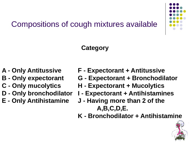
Compositions of cough mixtures available Category A — Only Antitussive F — Expectorant + Antitussive B — Only expectorant G — Expectorant + Bronchodilator C — Only mucolytics H — Expectorant + Mucolytics D — Only bronchodilator I — Expectorant + Antihistamines E — Only Antihistamine J — Having more than 2 of the A, B, C, D, E. K — Bronchodilator + Antihistamine
Postural drainage: There is no evidence for the use of a head-down position for postural drainage. Nebulized bronchodilators: Nebulized bronchodilators or saline do not improve the outcome of CAP. Cortocosteroids: There is no evidence to support the use of oral or inhaled corticosteroids in CAP.
Electrophoresis
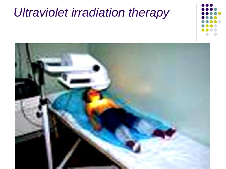
Ultraviolet irradiation therapy
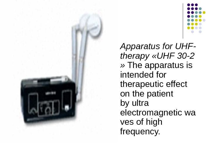
Apparatus for UHF — therapy « UHF 30 -2 » The apparatus is intended for therapeutic effect on the patient by ultra electromagnetic wa ves of high frequency.
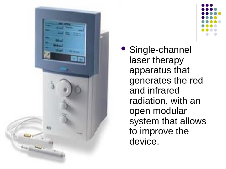
Single-channel laser therapy apparatus that generates the red and infrared radiation, with an open modular system that allows to improve the device.
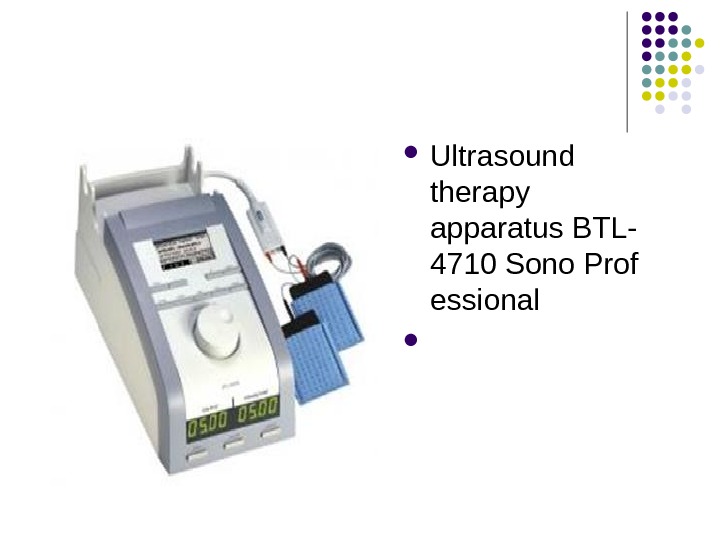
Ultrasound therapy apparatus BTL- 4710 Sono Prof essional
Complication of pneumonia
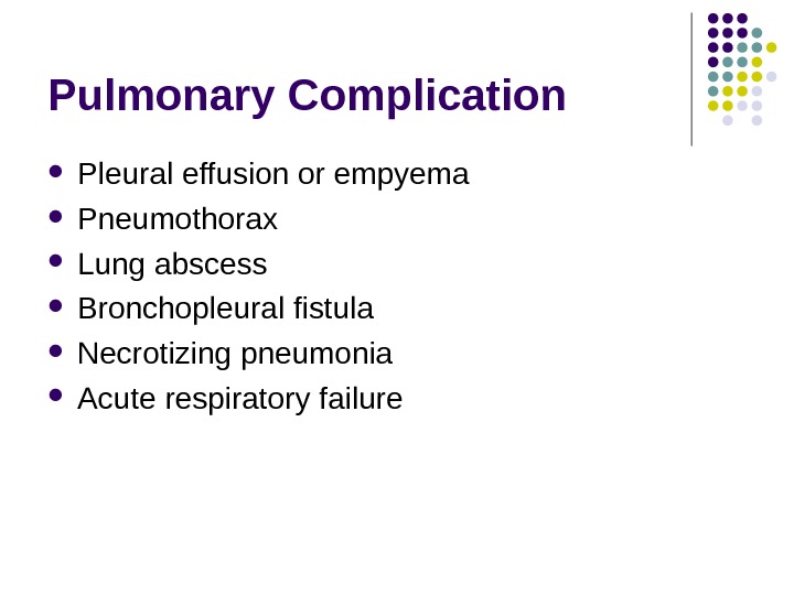
Pulmonary Complication Pleural effusion or empyema Pneumothorax Lung abscess Bronchopleural fistula Necrotizing pneumonia Acute respiratory failure
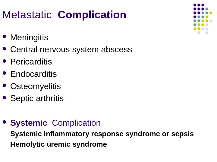
Metastatic Complication Meningitis Central nervous system abscess Pericarditis Endocarditis Osteomyelitis Septic arthritis Systemic Complication Systemic inflammatory response syndrome or sepsis Hemolytic uremic syndrome
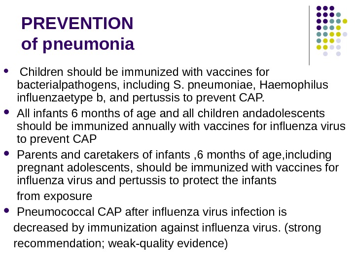
PREVENTION of pneumonia Children should be immunized with vaccines for bacterialpathogens, including S. pneumoniae, Haemophilus influenzaetype b, and pertussis to prevent CAP. All infants 6 months of age and all children andadolescents should be immunized annually with vaccines for influenza virus to prevent CAP Parents and caretakers of infants , 6 months of age, including pregnant adolescents, should be immunized with vaccines for influenza virus and pertussis to protect the infants from exposure Pneumococcal CAP after influenza virus infection is decreased by immunization against influenza virus. (strong recommendation; weak-quality evidence)