
PEPTIC ULCER DISEASE Lecture by MD Galina Pobedyonnaya


- Размер: 939 Кб
- Количество слайдов: 53
Описание презентации PEPTIC ULCER DISEASE Lecture by MD Galina Pobedyonnaya по слайдам
PEPTIC ULCER DISEASE Lecture by MD Galina Pobedyonnaya
• History of epigastric pain present in 80 -90 % of patient, but is nonspecific. • Burning epigastric pain exacerbated by fasting and improved with meals is a symptom complex associated with peptic ulcer disease (PUD). • Acid peptic disorders are very common in the United States, with 4 million individuals (new cases and recurrences) affected per year. Lifetime prevalence of PUD in the United States is ~12% in men and 10% in women. Moreover, an estimated 15, 000 deaths per year occur as a consequence of complicated PUD. The financial impact of these common disorders has been substantial, with an estimated burden on direct and indirect health care costs of ~$10 billion per year in the United States. • Why does the ulcer appear ? Let remember the physiology of gastric s ecretion.
PEPTIC ULCER DISEASE • The term ‘peptic ulcer’ refers to an ulcer in the lower oesophagus, stomach or duodenum, in the jejunum after surgical anastomosis to the stomach, or, rarely, in the ileum adjacent to a Meckel’s diverticulum. • Ulcers in the stomach or duodenum may be acute or chronic; both penetrate the muscularis mucosae but the acute ulcer shows no evidence of fibrosis. Erosions do not penetrate the muscularis mucosae.
• Ulcers are defined as a break in the mucosal surface >5 mm in size, with depth to the submucosa.
Why does the ulcer appear ? Let’s remember the physiology of gastric s ecretion. • Hydrochloric acid and pepsinogen are the two principal gastric secretory products capable of inducing mucosal injury. Acid secretion should be viewed as occurring under basal and stimulated conditions. Basal acid production occurs in a circadian pattern, with highest levels occurring during the night and lowest levels during the morning hours. Cholinergic input via the vagus nerve and histaminergic input from local gastric sources are the principal contributors to basal acid secretion. Stimulated gastric acid secretion occurs primarily in three phases based on the site where the signal originates (cephalic, gastric, and intestinal). • — Sight, smell, and taste of food are the components of the cephalic phase , which stimulates gastric secretion via the vagus nerve. • — The gastric phase is activated once food enters the stomach. This component of secretion is driven by nutrients (amino acids and amines) that directly stimulate the G cell to release gastrin, which in turn activates the parietal cell via direct and indirect mechanisms. Distention of the stomach wall also leads to gastrin release and acid production.
Control of acid secretion
• The gastric epithelium is under a constant assault by a series of endogenous noxious factors including HCl, pepsinogen/pepsin, and bile salts. In addition, a steady flow of exogenous substances such as medications, alcohol, and bacteria encounter the gastric mucosa. A highly intricate biologic system is in place to provide defense from mucosal injury and to repair any injury that may occur.
• The mucosal defense system can be envisioned as a three-level barrier, composed of preepithelial, and subepithelial elements. The first line of defense is a mucus-bicarbonate layer, which serves as a physicochemical barrier to multiple molecules including hydrogen ions. Mucus is secreted in a regulated fashion by gastroduodenal surface epithelial cells. It consists primarily of water (95%) and a mixture of lipids and glycoproteins. Mucin is the constituent glycoprotein that, in combination with phospholipids (also secreted by gastric mucous cells), forms a hydrophobic surface with fatty acids that extend into the lumen from the cell membrane. The mucous gel functions as a nonstirred water layer impeding diffusion of ions and molecules such as pepsin. Bicarbonate, secreted by surface epithelial cells of the gastroduodenal mucosa into the mucous gel, forms a p. H gradient ranging from 1 to 2 at the gastric luminal surface and reaching 6 to 7 along the epithelial cell surface. Bicarbonate secretion is stimulated by calcium, prostaglandins, cholinergic input, and luminal acidification.
• Surface epithelial cells provide the next line of defense through several factors, including mucus production, epithelial cell ionic transporters that maintain intracellular p. H and bicarbonate production, and intracellular tight junctions. If the preepithelial barrier were breached, gastric epithelial cells bordering a site of injury can migrate to restore a damaged region (restitution). This process occurs independent of cell division and requires uninterrupted blood flow and an alkaline p. H in the surrounding environment. Several growth factors including epidermal growth factor (EGF), transforming growth factor (TGF) α , and basic fibroblast growth factor (FGF) modulate the process of restitution.
• Prostaglandins play a central role in gastric epithelial defense/repair. The gastric mucosa contains abundant levels of prostaglandins. These metabolites of arachidonic acid regulate the release of mucosal bicarbonate and mucus, inhibit parietal cell secretion, and are important in maintaining mucosal blood flow and epithelial cell restitution. • An ulcer forms when there is an imbalance between aggressive factors, i. e. the digestive power of acid and pepsin, and defensive factors, i. e. the ability of the gastric and duodenal mucosa to resist this digestive power. This mucosal resistance constitutes the gastric mucosal barrier.
Gastroduodenal mucosal protection
GASTRIC AND DUODENAL ULCER • Although the prevalence of peptic ulcer is decreasing in many Western communities, it still affects approximately 10% of all adults at some time in their lives. The male to female ratio for DU varies from 5: 1 to 2: 1, whilst that for GU is 2: 1 or less.
E tiology : Helicobacter pylori • In the industrialised world the prevalence of H. pylori infection in the general population rises steadily with age, and in the UK approximately 50% of those over the age of 50 years are infected. In many parts of the underdeveloped world infection is much more common and is often acquired in childhood. Up to 90% of the population are infected by adult life in some countries. The vast majority of colonised people remain healthy and asymptomatic and only a minority develop clinical disease. Around 90% of duodenal ulcer patients and 70% of gastric ulcer patients are infected with H. pylori ; the remaining 30% of gastric ulcers are due to NSAIDs.
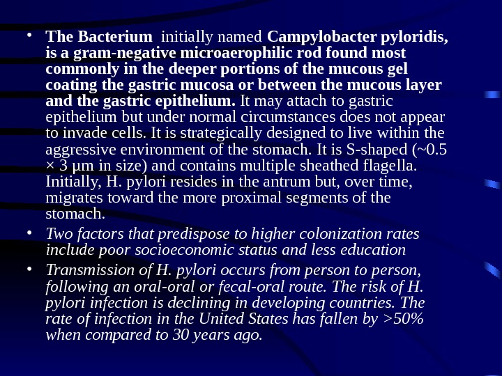
• The Bacterium initially named Campylobacter pyloridis, is a gram-negative microaerophilic rod found most commonly in the deeper portions of the mucous gel coating the gastric mucosa or between the mucous layer and the gastric epithelium. It may attach to gastric epithelium but under normal circumstances does not appear to invade cells. It is strategically designed to live within the aggressive environment of the stomach. It is S-shaped (~0. 5 × 3 µm in size) and contains multiple sheathed flagella. Initially, H. pylori resides in the antrum but, over time, migrates toward the more proximal segments of the stomach. • Two factors that predispose to higher colonization rates include poor socioeconomic status and less education • Transmission of H. pylori occurs from person to person, following an oral-oral or fecal-oral route. The risk of H. pylori infection is declining in developing countries. The rate of infection in the United States has fallen by >50% when compared to 30 years ago.
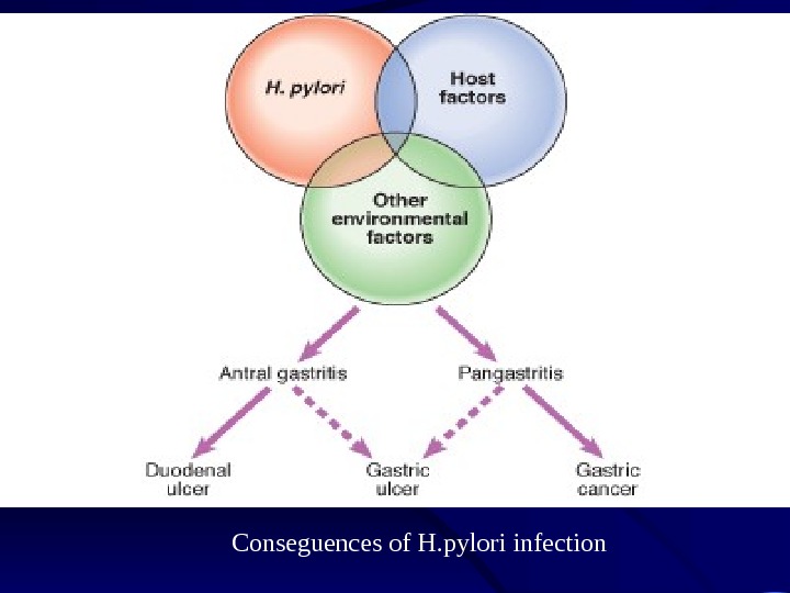
Conseguences of H. pylori infection
Pathogenesis and pathophysiology of infection
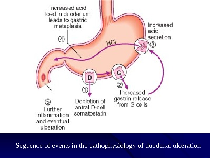
In most people H. pylori causes antral gastritis associated with depletion of somatostatin (from D cells) and gastrin release from G cells. The subsequent hypergastrinaemia stimulates acid production by parietal cells, but in the majority of cases this has no clinical consequences. In a minority of patients (perhaps those who inherit a large parietal cell mass) this effect is exaggerated, leading to duodenal ulceration. The role of H. pylori in the pathogenesis of GU is less clear but H. pylori probably acts by reducing gastric mucosal resistance to attack from acid and pepsin.
In approximately 1% of infected people, H. pylori causes a pangastritis leading to gastric atrophy and hypochlorhydria. This allows bacteria to proliferate within the stomach; these may produce mutagenic nitrites from dietary nitrates, predisposing to the development of gastric cancer. The reasons for different outcomes are unclear but bacterial strain differences and host genetic factors are both likely.
MISCELLANEOUS PATHOGENETIC FACTORS IN PEPTIC ULCER DISEASE • Cigarette smoking • Genetic predisposition • Psychological stress • Diet • NSAI
• Cigarette smoking • has been implicated in the pathogenesis of PUD. Not only have smokers been found to have ulcers more frequently than do nonsmokers, but smoking appears to decrease healing rates, impair response to therapy, and increase ulcer-related complications such as perforation. The mechanism responsible for increased ulcer diathesis in smokers is unknown. Theories have included altered gastric emptying, decreased proximal duodenal bicarbonate production, increased risk for H. pylori infection, and cigarette-induced generation of noxious mucosal free radicals. Acid secretion is not abnormal in smokers. Despite these interesting theories, a unifying mechanism for cigarette-induced peptic ulcer diathesis has not been established.
Genetic predisposition • has also been considered to play a role in ulcer development. First-degree relatives of DU patients are three times as likely to develop an ulcer; however, the potential role of H. pylori infection in contacts is a major consideration. Increased frequency of blood group O and of the nonsecretor status have also been implicated as genetic risk factors for peptic diathesis. However, H. pylori preferentially binds to group O antigens. Therefore, the role of genetic predisposition in common PUD has not been established.
• Psychological stress • has been thought to contribute to PUD, but studies examining the role of psychological factors in its pathogenesis have generated conflicting results. Although PUD is associated with certain personality traits ( neuroticism) , these same traits are also present in individuals with nonulcer dyspepsia (NUD) and other functional and organic disorders. Although more work in this area is needed, no typical PUD personality has been found.
Diet • has also been thought to play a role in peptic diseases. Certain foods can cause dyspepsia, but no convincing studies indicate an association between ulcer formation and a specific diet. This is also true for beverages containing alcohol and caffeine. Specific chronic disorders have been associated with PUD. Those with a strong association are (1) systemic mastocytosis, (2) chronic pulmonary disease, (3) chronic renal failure, (4) cirrhosis, (5) nephrolithiasis, and (6) α 1 -antitrypsin deficiency. Those with a possible association are (1) hyper- parathyroidism, (2) coronary artery disease, (3) polycythemia vera, and (4) chronic pancreatitis.
NSAIDs • About 20, 000 patients die each year from serious gastrointestinal complications from NSAIDs. Unfortunately, dyspeptic symptoms do not correlate with NSAID-induced pathology. Over 80% of patients with serious NSAID-related complications did not have preceding dyspepsia. In view of the lack of warning signs, it is important to identify patients who are at increased risk for morbidity and mortality related to NSAID usage. Even 75 mg/d of aspirin may lead to serious gastrointestinal ulceration, thus no dose of NSAID is completely safe.
• Multiple factors play a role in the pathogenesis of PUD. The two predominant causes are H. pylori infection and NSAID ingestion. PUD not related to H. pylori or NSAIDs may be increasing. Independent of the inciting or injurious agent, peptic ulcers develop as a result of an imbalance between mucosal protection/repair and aggressive factors. Gastric acid plays an essential role in mucosal injury.
DUODENAL ULCERS • DUs are estimated to occur in 6 to 15% of the western population. The incidence of DUs declined steadily from 1960 to 1980 and has remained stable since then. The death rates, need for surgery, and physician visits have decreased by >50% over the past 30 years. The reason for the reduction in the frequency of DUs is likely related to the decreasing frequency of Helicobacter pylori. Before the discovery of H. pylori, the natural history of DUs was typified by frequent recurrences after initial therapy. Eradication of H. pylori has greatly reduced these • Chronic DU usually occurs in the first part of the duodenum just distal to the junction of pyloric and duodenal mucosa; 50% are on the anterior wall.
Seguence of events in the pathophysiology of duodenal ulceration
GASTRIC ULCERS • As in DUs, the majority of GUs can be attributed to either H. pylori or NSAID-induced mucosal damage. GUs that occur in the prepyloric area or those in the body associated with a DU or a duodenal scar are similar in pathogenesis to DUs. Gastric acid output (basal and stimulated) tends to be normal or decreased in GU patients. When GUs develop in the presence of minimal acid levels, impairment of mucosal defense factors may be present. • Chronic GU is usually single; 90% are situated on the lesser curve within the antrum or at the junction between body and antral mucosa. • G U and DU coexist in 10% of patients and more than one peptic ulcer is found in 10 -15% of patients. A chronic ulcer extends to below the muscularis mucosa and the histology shows four layers: surface debris, an infiltrate of neutrophils, granulation tissue and collagen.
Clinical features • Abdominal pain is common to many gastrointestinal disorders, including DU and GU, but has a poor predictive value for the presence of either DU or GU. Up to 10% of patients with NSAID-induced mucosal disease can present with a complication (bleeding, perforation, and obstruction) without antecedent symptoms. Despite this poor correlation, a careful history and physical examination are essential components of the approach to a patient suspected of having peptic ulcers.
• Epigastric pain described as a burning or gnawing discomfort can be present in both DU and GU. The discomfort is also described as an ill-defined, aching sensation or as hunger pain. The typical pain pattern in DU occurs 90 min to 3 h after a meal and is frequently relieved by antacids or food. Pain that awakes the patient from sleep (between midnight and 3 A. M. ) is the most discriminating symptom, with two-thirds of DU patients describing this complaint. Unfortunately, this symptom is also present in one-third of patients with NUD. • The pain pattern in GU patients may be different from that in DU patients, where discomfort may actually be precipitated by food. Nausea and weight loss occur more commonly in GU patients. In the United States, endoscopy detects ulcers in <30% of patients who have dyspepsia. Despite this, 40% of these individuals with typical ulcer symptoms had an ulcer crater, and 40% had gastroduodenitis on endoscopic examination.
• Variation in the intensity or distribution of the abdominal pain, as well as the onset of associated symptoms such as nausea and/or vomiting, may be indicative of an ulcer complication. • Dyspepsia that becomes constant , is no longer relieved by food or antacids, or radiates to the back may indicate a penetrating ulcer (pancreas). Sudden onset of severe, generalized abdominal pain may indicate perforation. Pain worsening with meals, nausea, and vomiting of undigested food suggest gastric outlet obstruction. Tarry stools or coffee ground emesis indicate bleeding.
Diagnostic Evaluation Including such methods as • Barium studies of the proximal gastrointestinal tract, • Endoscopy • Several biopsy urease tests • Serologic testing, • The 13 C- or 14 C-urea breath test (UBT), • and the fecal H. pylori antigen test
Investigations • The diagnosis can be made by double-contrast barium meal examination or by endoscopy. — Endoscopy is the preferred investigation because it is more accurate and has the enormous advantage that suspicious lesions and H. pylori status can be evaluated by biopsy. For those with a DU seen at barium meal, urea breath testing will accurately define H. pylori status. Very occasionally, a GU may be malignant; therefore endoscopy and biopsy are mandatory when a GU is detected on barium examination. I n GU endoscopy must be repeated after suitable treatment to confirm that the ulcer has healed and to obtain further biopsies if it has not. In contrast, it is not necessary to repeat endoscopy after treating DU.
Tests for H. pylori • Tests for H. pylori can be divided into two groups : invasive tests , which require upper gastrointestinal endoscopy and are based on the analysis of gastric biopsy specimens, and noninvasive tests.
• If endoscopy is performed, the most convenient biopsy-based test is the biopsy urease test, in which one large or two small antral biopsy specimens are placed into a gel containing urea and an indicator. The presence of H. pylori urease elicits a color change, which often occurs within minutes but can require up to 24 h. Histologic examination of biopsy specimens is accurate, provided that a special stain (e. g. , a modified Giemsa or silver stain) permitting optimal visualization of H. pylori is used. If biopsies from both antrum and corpus are obtained, histologic study yields additional information, including the degree and pattern of inflammation, atrophy, metaplasia, and dysplasia.
• The most consistently accurate test is the urea breath test. In this simple test, the patient drinks a labeled urea solution and then blows into a tube. The urea is labeled with either the nonradioactive isotope 13 C or a minute dose of the radioactive isotope 14 C. If H. pylori urease is present, the urea is hydrolyzed and labeled carbon dioxide is detected in breath samples. The stool antigen test is another simple assay that is dependent on the detection of H. pylori antigens in stool. It is more convenient and less expensive than the urea breath test but has been slightly less accurate in some comparative studies. The urea breath test, the stool antigen test, and biopsy-based tests can all be used to assess the success of treatment. • However, because these tests are dependent on H. pylori load, their use <4 weeks after treatment may lead to false-negative results. These tests are also unreliable if performed within 4 weeks of intercurrent treatment with antibiotics or bismuth compounds or within 2 weeks of the discontinuation of PPI treatment. In the assessment of treatment success, noninvasive tests are normally preferred; however, after gastric ulceration, endoscopy should be repeated to ensure healing and to exclude gastric carcinoma by further histologic sampling.
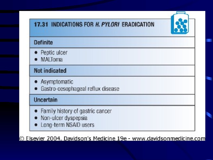
Management • The aims of management are : — to relieve symptoms, — induce ulcer healing in the short term, — cure the ulcer in the long term. H. pylori eradication is the cornerstone of therapy for peptic ulcers, as this will successfully prevent relapse and eliminate the need for long-term therapy in the majority of patients.
H. pylori eradication • First-line (triple) therapy : — IPP at standart dose (12 -hourly) for eg. — rabeprasol — Amoxicillin -1000 mg (12 -hourly) — Claritromycin – 500 mg (12 -hourly) for 7 days Good result is > 85% eradication!
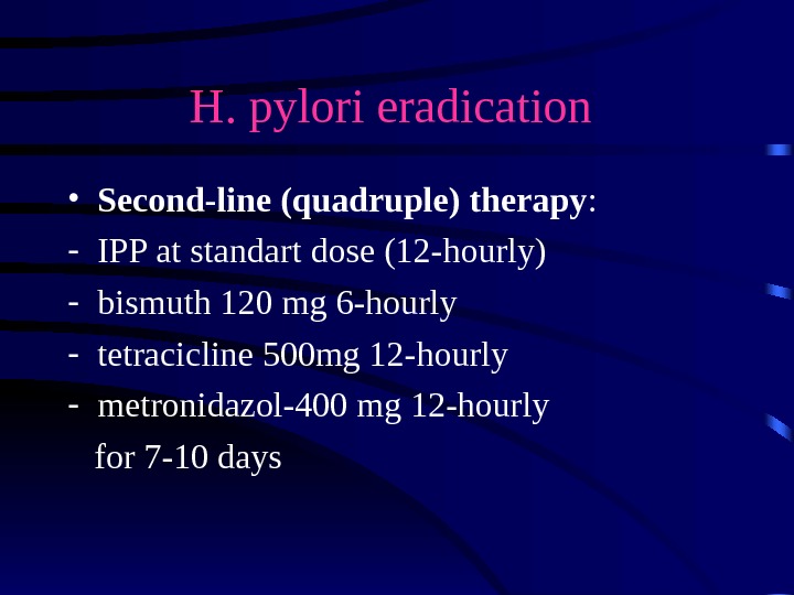
H. pylori eradication • Second-line (quadruple) therapy : — IPP at standart dose (12 -hourly) — bismuth 120 mg 6 -hourly — tetracicline 500 mg 12 -hourly — metronidazol-400 mg 12 -hourly for 7 -10 days
General measures • Cigarette smoking, aspirin and NSAIDs should be avoided. • Alcohol in moderation is not harmful and no special dietary advice is required.
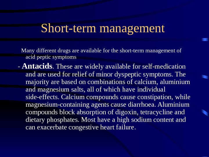
Short-term management Many different drugs are available for the short-term management of acid peptic symptoms — Antacids. These are widely available for self-medication and are used for relief of minor dyspeptic symptoms. The majority are based on combinations of calcium, aluminium and magnesium salts, all of which have individual side-effects. Calcium compounds cause constipation, while magnesium-containing agents cause diarrhoea. Aluminium compounds block absorption of digoxin, tetracycline and dietary phosphates. Most have a high sodium content and can exacerbate congestive heart failure.
Histamine H 2 -receptor antagonist drugs. These are competitive inhibitors of histamine at the H 2 -receptor on the parietal cell. Dyspeptic symptoms remit promptly, usually within days of starting treatment, and 80% of duodenal ulcerswill heal after 4 weeks. These drugs do not inhibit acid secretion to the same degree as the proton pump inhibitors but are useful for the short-term management of acid dyspeptic symptoms prior to investigation. They are moderately effective for the management of reflux disease.
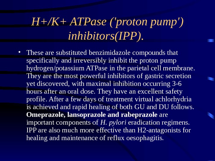
H+/K+ ATPase (‘proton pump’) inhibitors (IPP). • These are substituted benzimidazole compounds that specifically and irreversibly inhibit the proton pump hydrogen/potassium ATPase in the parietal cell membrane. They are the most powerful inhibitors of gastric secretion yet discovered, with maximal inhibition occurring 3 -6 hours after an oral dose. They have an excellent safety profile. After a few days of treatment virtual achlorhydria is achieved and rapid healing of both GU and DU follows. Omeprazole , lansoprazole and rabeprazole are important components of H. pylori eradication regimens. IPP are also much more effective than H 2 -antagonists for healing and maintenance of reflux oesophagitis.
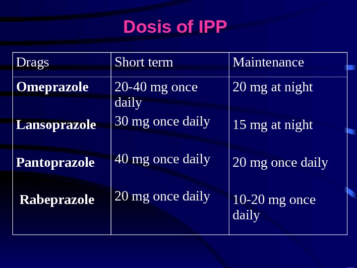
Drags Short term Maintenance Omeprazole Lansoprazole Pantoprazole Rabeprazole 20 -40 mg once daily 30 mg once daily 40 mg once daily 20 mg at night 15 mg at night 20 mg once daily 10 -20 mg once daily. Dosis of IPP
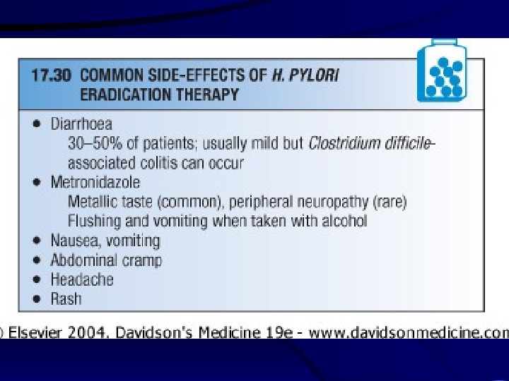
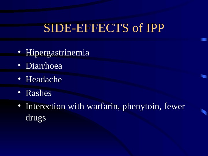
SIDE-EFFECTS of IPP • Hipergastrinemia • Diarrhoea • Headache • Rashes • Interection with warfarin, phenytoin, fewer drugs
Colloidal bismuth compounds. • Colloidal bismuth subcitrate (CBS) is an ammoniacal suspension of a complex colloidal bismuth salt. It has little, if any, effect on gastric acid secretion and its ulcer-healing effect is probably due to a combination of activity against H. pylori and enhancement of mucosal defence mechanisms.
Synthetic prostaglandin analogues (misoprostol). • Prostaglandins exert complex In low doses protect against injury induced by aspirin and NSAIDs by enhancing mucosal blood flow, and by stimulating mucus and bicarbonate secretion and epithelial cell proliferation. • At high doses acid secretion is inhibited. Misoprostol is effective for the prevention and treatment of NSAID-induced ulcers, but in clinical practice IPP are preferred, since they are at least as effective and have fewer side-effects.
Maintenance treatment • Continuous maintenance treatment should not be necessary after successful H. pylori eradication. For the minority who require maintenance treatment the lowest effective dose should be used.
INDICATIONS FOR SURGERY IN PEPTIC ULCER • EMERGENCY — Perforation — Haemorrhage ELECTIVE — Gastric outflow obstruction — Recurrent ulcer following gastric surgery
C OMPLICATION of peptic ulcer disease • perforation, • gastric outlet obstruction • bleeding.