
PEDIATRIC MALIGNANT SOLID TUMORS Iskra Christosova, Ognyan Brankov


PEDIATRIC MALIGNANT SOLID TUMORS Iskra Christosova, Ognyan Brankov Pediatric Oncohaematology Pediatric Surgery Sofia BULGARIA

Pediatric Cancer 2 nd leading cause of death in children 1/350 children diagnosed annually or the incidence per year would be 15-16 cases / 100 000 children per year/ 11 000 new cases in children under 20 years of age each year in the whole world. Considered in the past as hopeless diseases now 70% of children with cancer can be cured definitively

Cancerogenesis I. Exogenous Factors Radiation Exposure. Other Factors of the Surrounding. Enviroment - Chemical Cancerogens. Oncogenic Viruses.

II. Endogenous Factors Familial and Genetic Factor Cancer Malformation Syndromes Multiple Primary Tumors Second Malignant Neoplasms

Charactreristic Features of Childhood Cancer Child’s Organism is with forming Immune System and with Rapid Growth. Different Histological Types from those of Adults. Different Localizations from those of Adults. Higher Sensitiveness to Chemotherapy than Adults.
 : Solid Tumors = 1:1.")
Clasification of Pediatric Malignancies Systemic Neoplasms (Leukaemias and Lymphomas) : Solid Tumors = 1:1.

Systemic Neoplasms - 50% Leukaemias - 1/3 of Pediatric neoplasms - 35% Lymphomas (NHL 55% and Hodgkin’s Disease 45%) - 15%

Malignant Solid Tumors - 50% Embrional Tumors - 17% (Neurollastoma - 8%, Nephroblastoma - 7%, Retinollastoma - 1.5%, Hepatoblastoma - 0.5%) Brain Tumors - 17% (Astrocytoma,Medulloblastoma....)
 Bone Tumors - 4% (Sarcoma,")
Soft Tissue Sarcomas 8% (Rhabdomyosarcoma - 6%...) Bone Tumors - 4% (Sarcoma, Osteogenes, Sarcoma Ewing...) Germ Cell Tumors - 2% Epithelial Tumors - Carcinomas and other very Rare Tumors - 2% Malignant Solid Tumors - 50%
 Epidemiology - 7% of Neoplastic Diseases in Children Unfavonrable Histology")
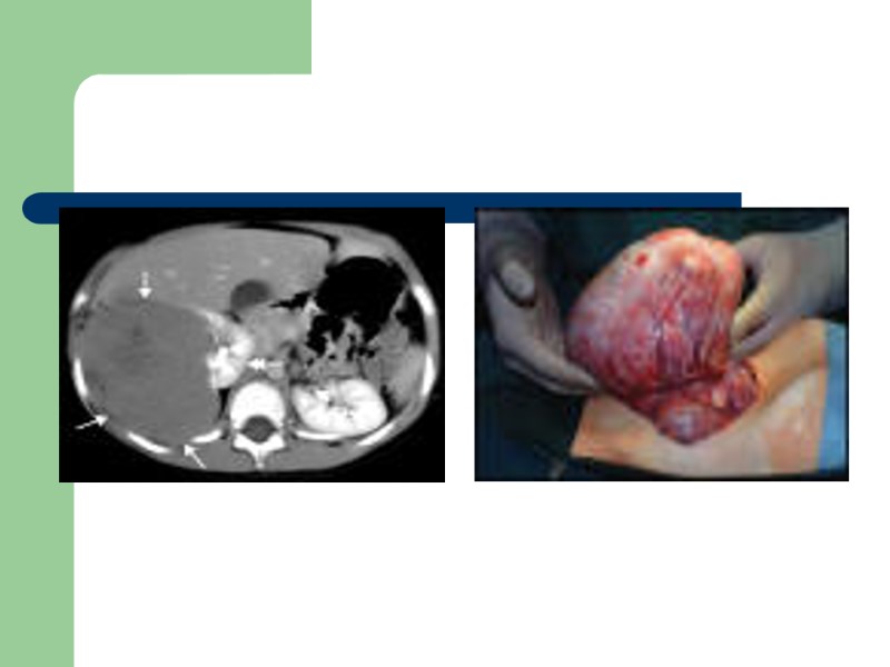
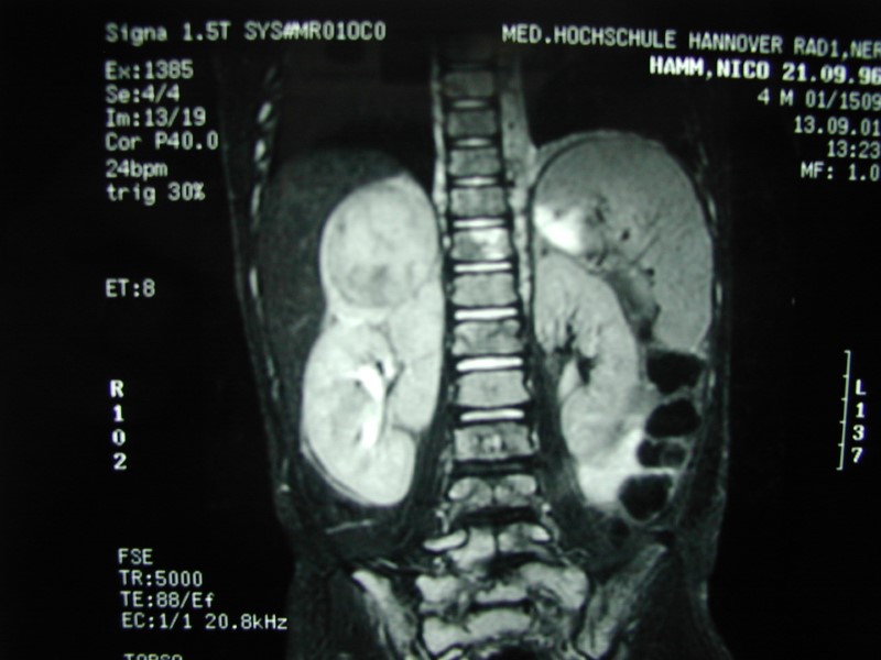
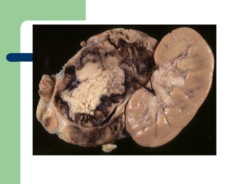
Wilms” Tumors (Nephroblastoma) Epidemiology - 7% of Neoplastic Diseases in Children Unfavonrable Histology (Focal or Diffuse Anaplasia) - 10% Favourable Histology (Multicystic and with Fibroadenomatous Structures) - 90% Gene Mutations - 11p 13 for WT1 and 11p 15 for WT2 Caner Malformation Syndromes

Incidence

Wilms Tumor: Histology: mixture of immature cells metanephric, stromal, tubular Cajaiba MM et al. (2006) Rhabdomyosarcoma, Wilms tumor, and deletion of the patched gene in Gorlin syndrome Nat Clin Pract Oncol 3: 575–580 doi:10.1038/ncponc0608

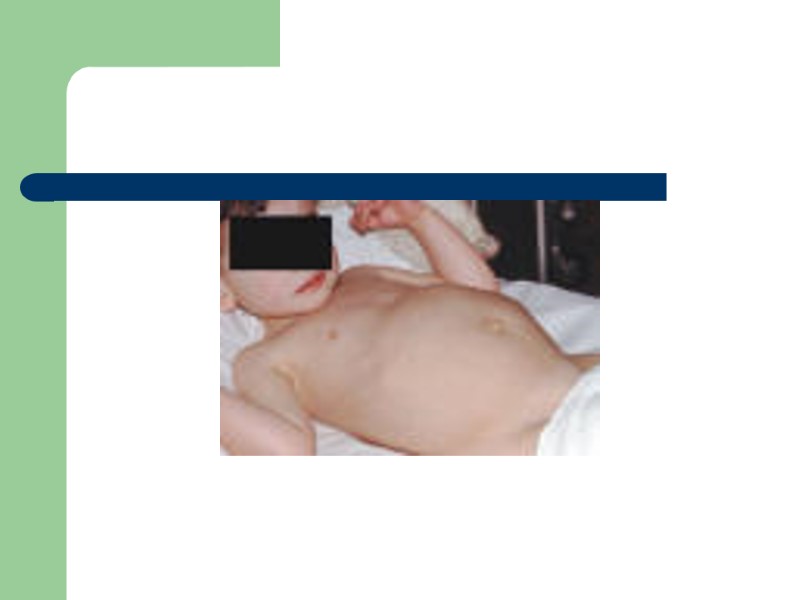
Clinical Manifestation Good Clinical State Haematuria - 25% Abdominal pain - 35% High blood pressure - 35% An abdominal Tumor mass - discovered accidentally

Clinical stages I. Tumor limited to the Kidney, in size - 5 cm. Intact Renal Capsule, Excised Completly. II. Tumor extends outside the Kidney in size - 10 cm, Excised Completly. III. Tumor over 10 cm in size. Infiltrated other Organs in Ablomen, without Hematogenous Mts, Complete Excision Impossible. IV. Tumor with Hematogenous Mts (Lungs - 10% liver - 1% ets.) V. Bilateral Renal Tumors

Laboratory and RadiologicalExaminations Hb, Plt, WBS, LDH, UrineanalysisCT and Abdominal UltrasoundChest X-ray Tumor Histology

Risk Factors Low Risk - Cases withl Favourable Histology, in I and II Stages, under 3 Year of Age High Risk - Cases with Unfavourable Histology, in III, IV and V Stages, over 3 Year of Age

Treatment Surgery - Nephrectomy, Lymphadenotomy, Excision when it is possibly of lung or liver Mts. In V stade -partial resection. Radiotherapy. No RT in I and II stage. In III and IV stage RT in Tumor Region with Dose 20-30 GY. In Mts regions - 15 GY

Chemotherapy L. R. Actinomycin D, Vincristine H. R. Act D, Vcr, Farmarubicin, VP16

Prognosis Low Risk - 85% survival High Risk - 40% survival
")
Neuroblastoma Epidemiology - 8% of Neoplastic Diseases in children Unfavourable Histology (Neuroblastoma) - 90% Favourable Histology (Ganglioneuroblastoma) - 10%
 15%")
Neuroblastoma Localization - 70% Abdomen (1/2 of Cases from suprarenal gl.) 15% Mediastinum, 3% Neck, 8% Paravertebral Region, 4% Other Rare Regions (Olfactory Region, Multiple Primary Tumors C.N.S ets)

Neuroblastoma “Small blue round cell” tumor Immunohistochemical stains: neurofilament proteins, synaptophysin, NSE Electron microscopy: neurosecretory granules, microtubules and filaments Chromosome 1 deletions or N-myc oncogene amplification From, Principles and Practice of Pediatric Oncology, Lippincott Williams & Wilkins, p 903.

Incidence

Clinical Manifestation Poor clinical State Symptoms of Primary Localization Symptoms of Metastatic Localization Paraneoplastic Symptoms

There are orbital and skull vault metastases, with associated enhancing soft-tissue masses. The skull lesions are extradural masses which deform the underlying brain. The right orbital lesion forms a superior extraconal mass, depressing the right globe.

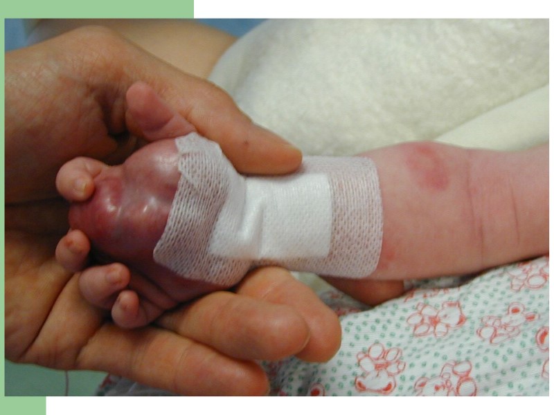
bilateral ecchymosis in a child with metastatic neuroblastoma.
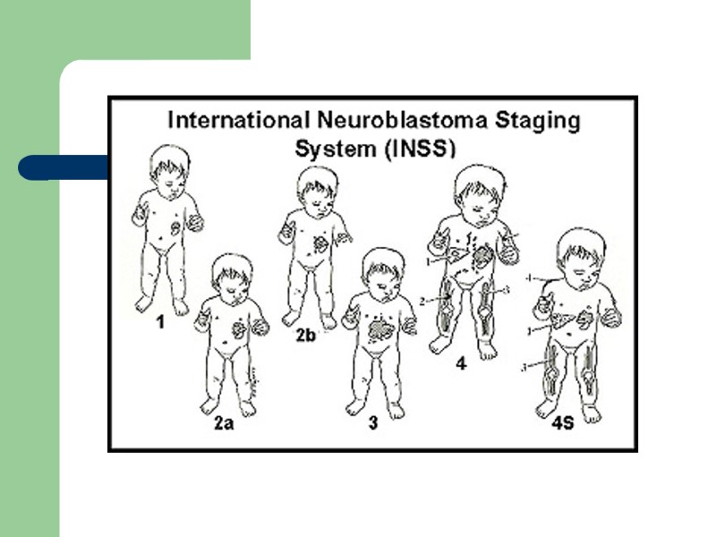
 I Tumor Limited to the Organ or Structure of")
Clinical Stages (Evans et al.) I Tumor Limited to the Organ or Structure of Origin. Excised Comletely. II Tumor with Regional Spread, not Crossing the Midline. III Tumor Crossing the Midline, Bilateral Lymph Nodes May by involved. Complete Excision imposible. IV Tumor with Distant Mts (Bone - 50% of cases, Lymph Nodes, Organs, Soft Tissues) IV-S Tumor in I and II Cl Stage, with Limited Dissemination to Liver, Skin and Bone Marrow (without Bones), Infants under 2 Years of Age, especially under 1 Year of Age.

Laboratory and Radiological Examinations Hb, Plt, WBC, LDH, Bone Marrow Aspiration Urinanalysis CT and Abdominal Ultrasound Bone Isotope Scanning, Scanning with 131I-MIBG Chest X-ray Tumor Markers - N-myc, Ferritin, NSE, Cantecholamines’ Metabolites Tumor Histology

Risk Factors Low Risk - Cases with Ganglioneuroblastoma, in I, II and IV-S Stages, under 1 Year of age, with Neck and Mediastinum Localisation High Risk - Cases with Neuroblastoma, in III and IV Stages (Bone Mts), over 2 Years of age, with High Levels of LDH, NSE and Fevritin, with Abdominal and Paravertebral Localisation, with Amplification of N-myc.

Treatment Surgery - Survival is better when Radical Excision is done. Radiotherapy - No RT in I, II and IV-S Stages In III and IV Stages RT in Tumor and Mts Redions with Dose 15-35 GY.

Chemotherapy L. R. - Vcr, Endoxan, Farmarubicin H. R. - Vcr, Endoxan, VP16, Cisplatin, Carboplatin, Holoxan, Farmarubicin

Prognosis Low Risk - 80% survival High Risk - 35% survival

Rhabdomyosarcoma Epidemiology - 5% of NeoplasticDiseases in Children Histology - Embrional and Botroid - 75%; Alveolar + Pleomorphic - 20%; Undifferentiated - 5%. Mts - Lungs, Bones, Lymph Nodes, Brain.

Incidence

Rhabdomyosarcoma Localization Head and Neck - 40%; Pelvis + Urinary Tract - 25%; Limbs - 20%; Other Rare Localizations - 15%; (Diapharagm Thorax and Abdominal walls, Viscera and every Region originated from Mesenchyme arising in Striated Muscle.)

Rhabdomyosarcoma Clinical Manifestation depends from Primary Localisation.

Nasopharyngeal Rhabdomyosarcoma
 or t(1;13) Gene amplification")
Rhabdomyosarcoma Alveolar 20% of pediatric cases Chromosomal translocation: t(2;13) or t(1;13) Gene amplification Tetraploid DNA From, Surgical Pathology of the Head and Neck, Lippincott Williams & Wilkins, p 157.

Rhabdomyosarcoma Botryoid 5-10% of pediatric cases Grape-like tumor masses Pleomorphic Rare in children From, Diagnostic Surgical Pathology of the Head and Neck, W.B.Saunders, p 554.

Clinical Stage I Limited Tumor Excised Comletely. II Grossly Removed Tumor with microscopic residual disease. III Incomplete Removal or only Biopsy with Gross Residual Tumor. IV Metastatic Disease at Diagnosis.

Laboratory and Radiological Examinations Hb, Plt, WBC, LDH CT and MRI of Primary Tumor Chest X-ray and CT Bone Scan Tumor Histology

Risk Factors Low Risk - I and II stages - 70% survival High Risk - III and IV stages, Parameningial Localization, Alveolar Histology - 30% survival

Treatment Surgery Radiotherapy Chemotherapy - Vcr, Holoxan Farmarubicin, VP16, Endoxan Carloplatin, Actinomycin D.
15468-pediatric_malignant_solid_tumors__-christosova,brankov.ppt
- Количество слайдов: 50