Описание презентации Occult With No Classic CNV Red-free Photograph по слайдам
 Occult With No Classic CNV
 Red-free Photograph Image courtesy of J Slakter
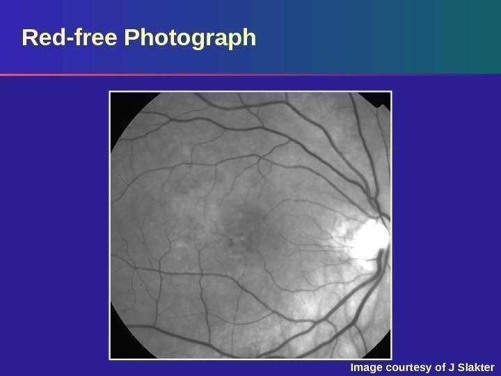
 Red-free Photograph Red-free photograph: shallow subretinal fluid and drusen are seen – There was a small pigment epithelial detachment (PED) noted on clinical examination
 Early-phase Fluorescein Angiogram Image courtesy of J Slakter
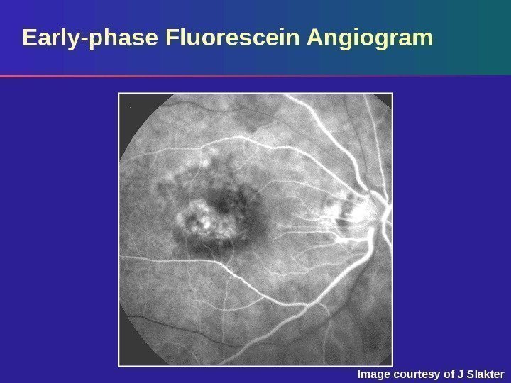
 Early-phase Fluorescein Angiogram Early phase: an area of irregular hyperfluorescence noted centrally with some surrounding areas of hypofluorescence – The margins of this region are fairly well defined
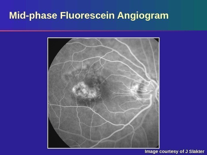 Mid-phase Fluorescein Angiogram Image courtesy of J Slakter
 Mid-phase Fluorescein Angiogram Mid (venous) phase: the area of hyperfluorescence stands out in better contrast to the more homogenous background fluorescence Note: there is no increased hyperfluorescence of the lesion typically seen with classic CNV
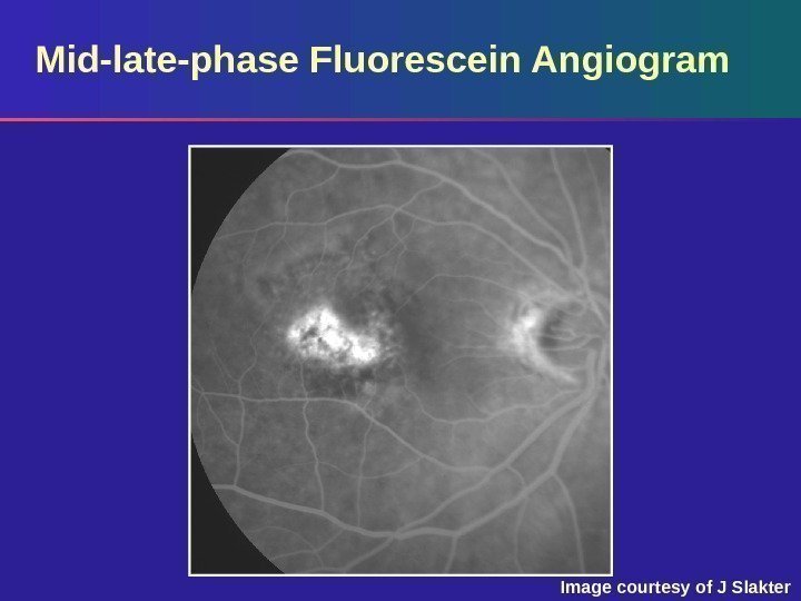 Mid-late-phase Fluorescein Angiogram Image courtesy of J Slakter
 Mid-late-phase Fluorescein Angiogram Mid-late phase: there is slightly more hyperfluorescence from the lesion at this point
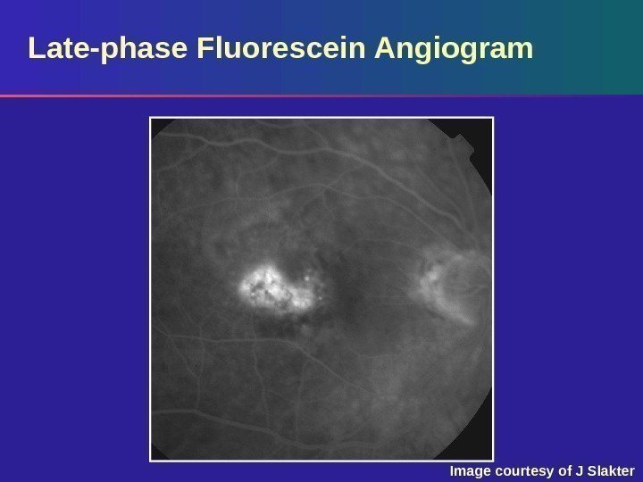 Late-phase Fluorescein Angiogram Image courtesy of J Slakter
 Late-phase Fluorescein Angiogram Late phase: the lesion remains unchanged throughout the later phases of the angiogram, without the increased hyperfluorescence and leakage beyond the margins of the CNV seen with classic CNV – This is an example of occult CNV, likely representing a fibrovascular P


