Общая хар. бактерий.ppt
- Количество слайдов: 158


Общая характеристика бактерий

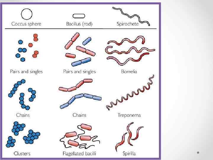
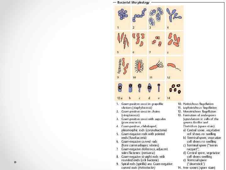
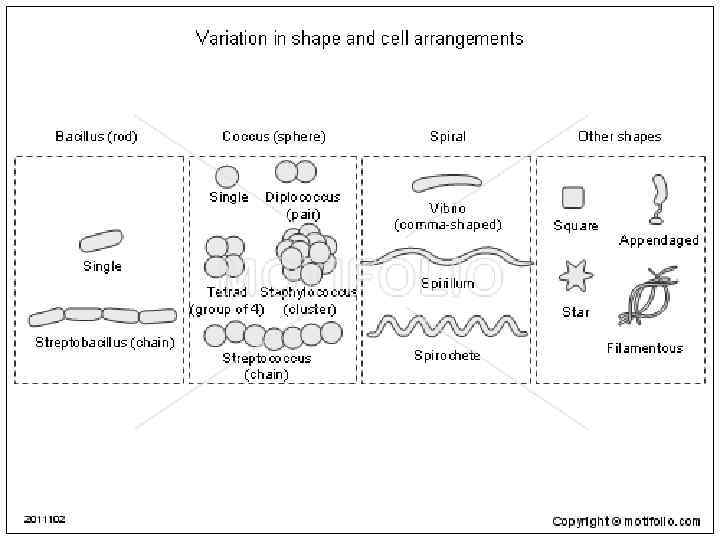
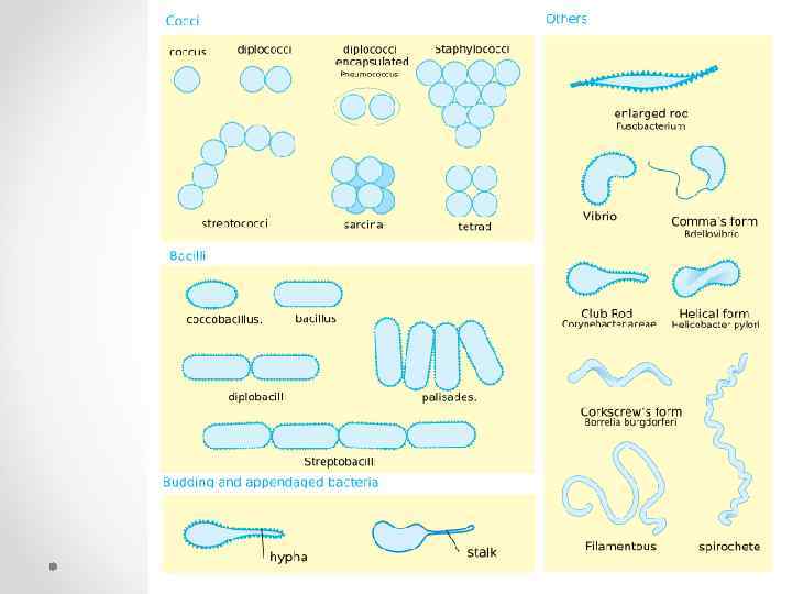
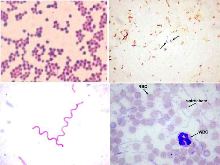
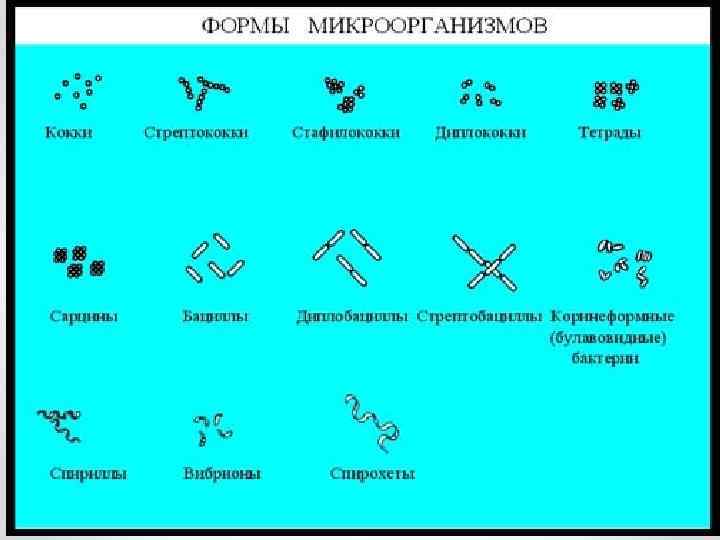
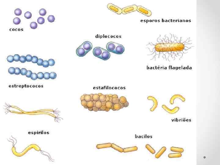
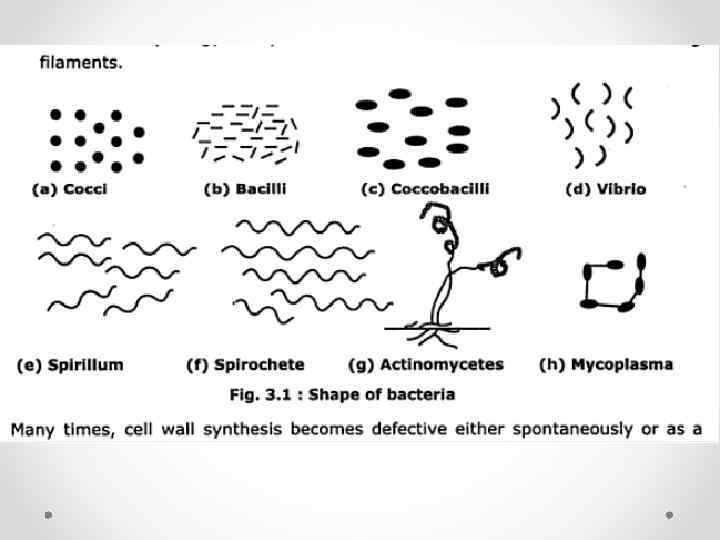
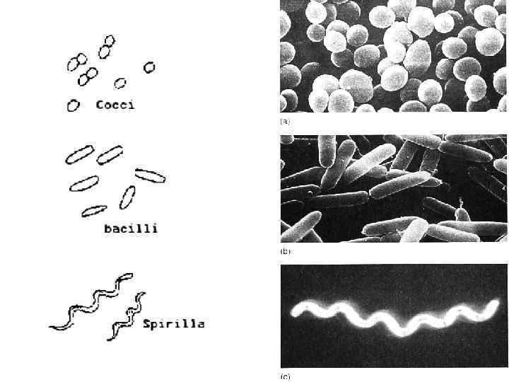
Форма бактерий

http: //accessmedicine. net/search. AM Result. Img. aspx? search. Str=bilirubin+in+urine &full. Text. Str=bilirubin+in+urine&search. Type=
")
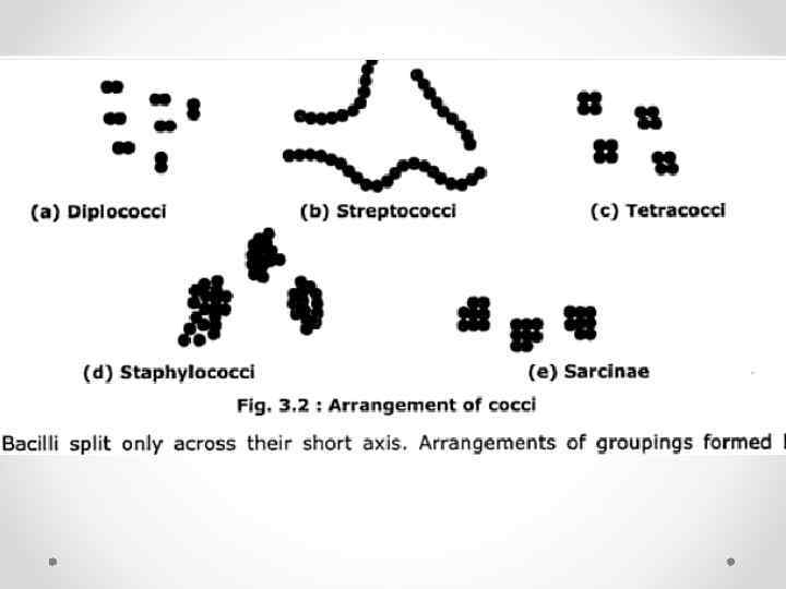
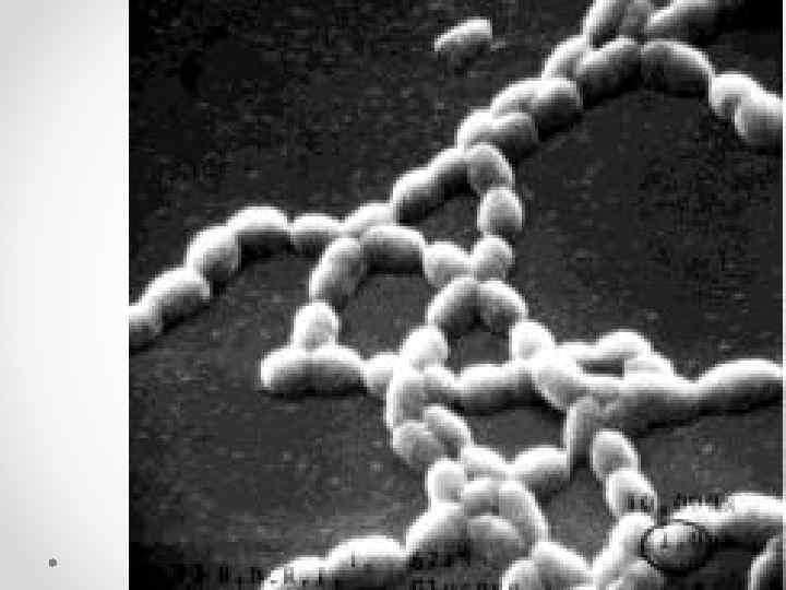
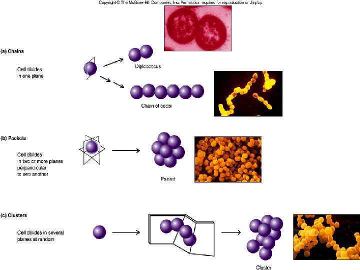
ШАРОВИДНЫЕ БАКТЕРИИ (КОККИ)

ТЕТРАКОККИ

Сарцины
 В ЧИСТОЙ КУЛЬТУРЕ")
(МЕНИНГОКОККИ) В ЧИСТОЙ КУЛЬТУРЕ
")
Диплококки (гонококки)
")
ДИПЛОКОККИ (ГОНОКОККИ)
")
Диплококки (ПНЕВМОКОККИ)

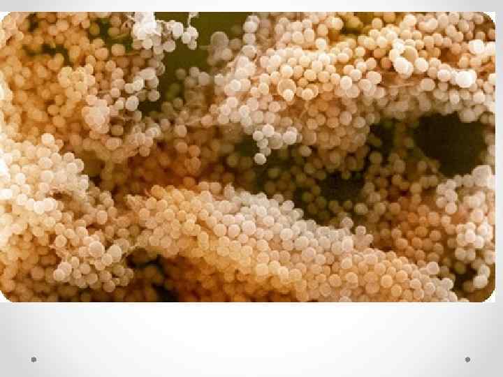
Стафилококки

Стафилококки

Микрококки

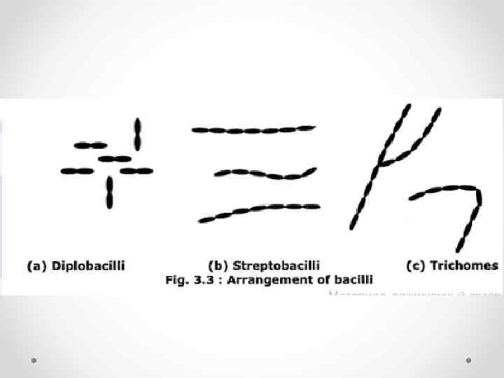
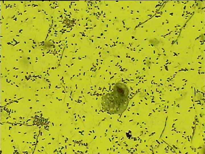
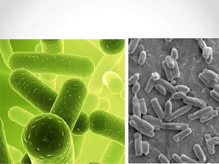
Палочковидн ые микроорганизм ы
")
НЕПРАВИЛЬНОЙ ФОРМЫ Коринебактерии (ГРАМПОЛОЖИТЕЛЬНЫЕ)

ПАЛОЧКОВИДНЫЕ МИКРООРГАНИЗМЫ НЕПРАВИЛЬНОЙ ФОРМЫ Микобактерии

ПАЛОЧКОВИДНЫЕ МИКРООРГАНИЗМЫ НЕПРАВИЛЬНОЙ ФОРМЫ Бифидобактерии

ПАЛОЧКОВИДНЫЕ МИКРООРГАНИЗМЫ НЕПРАВИЛЬНОЙ ФОРМЫ Вибрионы
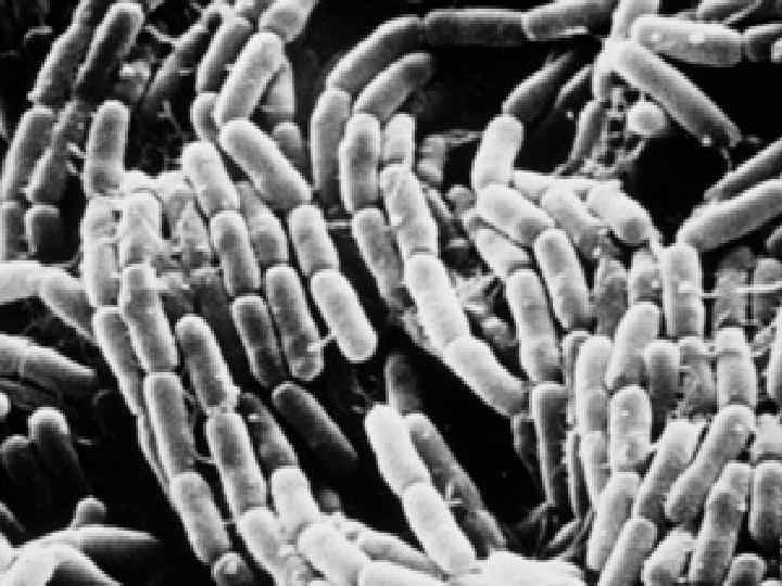

Стрептобактери и

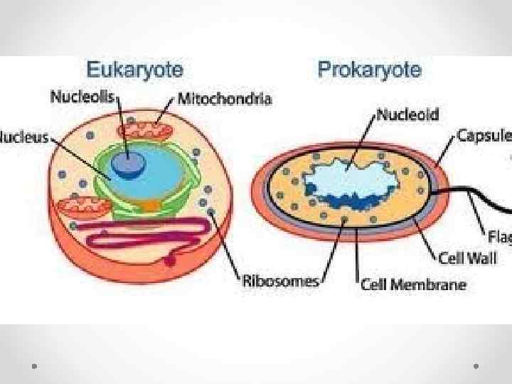
Comparison of eukaryotes and "eubacterial" prokaryotes

Клетки животных

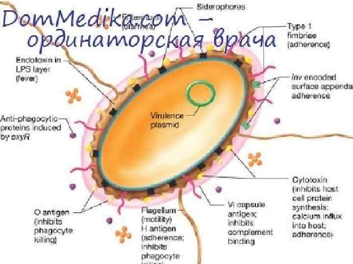
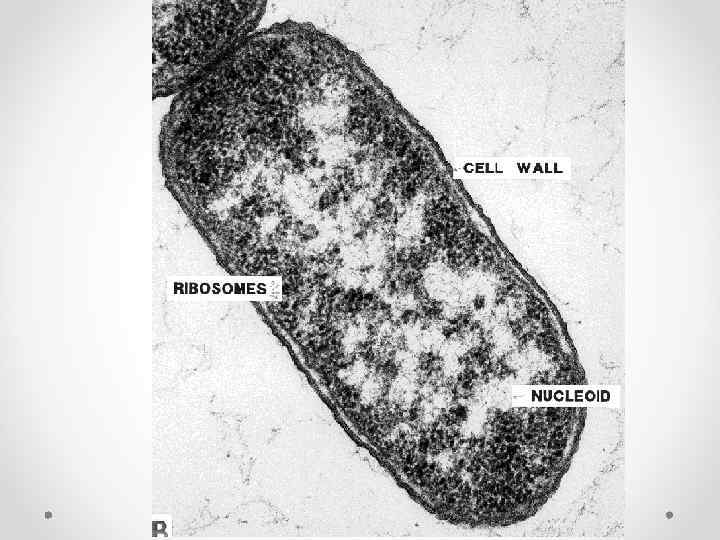
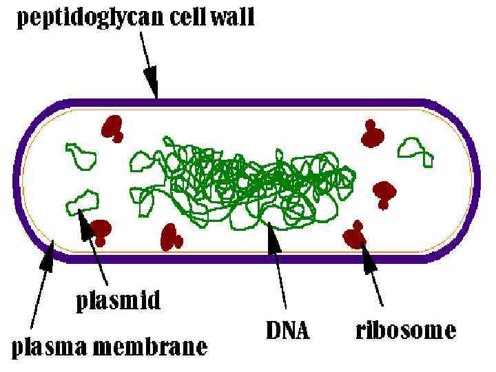
Бактерия

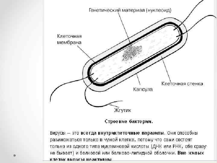
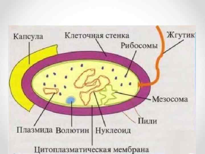
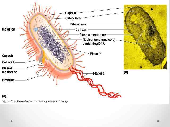
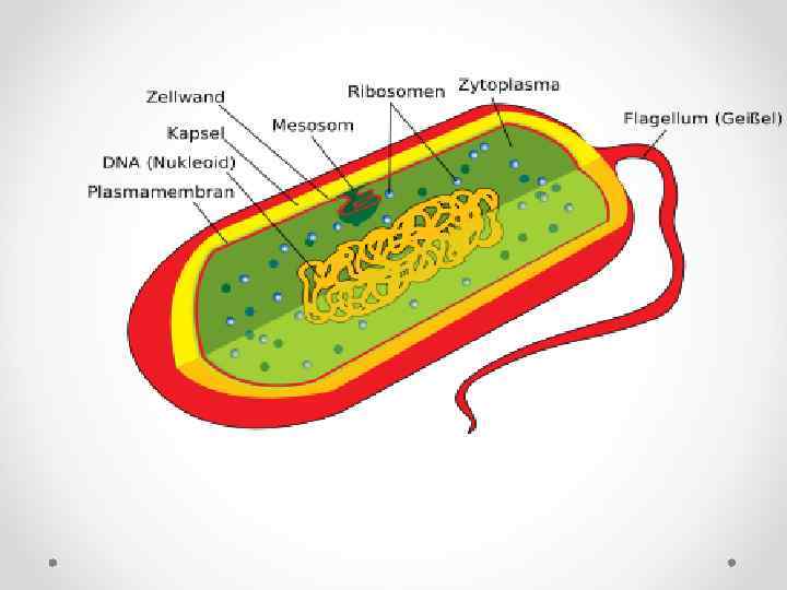
бактериальной клетки

http: //www. slideshare. net/josemanuel 7160/tema-16 -microbiologa
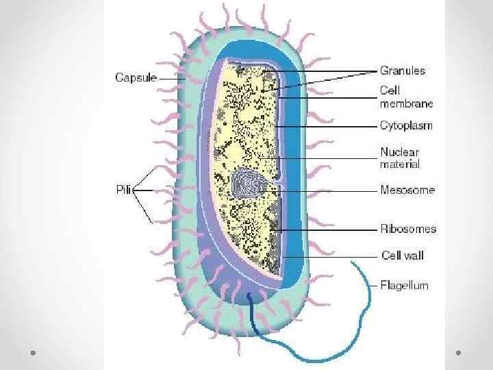

Строение бактериальной клетки

Хроматин

Плазмиды кишечной палочки
 Eukaryotic")
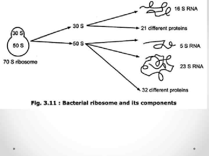
Рибосомы Comparison of Ribosome Structure in Bacteria, Eukaryotes, and Mitochondria Bacterial (70 S) Eukaryotic (80 S) Mitochondrial (55 S) r. RNAs (1 of each) 50 S 60 S 39 S 23 S (2904 nts) Large Subunit 28 S (4700 nts) 16 S (1560 nts) 5 S (120 nts) 5. 8 S (160 nts) Proteins 33 ~49 48 Small Subunit 30 S 40 S 28 S r. RNA 16 S (1542 nts) 18 S (1900 nts) 12 S (950 nts) Proteins 20 ~33 29

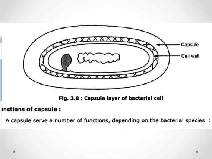
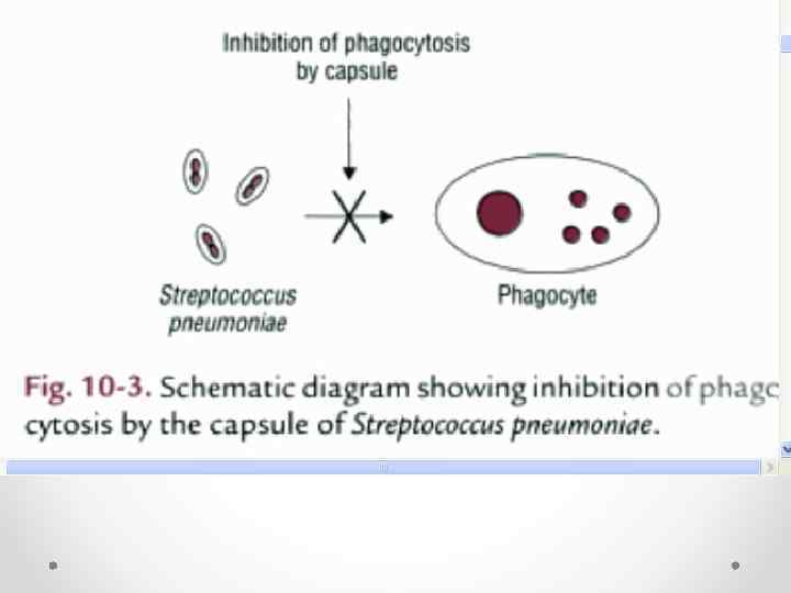
Капсула

Capsule-producing bacillus-shaped bacteria. The capsule is composed of polysaccharides and polyproteins. Capsules have a role in adherence, virulence, protection, securing nutrients, and cell-to-cell recognition. Capsules vary in thickness and can easily be 2 times the volume of the organism. In a capsule stain, the background is stained grayish blue and the cells are stained red. The capsule is unstained and appears as a halo around the cell

http: //www. slideshare. net/josemanuel 7160/tema-16 -microbiologa

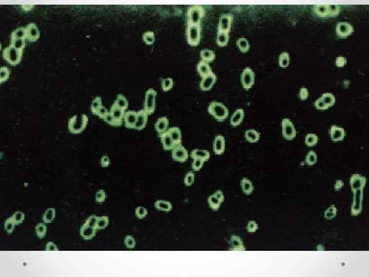

Bacterial capsules outlined by India ink viewed by light microscopy. This is a true capsule, a discrete layer of polysaccharide surrounding the cells. Sometimes bacterial cells are embedded more randomly in a polysaccharide matrix called a slime layer or biofilm. Polysaccharide films that may inevitably be present on the surfaces of bacterial cells, but which cannot be detected visually, are called glycocalyx.

http: //www. ncbi. nlm. nih. gov/pmc/articles/ PMC 3222727/figure/F 2/

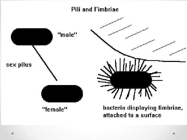
Реснички
 Whole cells were incubated with anti-fimbria")
Streptococcus salivarius cells (ФИМБРИИ /12 нм в диаметре/) Whole cells were incubated with anti-fimbria polyclonal antibodies prior to labelling with 12 nm diameter colloidal gold-conjugated donkey antirabbit immunoglobulin G.

Жгутики
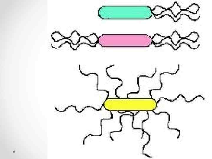

Жгутики

Жгутики
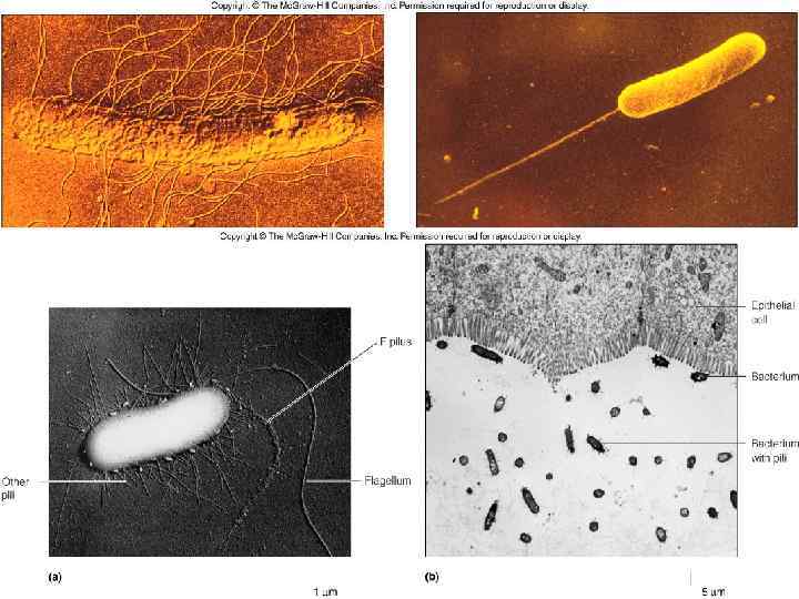
 and flagella on the surface of bacterial cells. Left: dividing Shigella")
Fimbriae (common pili) and flagella on the surface of bacterial cells. Left: dividing Shigella enclosed in fimbriae. The structures are probably involved in the bacterium's ability to adhere to the intestinal surface. Right: dividing pair of Salmonella displaying both its peritrichous flagella and its fimbriae. The fimbriae are much shorter and slightly smaller in diameter than flagella. Both Shigella and Salmonella are enteric bacteria that cause different types of intestinal diarrheas. The bacteria can be differentiated by a motility test. Salmonella is motile; Shigella is nonmotile.

Flagellum of Gram-negative Bacteria
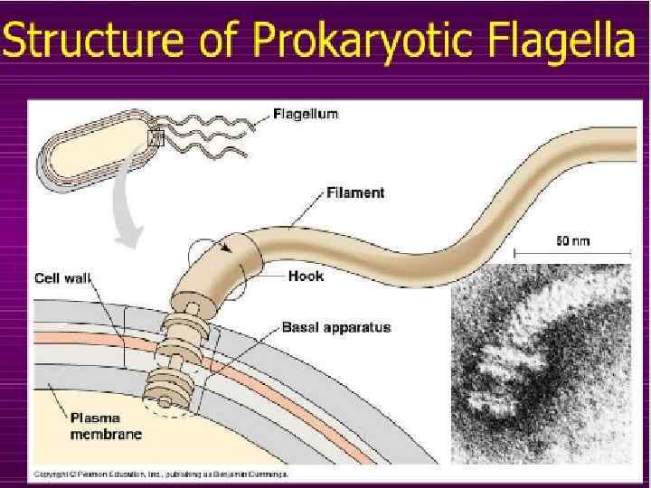
. Measurements are in nanometers. The")
The ultrastructure of a bacterial flagellum (after J. Adler). Measurements are in nanometers. The flagellum of E. coli consists of three parts, filament, hook and basal body, all composed of different proteins. The basal body and hook anchor the whip-like filament to the cell surface. The basal body consists of four ring-shaped proteins stacked like donuts around a central rod in the cell envelope. The inner rings, associated with the plasma membrane, are the flagellar powerhouse for activating the filament. The outer rings in the peptidoglycan and outer membrane are support rings or "bushings" for the rod. The filament rotates and contracts which propels and steers the cell during movement.

Cross section of an axoneme The "9+2" structure is visible in this cross-section micrograph of axoneme

Schema eines Querschnitts durch eine Eukaryoten-Geißel, 1 A + 1 B - Doppelmikrotubuli an der Peripherie, 2 - zwei einfache Mikrotubuli in der Mitte, 3 Dyneinarme, 4 - Speichen, 5 - Nexin-Verbindungen, 6 - Zellmembran

Модель жгутика
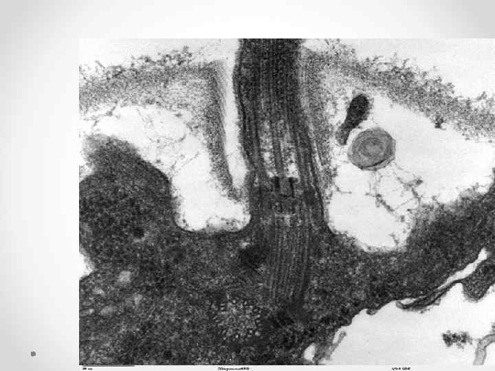
, 4 -Basal")
Eukaryotic flagella 1 -axoneme, 2 -cell membrane, 3 -IFT (Intra. Flagellar Transport), 4 -Basal body, 5 -Cross section of flagella, 6 -Triplets of microtubules of basal body. Longitudinal section through the flagella area in Chlamydomonas reinhardtii. In the cell apex is the basal body that is the anchoring site for a flagella. Basal bodies originate from and have a substructure similar to that of centrioles, with nine peripheral microtubule triplets (see structure at bottom center of image).

and cilia

form bundles that rotate counter-clockwise, creating a torque that causes the bacterium to rotate clockwise.

; suoirci. D h er P t


Bacterial cultures grown in motility test medium. The tube on left is a non motile organism; the tube on right is a motile organism. Motility test medium is a semi-soft medium that is inoculated with a straight needle. If the bacteria are motile, they will swim away from the line of inoculation in order to find nutrients, causing turbidity or cloudiness throughout the medium. If they are non motile, they will only grow along the line of inoculation. www. jlindquist. net/generalmicro/dfmotility. html.

Иные виды подвижности


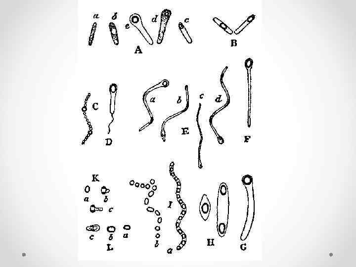
СПОРЫ

Оболочки спор Atomic force microscopy image of the crystalline rodlet layer on the outer spore coat of Bacillus atrophaeus spore. The image shown on the cover is approximately 250 x 700 nm.

Строение оболочки спор у различных видов анаэробных бактерий: Э — экзоспориум, О — оболочка споры. 1, 2, 3 — слои споровой оболочки

Спорообразование


Эндоспоры


Spores of Vairimorpha

infection in the Indian meal moth, Plodia interpunctella.

Включения

Метахроматин в клетках возбудителя дифтерии
 с зёрнами метахроматина")
Крупные изогнутые клетки (спириллы) с зёрнами метахроматина
 в клетках анаэробной бактерии")
Включения запасного вещества — гранулёзы (окрашивается иодом в синий цвет) в клетках анаэробной бактерии во время образования ею спор.

бактерий с зёрнами метахроматина

бактерии с крупными каплями жира

inclusions A variety of bacterial . a. PHB granules; b. b. a parasporal BT crystal in the sporangium of Bacillus thuringiensis; c. carboxysomes in Anabaena viriabilis, showing their polyhedral shape; d. d. sulfur globules in the cytoplasm of Beggiatoa


БАКТЕРИЯ ВНУТРИ КЛЕТКИ

What is Microbiology? • Micro - too small to be seen with the naked eye • Bio - life • ology - study of

• • Organisms included in the study of Microbiology Bacteriology Protozoology Phycology Parasitology • Mycology • Virology • • • 1. Bacteria 2. Protozoans 3. Algae 4. Parasites 5. Yeasts and Molds o Fungi • 6. Viruses Microorganisms - Microbes - Germs

• • • Bacteria - what comes to mind? Diseases Infections Epidemics Food Spoilage 1% of all known bacteria cause human diseases 4% of all known bacteria cause plant diseases 95% of known bacteria are non-pathogens

Microbes Benefit Humans 1. Bacteria are primary decomposers – recycle nutrients back into the environment (sewage treatment plants) 2. Microbes produce various food products o cheese, pickles, sauerkraut, green olives o yogurt, soy sauce, vinegar, bread o Beer, Wine, Alcohol

3. Microbes are used to produce Antibiotics • Penicillin • Mold o Penicillium notatum 1928 Alexander Fleming

4. Bacteria synthesize chemicals that our body needs, but cannot synthesize • Example: E. coli o B vitamins - for metabolism o Vitamin K - blood clotting • Escherichia coli o Dr. Escherich o Colon (intestine)

Microbial Antagonism Our normal microbial flora prevents potential pathogens from gaining access to our body

Gene Therapy Genetic Engineering • Bacteria can be manipulated to produce enzymes and proteins they normally would not produce o Insulin o Human Growth Hormone o Interferon

Microbes do benefit us, but they are also capable of causing many diseases • • Pneumonia Botulism Cholera Syphilis Chlamydia Meningitis Strep Throat Black Plague Whooping Cough Typhoid Fever Measles Scarlet Fever Mumps Gonorrhea Herpes 1 Tuberculosis Herpes 2 Tetanus RMSV Lyme Disease AIDS Diarrhea Gangrene


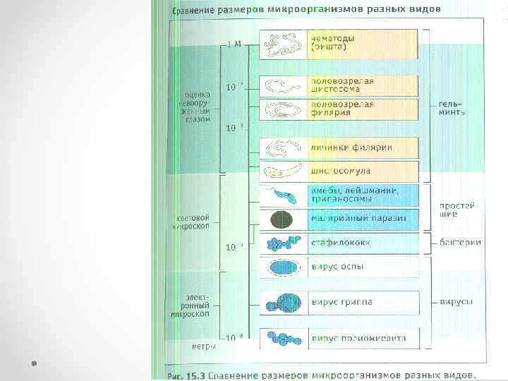
• Прокариоты = Monerans ранние организмы жили и развивались только на земле в течение 2 х 10 9 лет наименьший независимо живых намного меньше, чем одноклеточных эукариот Типичные бактерии = 2 μ Средняя эукариотической клетки = 50 -200 μ превышает число всех эукариот в сочетании населяют более рот, чем общее количество людей, которые когда-либо жили! • 3. Разнообразие и классификация 2 филиала Archebacteria Прикованный к экстремальных условиях, похожих на ранней Земле более тесно связаны с эукариот, чем к современным бактериям только несколько родов: Метаногены уменьшить CO 2 в CH 4 Extreme галофилы - соль любящий Thermoacidophiles эубактерий самых современных бактерий очень разнообразны

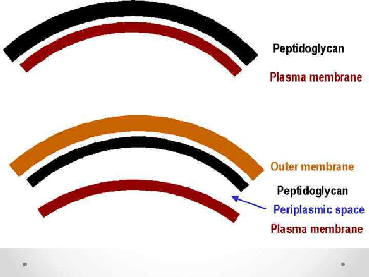
• • 5. Прокариоты против эукариот нет митохондрий, хлоропластов и других органелл мембраносвязанных Большинство из них являются одноклеточными и намного меньше, проще ДНК геномов не расположены в клеточной стенке хромосомы отличаются от растений, грибов, простейших содержит муреин / peptidoglcan (азот, содержащий полисахарид) отличаются В механизмы генетической репликации, выражения, и рекомбинации 6. Прокариот и эукариотических клеток 7. Функции и взаимодействие только болезнь меньшинств причиной Многие имеют важное значение для жизни на Земле Decomposers необходимо химические циклы часто живут в симбиотические отношения с другими организмами 8. Формы и функции одноклеточных некоторые совокупности в 2 одноклеточные или нескольких групп одноклеточных некоторое разнообразие форм колоний формы 3 наиболее распространенных: сферы = кокки стержней = спирали бациллы = spirilla
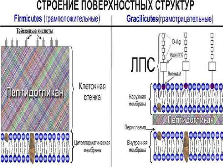
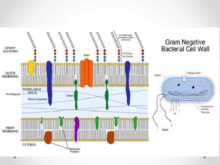
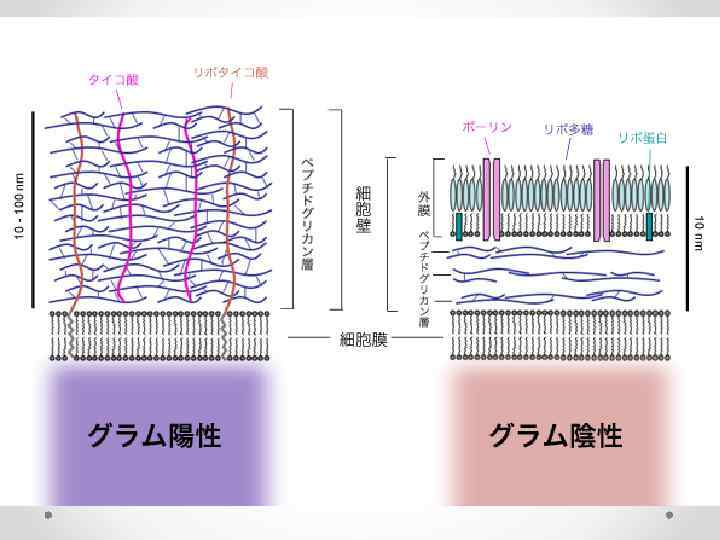
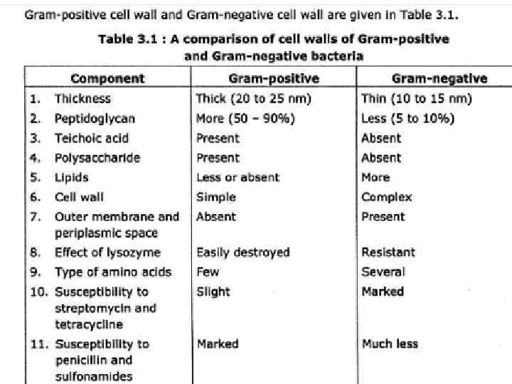
 не окрашиваются более сложные клеточные стенки с меньшим пептидогликана клеточной")
• Gram (-) не окрашиваются более сложные клеточные стенки с меньшим пептидогликана клеточной стенки содержат липополисахариды, скорее всего, быть патогенными (причина болезни) более устойчивы к антибиотикам

• Капсула Многие бактерии выделяют липкое вещество, которое образует защитный слой, другой, капсула Вне клеточной стенки помогает им придерживаться вещи Обеспечивает защиту • 17. Подвижность Около половины всех monerans способны направленного движения. 3 механизмов: жгутиков - отличаются от эукариотов спиральные бактерии (спирохеты) имеют нити, что спирали вокруг клетки под внешней оболочкой приводит к клетке двигаться, как штопор некоторые бактерии вырабатывают слизистый химических и скольжение Такси движение к или от стимула много бактерии проявляют эту форму движения

 • http: //www. nature. com/nrmicro/archive/index. html")
Обзоры по микробиологии (ин. журнал – архив; резюме) • http: //www. nature. com/nrmicro/archive/index. html


http: //www. slideshare. net/josemanuel 7160/tema-16 -microbiologa

http: //www. slideshare. net/ardiansyahrohis/eubacteria-

http: //www. slideshare. net/ardiansyahrohis/eubacteria-

http: //www. slideshare. net/ardiansyahrohis/eubacteria-

http: //www. ncbi. nlm. nih. gov/books/NBK 26810/? redirect-on-
Общая хар. бактерий.ppt