

4c76ff7b6caf88a05b79926be735278d.ppt
- Количество слайдов: 61
 Normalisation des données Oury Monchi, Ph. D. Centre de Recherche, Institut Universitaire de Gériatrie de Montréal & Université de Montréal

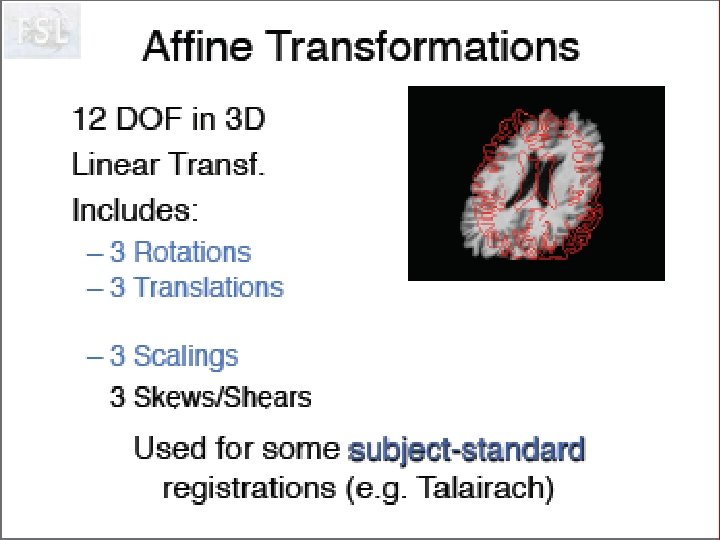
 Stereotaxic Space J. Talairach and P. Tournoux, Co-planar stereotactic atlas of the human brain: 3 -Dimensional proportional system: an approach to cerebral imaging, Stuttgart, Georg Thieme Verlag, 1988 • based on anatomical landmarks (anterior and posterior commissures) • originally used to guide blind stereotaxic neurosurgical procedures (thalamotomy, pallidotomy) • now used by Neuro. Scientific community for interpretation and comparison of results
 AC-PC line posterior commissure AC-PC line anterior commissure VAC
 Stereotaxic Space J Talairach & P Tournoux, Co-planar stereotaxic atlas of the human brain, Georg Thieme, 1988
 Stereotaxic Space
 Anatomical variability remains Talairach & Tournoux Atlas, 1988 variability of central sulcus from 20 subjects
 Not Registered Data Images courtesy A. Zijdenbos, MNI
 Registered Data
 Registration to Stereotaxic Space Advantages for anatomical/structural imaging: • facilitates comparisons across – time points – subjects – groups – sites • permits averaging between subjects to S/N • Allows the use of spatial masks for post-processing (anatomically driven hypothesis testing) • allows the use of spatial priors (classification) • allows the use of anatomical models (segmentation) • provides a framework for statistical analysis with wellestablished random field models • Allows the rapid re-analysis using different criteria
 Registration to Stereotaxic Space Advantages for functional imaging: • Provides a conceptual framework for the completely automated, 3 D analysis across subjects. • Facilitate intra/inter-subject comparisons across – time points, subjects, groups, sites • Extrapolate findings to the population as a whole • Increase activation signal above that obtained from single subject • Increase number of possible degrees of freedom allowed in statistical model • Enable reporting of activations as co-ordinates within a known standard space – e. g. the space described by Talairach & Tournoux
 Talairach Atlas Drawbacks for functional imaging: • is derived from an unrepresentative single 60 -yr old female cadaver brain (when most functional activation studies are done on young living subjects!) • ignores left-right hemispheric differences • has variable slice separation, up to 4 mm • while it contains transverse, coronal and sagittal slices, it is not contiguous in 3 D
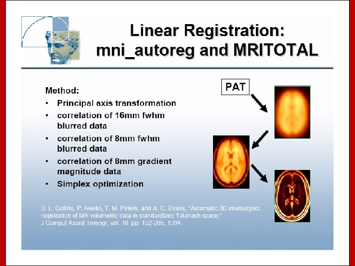
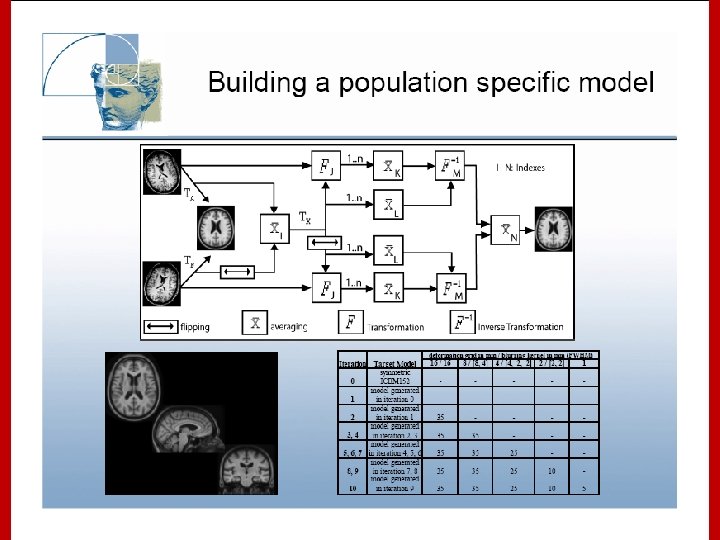
 Stereotaxic Space However, the space and the stereotaxic concept are still worthwhile: • Provides a conceptual framework for the completely automated, 3 D analysis across subjects. • Collins, L. , Evans A. , et al. have created a replacement target volume for stereotaxic mapping to address weaknesses of the Talairach atlas
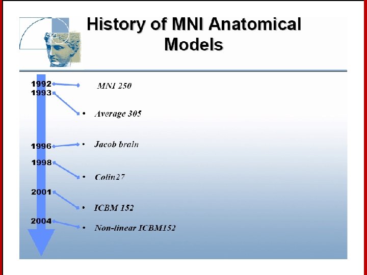
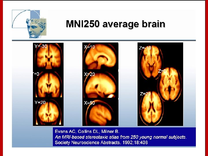
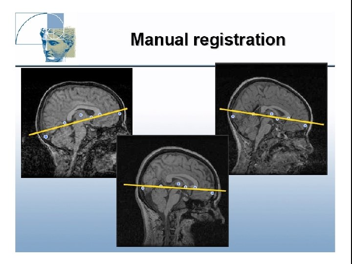
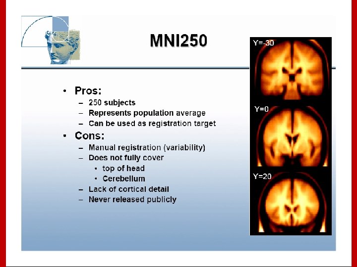
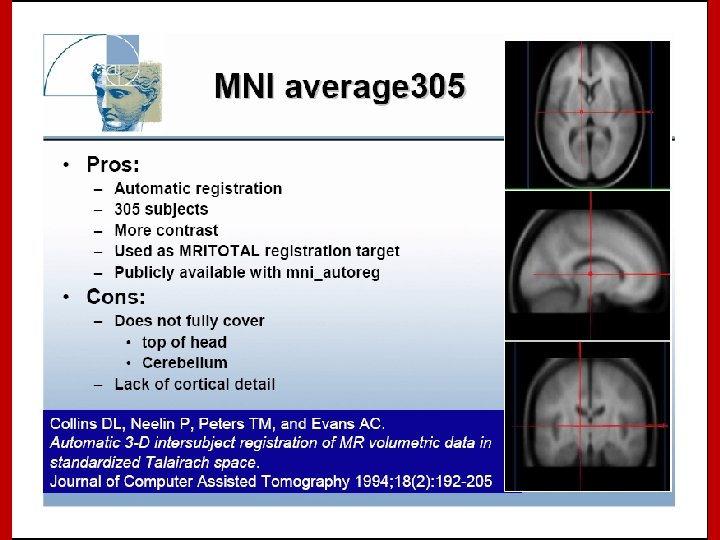
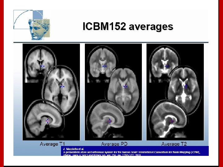
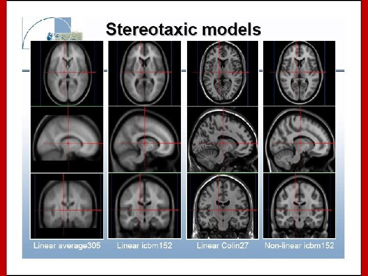
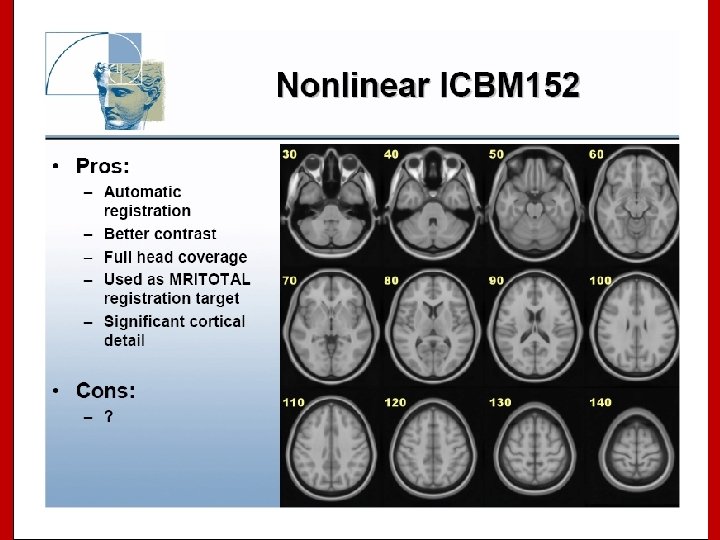
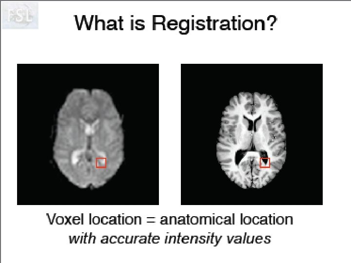
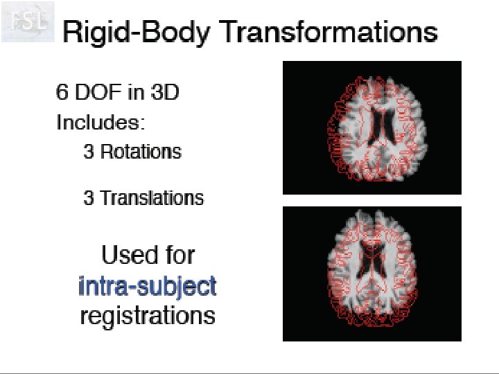
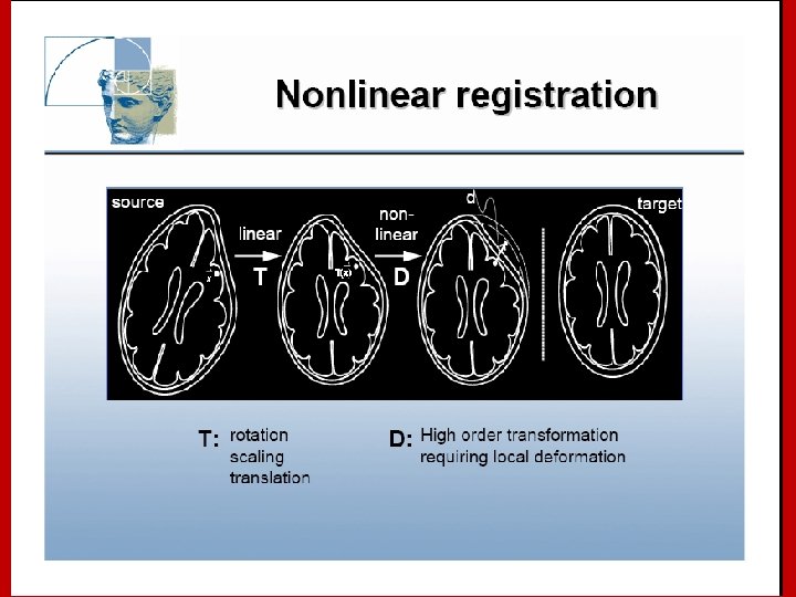
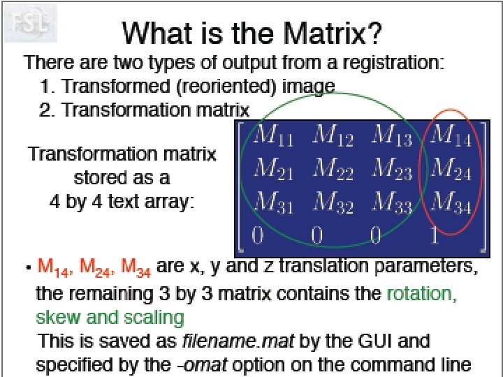
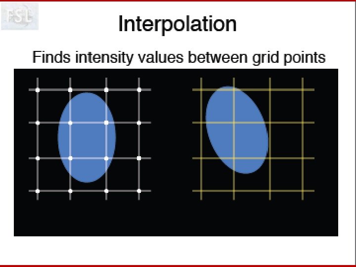
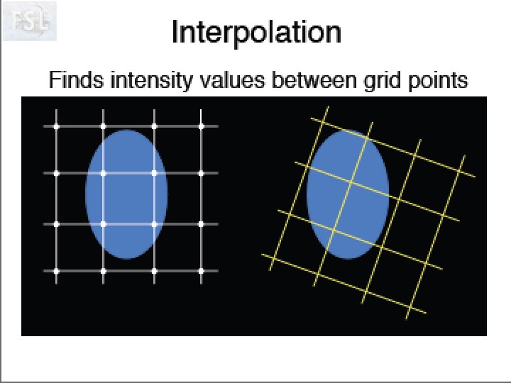
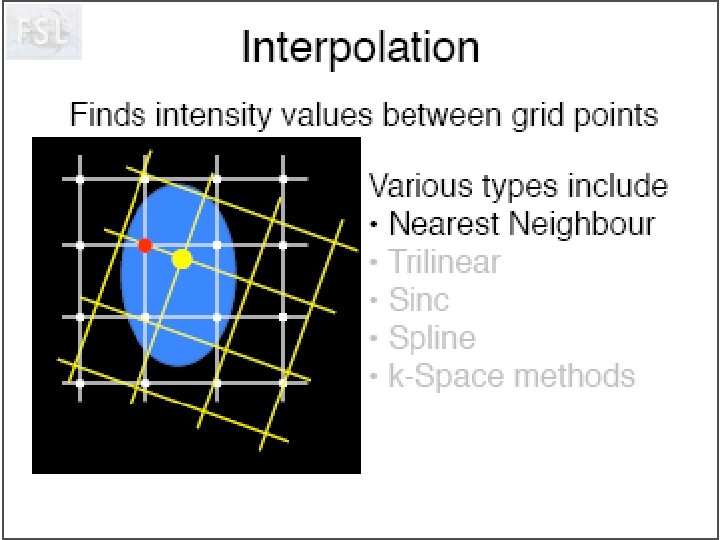
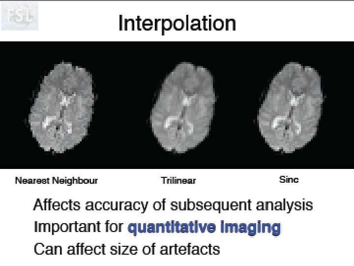
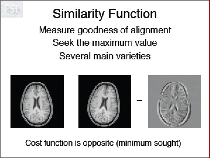
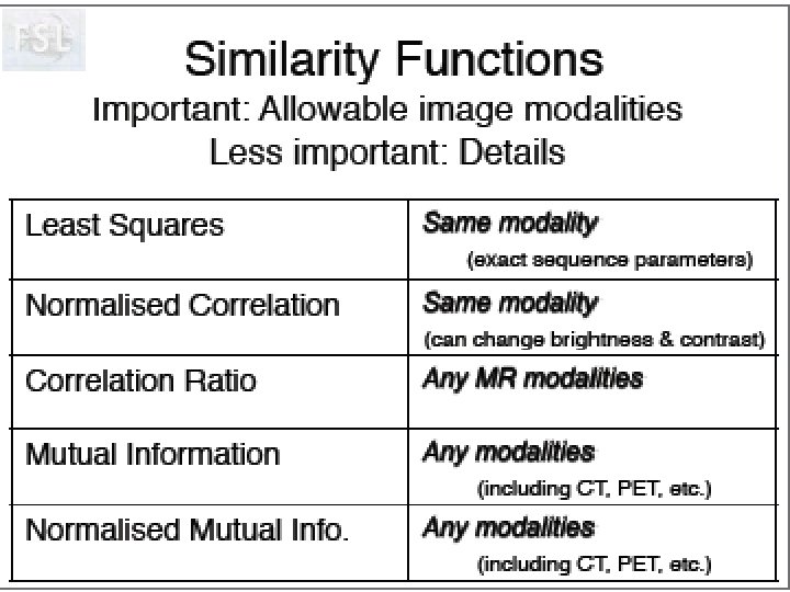
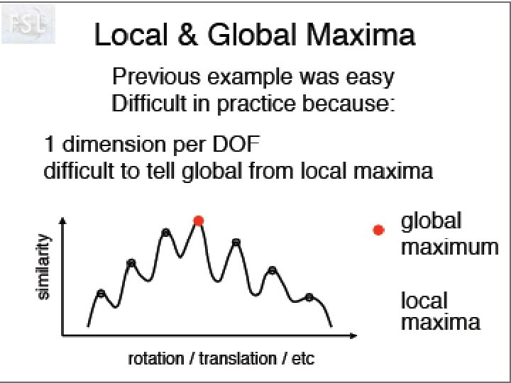
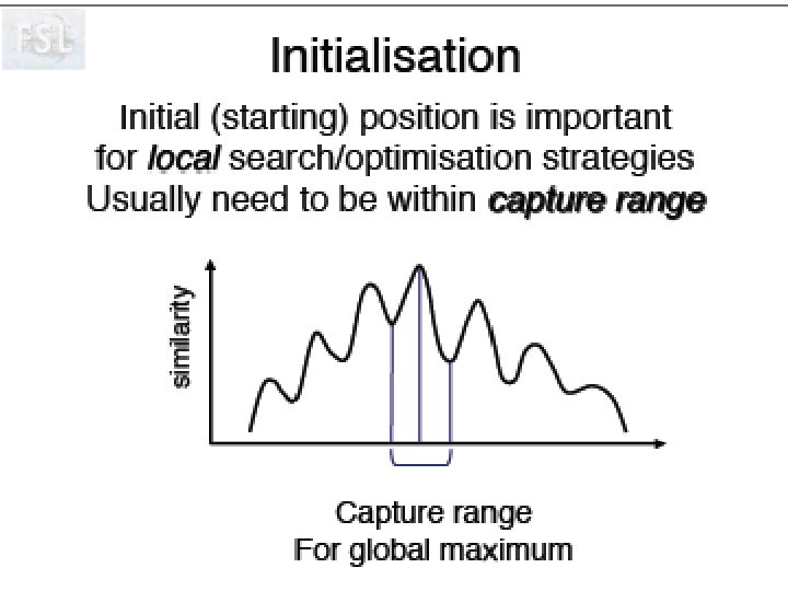
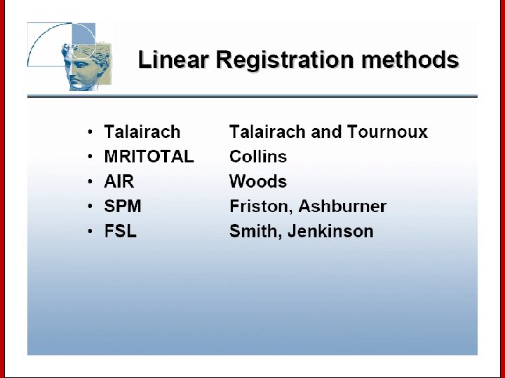
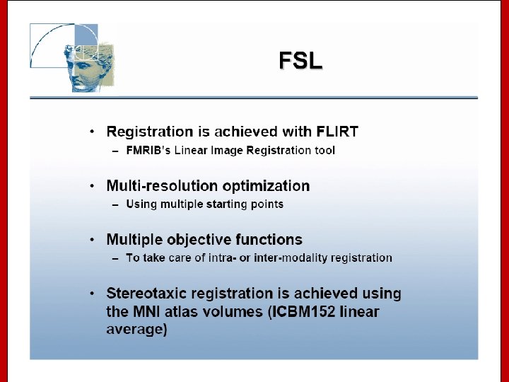
 Image Registration • Registration - i. e. Optimise the parameters that describe a spatial transformation between the source and reference (template) images mritotal: créer la matrice de transformation. xfm • Transformation - i. e. Re-sample according to the determined transformation parameters p. ex: mincresample ou resample_tal: appliquer la transformation aux données

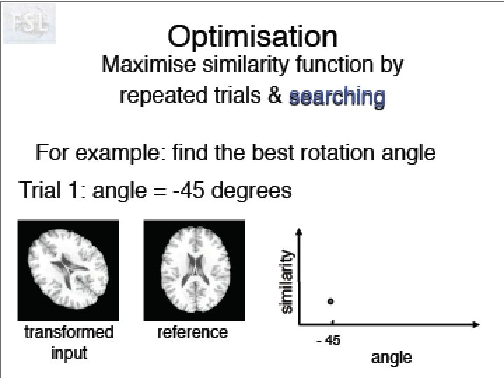
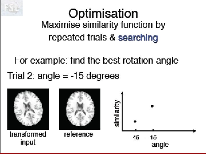
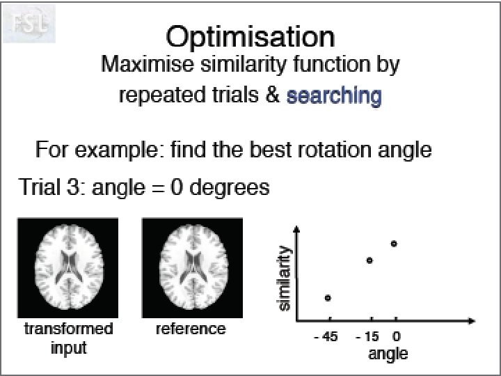
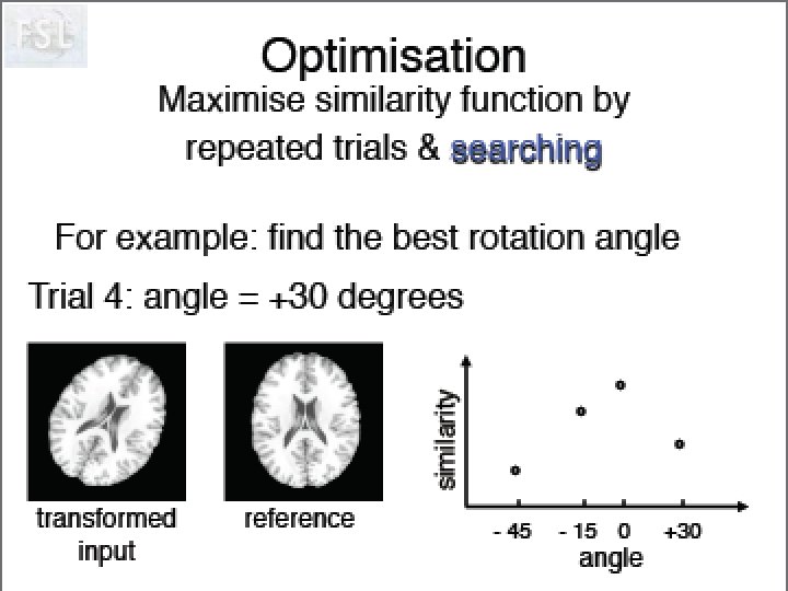
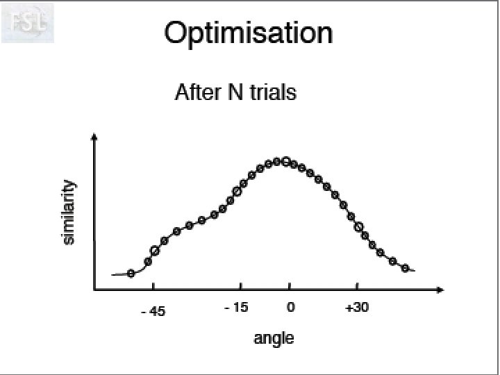
 Idée de Neurolens Pourquoi normaliser des données fonctionnelles sur un ‘template’ anatomique. Création d’un template T 2* Visualisation du processus d’optimisation!
 Slides Aknowledgements Louis Collins, Montreal Neurological Institute Andrew Janke, Montreal Neurological Institute FSL & Free. Surfer Course, f. MRIb, Oxford