

e8642fd3ba9c135c522173d2f57e713a.ppt
- Количество слайдов: 81
 Neurotrauma James F. Holmes, MD, MPH Professor, Department of Emergency Medicine University of California at Davis - School of Medicine Director, EM Research Fellowship Director, Department of EM Journal Club Davis, California
 • Diagnosis of patients") Objectives • Epidemiology • Types of traumatic brain injuries (TBI) • Diagnosis of patients with TBI • Treatment of TBI
Objectives • Epidemiology • Types of traumatic brain injuries (TBI) • Diagnosis of patients with TBI • Treatment of TBI
 Essentials of TBI • Most traumatic deaths secondary to TBI • Secondary brain injury may cause morbidity and mortality but is often preventable • Initial Resuscitation and care: – Maintain oxygenation – Appropriate ventilation – Maintain blood pressure (prevent hypotension) • Cranial CT scanning is the diagnostic test of choice
 brain injury – Structural damage from the initial impact –") Pathophysiology • Primary (initial) brain injury – Structural damage from the initial impact – Prevention strategies to minimize the number of patients sustaining head injuries • Example: motorcycle helmets
Pathophysiology • Primary (initial) brain injury – Structural damage from the initial impact – Prevention strategies to minimize the number of patients sustaining head injuries • Example: motorcycle helmets
 Pathophysiology • Secondary brain injury – Tissue injury occurring after the initial injury • Usually in the initial 24 hours • Hypoxia • Mass effect • Limited blood flow • Infection • Hyperthermia – Treatment goal: prevent these types of insults with appropriate treatment
 • Goal") Pathophysiology • CPP = MAP - ICP – Cerebral perfusion pressure (CPP) • Goal is > 70 mm. Hg – Mean Arterial Pressure (MAP) – Intracranial Pressure (ICP) • Intracranial pressure may be with: – mass effect of a bleed – generalized brain edema • Cellular dysfunction occurs with decreased perfusion to the brain cells
Pathophysiology • CPP = MAP - ICP – Cerebral perfusion pressure (CPP) • Goal is > 70 mm. Hg – Mean Arterial Pressure (MAP) – Intracranial Pressure (ICP) • Intracranial pressure may be with: – mass effect of a bleed – generalized brain edema • Cellular dysfunction occurs with decreased perfusion to the brain cells
 Types of Injuries • • Skull Fracture Concussion Contusion Intracranial hematoma – Epidural, Subdural, Epidural, Intracerebral • Diffuse axonal injury • Intraventricular and Subarrachnoid hemorrhages • Penetrating injury
 Skull Fracture • Multiple types: – Linear, depressed, and basilar • CT must be viewed on bone windows • Most important feature is injury occurring to the brain at the site of the skull fracture • Linear skull fracture: – If closed, can simply be observed
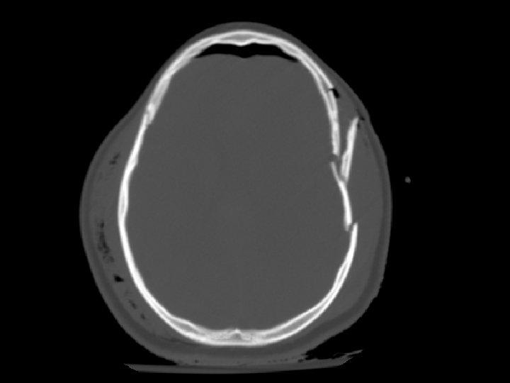
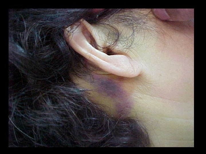
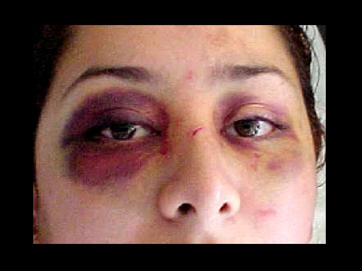
 Skull Fracture • Basilar skull fracture: – Most often involving the temporal bone – Clinical diagnosis (CT often fails to show fracture) • Raccoon’s (Panda’s) eyes, hemotympanum, Battle’s sign (mastoid ecchymosis), CSF rhinorrhea • Depressed skull fracture: – Surgical elevation: • if greater than width of skull or • > 5 mm
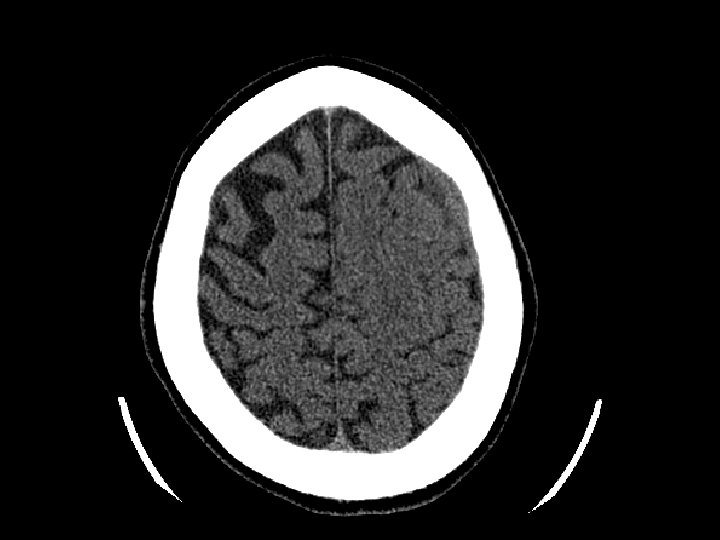
 Concussion • Brief change in brain function usually with loss of consciousness/amnesia • Head CT often normal • Post-concussive syndrome: – Headache, confusion, difficulty concentrating, memory problems, nausea – May last hours to months • Treatment – Simple observation – Limit activity that may reinjure patient’s head • Avoid “repeat concussion”
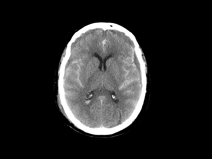
 Cerebral Contusion • Bruising to the brain • Most common traumatic finding on CT • Large contusions may have significant bleeding (hemorrhagic contusion or intracerebral hematoma), edema, or cause seizures • Location: Frontal and Temporal lobes – Due to irregularity of the skull base • Treatment: close observation
 • Hemorrhage “above” the dura – Usually arterial (middle meningeal artery)") Epidural Hematoma (EDH) • Hemorrhage “above” the dura – Usually arterial (middle meningeal artery) but can also be from venous bleeding • CT: convex (lens shaped) area of blood – Bleeding restricted by the dura’s attachment to the skull • 5 -15% of patients with TBI on CT have an EDH
Epidural Hematoma (EDH) • Hemorrhage “above” the dura – Usually arterial (middle meningeal artery) but can also be from venous bleeding • CT: convex (lens shaped) area of blood – Bleeding restricted by the dura’s attachment to the skull • 5 -15% of patients with TBI on CT have an EDH
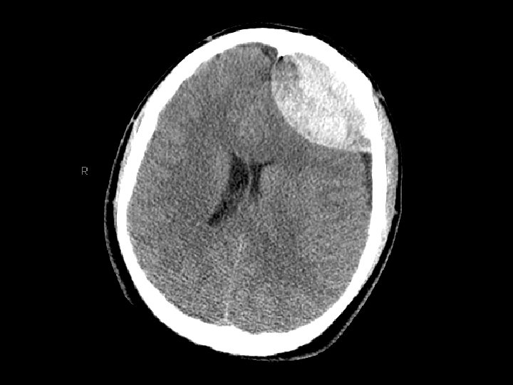
 • Potentially rapidly expanding with mass effect and uncal herniation (fixed/dilated") Epidural Hematoma (EDH) • Potentially rapidly expanding with mass effect and uncal herniation (fixed/dilated pupil) – “Awake and then die” • Most EDHs can be observed • EDHs with mass effect require surgical drainage
Epidural Hematoma (EDH) • Potentially rapidly expanding with mass effect and uncal herniation (fixed/dilated pupil) – “Awake and then die” • Most EDHs can be observed • EDHs with mass effect require surgical drainage
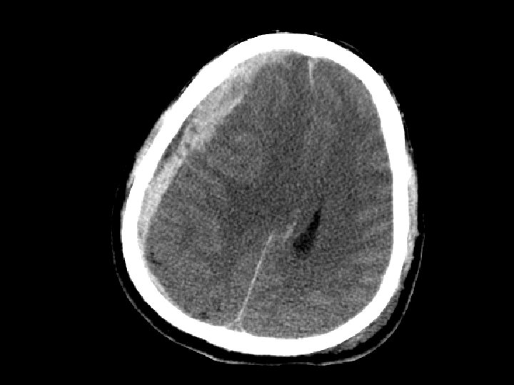
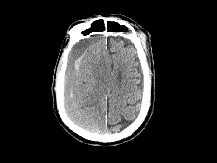
 • Hemorrhage between the dura and the brain – More common") Subdural Hematoma (SDH) • Hemorrhage between the dura and the brain – More common than EDH – 30% of patients with TBI have a SDH • CT: Crescent shaped area of blood • Can be acute, subacute or chronic – Subacute or chronic presents days/weeks after injury
Subdural Hematoma (SDH) • Hemorrhage between the dura and the brain – More common than EDH – 30% of patients with TBI have a SDH • CT: Crescent shaped area of blood • Can be acute, subacute or chronic – Subacute or chronic presents days/weeks after injury
 • Elderly at risk (less severe mechanisms) – Brain shrinks with") Subdural Hematoma (SDH) • Elderly at risk (less severe mechanisms) – Brain shrinks with age resulting in stretching of the bridging veins in the subdural space • May gradually enlarge (slower than EDH) • Surgical drainage for lesions resulting in masse effect and shift
Subdural Hematoma (SDH) • Elderly at risk (less severe mechanisms) – Brain shrinks with age resulting in stretching of the bridging veins in the subdural space • May gradually enlarge (slower than EDH) • Surgical drainage for lesions resulting in masse effect and shift
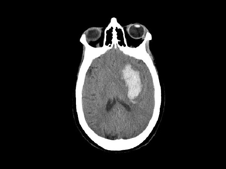
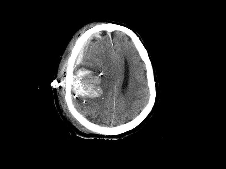
 Intracerebral Hematoma • Similar to cerebral contusions – More severe type injury – Bleeding within the brain itself • Frontal and temporal lobes • Close observation for progression of bleeding • May cause mass effect that results in herniation
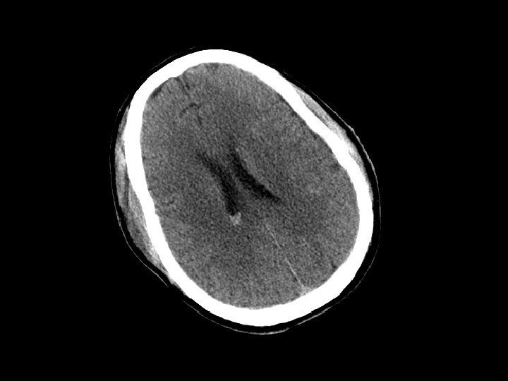
 • Collection of blood in the ventricles • Isolated IVH usually") Intraventricular Hemorrhage (IVH) • Collection of blood in the ventricles • Isolated IVH usually not problematic and requires no specific treatment
Intraventricular Hemorrhage (IVH) • Collection of blood in the ventricles • Isolated IVH usually not problematic and requires no specific treatment
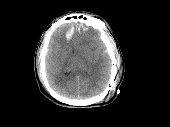
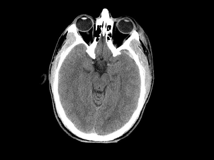
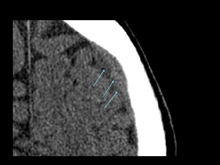
 • SAH and contusion are most common injuries on CT after") Subarrachnoid Hemorrhage (SAH) • SAH and contusion are most common injuries on CT after blunt trauma • CT: blood in the sulci and basal cisterns • Often associated with intracranial hematomas • Must consider that the SAH caused the trauma (spontaneous bleed and then fall) • Complications: – Arterial vasospasm: 2 -3 days after injury
Subarrachnoid Hemorrhage (SAH) • SAH and contusion are most common injuries on CT after blunt trauma • CT: blood in the sulci and basal cisterns • Often associated with intracranial hematomas • Must consider that the SAH caused the trauma (spontaneous bleed and then fall) • Complications: – Arterial vasospasm: 2 -3 days after injury
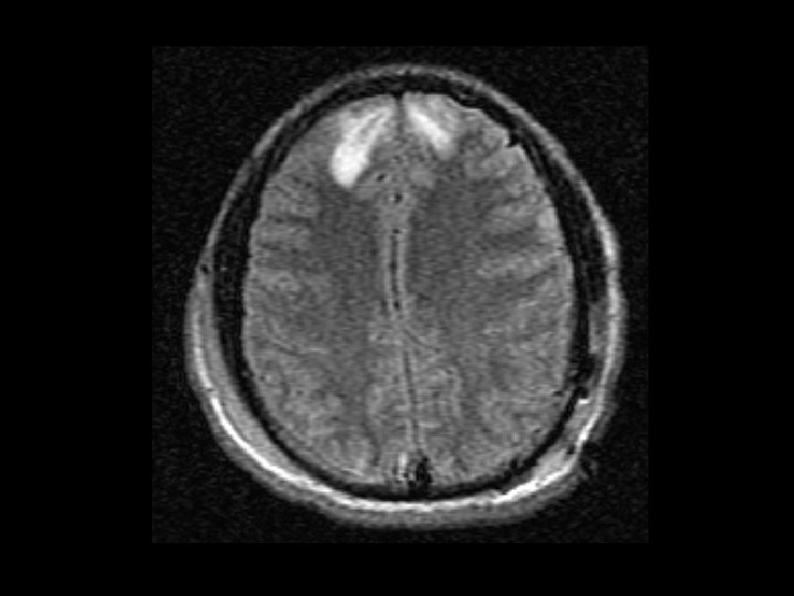
 • Shearing and rotational forces disrupt the axonal network •") Diffuse Axonal Injury (DAI) • Shearing and rotational forces disrupt the axonal network • Mechanism: rapid acceleration/deceleration injury • Imaging – Not well visualized by CT – MRI can better define the extent of injury • No specific therapy • Likely results in the persistent neurologic deficits in patients with normal CT scans but substantial injury
Diffuse Axonal Injury (DAI) • Shearing and rotational forces disrupt the axonal network • Mechanism: rapid acceleration/deceleration injury • Imaging – Not well visualized by CT – MRI can better define the extent of injury • No specific therapy • Likely results in the persistent neurologic deficits in patients with normal CT scans but substantial injury
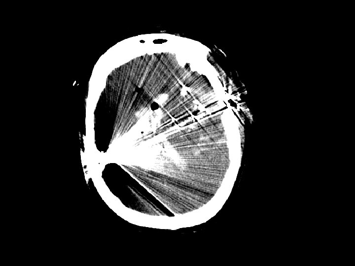
 but also from stab wounds (SWs) •") Penetrating Injury • Primarily gunshot wounds (GSWs) but also from stab wounds (SWs) • Higher rate in urban areas • Injuries that cross the midline have very poor outcomes • Periorbital/nasal region risk of infection
Penetrating Injury • Primarily gunshot wounds (GSWs) but also from stab wounds (SWs) • Higher rate in urban areas • Injuries that cross the midline have very poor outcomes • Periorbital/nasal region risk of infection
 Epidemiology of Head Injury • Varies by country, but in general: – Most common in males (ages 15 -30 years) – Highest rates of TBI on CT in: • elderly (>65 years) • young (<10 years) • Mechanisms of injury: – Motor vehicle crash (including pedestrian/bike) – Falls (especially the young and elderly) – Assaults • Alcohol frequently involved
 Classification of Neurotrauma • Multiple different classification schemes • Minimal – GCS = 15, No loss of consciousness – Normal alertness, memory and neuro exam – No evidence of skull fracture • Mild – – Brief loss of consciousness, amnesia GCS = 14 Impaired alertness, memory No evidence of skull fracture
") Classification of Neurotrauma • Moderate or potentially severe – – – Prolonged (>5 minutes) loss of consciousness GCS 9 -13 Focal neurological deficit Post-traumatic seizure Intracranial lesion on CT scan Palpable depressed skull fracture • Severe: – GCS < 8 – Requires airway control
Classification of Neurotrauma • Moderate or potentially severe – – – Prolonged (>5 minutes) loss of consciousness GCS 9 -13 Focal neurological deficit Post-traumatic seizure Intracranial lesion on CT scan Palpable depressed skull fracture • Severe: – GCS < 8 – Requires airway control
 Prehospital Care • Initial GCS score • Spine immobilization • Airway management for moderate/severe head injury – Bag valve mask with oxygen to assist with ventilation – Endotracheal intubation not shown to help • Suggests harm in multiple studies
 History • Mechanism of injury – any seizure after the event? • Prehospital care – Medications given (and times) – Spine immobilization • Medical history • Allergies • Medications – Especially anticoagulant medications • Tetanus
 • Assign a GCS") Primary Survey • Standard ATLS (A, B, C, D, E) • Assign a GCS score to the patient – May use AVPU scale • Alert • Verbal stimuli • Painful stimuli • Unresponsive • Cervical spine protection • Movement in all extremities if patient to be paralyzed for airway management
Primary Survey • Standard ATLS (A, B, C, D, E) • Assign a GCS score to the patient – May use AVPU scale • Alert • Verbal stimuli • Painful stimuli • Unresponsive • Cervical spine protection • Movement in all extremities if patient to be paralyzed for airway management
 Primary Survey: Airway Managment • Most common reason for intubation in trauma • Rapid Sequence Intubation is standard in USA – Premedications • Lidocaine: 1 -2 mg/kg IV 3 -4 minutes prior, potentially protects the rise in ICP with intubation – Sedatives • Etomidate: 0. 3 mg/kg, no effects on blood pressure • Benzodiazepines/Thiopental: volume status must be normal – Paralytics • Succinylcholine: 1 -1. 5 mg/kg, paralysis for 5 minutes • Rocuronium: 1 mg/kg, paralysis for 20 -30 minutes
 score Standard GCS Pediatric GCS Spontaneous To Voice To Pain") Glasgow Coma Scale (GCS) score Standard GCS Pediatric GCS Spontaneous To Voice To Pain None 4 3 2 1 Verbal Response Oriented Confused Inappropriate words Incomprehensible sounds None 5 4 3 2 1 Coos/babbles Irritable/cries Cries to pain Moans None 5 4 3 2 1 Motor Response Follows commands Localizes pain Withdraws to pain Abnormal flexure posturing Abnormal extension posturing None 6 5 4 3 2 1 Spontaneous movement Withdraws to touch Withdraws to pain Abnormal flexure posturing Abnormal extension posturing None 6 5 4 3 2 1 Eye Opening
Glasgow Coma Scale (GCS) score Standard GCS Pediatric GCS Spontaneous To Voice To Pain None 4 3 2 1 Verbal Response Oriented Confused Inappropriate words Incomprehensible sounds None 5 4 3 2 1 Coos/babbles Irritable/cries Cries to pain Moans None 5 4 3 2 1 Motor Response Follows commands Localizes pain Withdraws to pain Abnormal flexure posturing Abnormal extension posturing None 6 5 4 3 2 1 Spontaneous movement Withdraws to touch Withdraws to pain Abnormal flexure posturing Abnormal extension posturing None 6 5 4 3 2 1 Eye Opening
 GCS score • Pupil examination • General neurological examination") Secondary Survey • Formal (repeat) GCS score • Pupil examination • General neurological examination – – Cranial nerves Motor exam Sensory exam Reflexes
Secondary Survey • Formal (repeat) GCS score • Pupil examination • General neurological examination – – Cranial nerves Motor exam Sensory exam Reflexes
 Secondary Survey • Head examination: – Lacerations and possible skull fracture – Evaluation for Basilar skull fracture • Hemotympanum • Raccoon’s (panda’s) eyes • Battle’s sign – mastoid ecchymosis, often takes > 12 hours to develop • CSF from the nose: – Halo test: drop of fluid onto a towel, central spot bloody with surrounding tint (CSF) – CSF contains glucose, nasal secretions do not
 Diagnostic Testing • CT scan: – – Primary screening tool Has decreased morbidity and mortality Images viewed in bone and brain windows Images rapidly obtained (<1 minute on fast CT scanners) – Exposes the patient to large amounts of radiation – Costly
 Diagnostic Testing • CT scan: – Alert patients with normal CT scans are at very low risk for neurologic deterioration • Safe to discharge home from the ED – CT limitations: • Ability to detect diffuse axonal injury • No information on blood flow to the brain
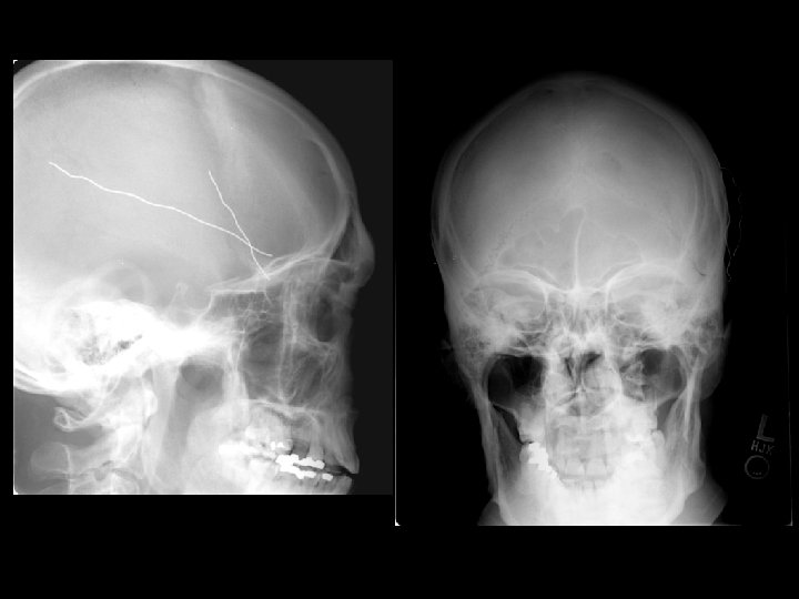
 Diagnostic Testing • Skull radiographs: – May localize a foreign body – low utility and generally not performed in those at risk for brain injury – Consider in very young children (<1 year) who appear well but only have a hematoma to head
 Diagnostic Testing • MRI: – Better than CT – May show diffuse axonal injury and injuries not identified by CT scan – Not used as a routine screening test • 30 – 45 minutes to obtain images • Very costly and not always available
 Evaluation – Mild Head Injury • Diagnostic evaluation with cranial CT imaging when appropriate – GCS 14 -15 with LOC: • TBI on CT in 4 -15% • Neurosurgery 1 -3% • Determining appropriate indications to obtain CT is difficult – GCS = 14: patient should get a CT scan – GCS = 15: not all patients need a CT scan • Several rules to help determine when CT should be performed have been published
 GCS = 15: Indications for CT • • Canadian Cranial CT Rule Stiell I, Lancet, 2001 New Orleans Rule Haydel, New England Journal Medicine, 2000 NEXUS Cranial CT Rule Mower W, Journal Trauma, 2005 NICE Head Injury Guidelines – http: //www. nice. org. uk/nicemedia/live/11836/36259. pdf • These rules are designed to more appropriately utilize CT in those with GCS = 15 but these rules may actually increase CT use
 – –") Canadian Cranial CT Rule • High Risk (to identify those needing neurosurgery) – – Failure to reach GCS=15 within 2 hours of injury Open or basilar skull fracture Vomiting more than once Age > 65 years • Moderate Risk (to identify those with brain injury on CT) – Amnesia >30 minutes before accident – High risk mechanism: auto v. ped, ejection from a motor vehicle, fall over 3 feet/5 stairs
Canadian Cranial CT Rule • High Risk (to identify those needing neurosurgery) – – Failure to reach GCS=15 within 2 hours of injury Open or basilar skull fracture Vomiting more than once Age > 65 years • Moderate Risk (to identify those with brain injury on CT) – Amnesia >30 minutes before accident – High risk mechanism: auto v. ped, ejection from a motor vehicle, fall over 3 feet/5 stairs
 New Orleans Head CT Rule • • Short-term memory deficits: 5/9 patients Intoxication: 19/176 patients Age > 60 years: 4/26 patients Seizure: 2/16 patients Headache: 4/81 patients Vomiting: 1/15 patients Above clavicle trauma: 1/197 patients
 NEXUS Head CT Rule • Age < 65 • No evidence of skull fracture or scalp hematoma • No neurologic deficit • No abnormal alertness • No abnormal behavior • No coagulopathy • No persistent vomiting • If negative for all, then no need for CT
 NICE Head Injury Guidelines for CT • • • GCS < 13 on initial assessment GCS < 15 at 2 hours after the injury on assessment Suspected open, depressed, or basilar skull fracture Post-traumatic seizure Focal neurological deficit More than one episode of vomiting in adults Coagulopathy Amnesia for events > 30 minutes before impact Age > 65 years with loss of consciousness or amnesia Dangerous mechanism of injury (pedestrian struck by a motor vehicle, an occupant ejected from a motor vehicle or a fall from a height of greater than 1 m or five stairs) with loss of consciousness or amnesia
 GCS = 15: Indications for CT • Risk factors for injury on CT from these prior studies include: – – – – Elderly: > 60 or 65 years of age Vomiting Altered Mental status/abnormal neurologic exam Skull fracture: depressed, basilar, linear LOC with symptoms (headache, nausea, etc) Post-traumatic seizure Coagulopathy (on blood thinning medications)
 GCS = 15: CT caveats • If CT is not available, consider: – Transfer to center with CT capabilities – Observation in ED or prolonged observation in hospital depending on resources
 Pediatric: Indications for CT • Children different than adults in many aspects • Indications for cranial CT in children different than adults • Risk of radiation from the CT scan is greatest in children • Recent large pediatric study to evaluate indications for cranial CT in children – 42, 412 children with GCS = 14 -15 – Kuppermann, Lancet; 2009; 374: 1160
 Suggested CT algorithm for patients >2 years old with GCS 14 -15 after head trauma GCS = 14 or other signs of altered mental status, or Palpable skull fracture No Yes 14. 0% of population 4. 3% risk of clinically important TBI Yes History of LOC, or vomiting, or Severe mechanism of injury, or Severe Headache No 27. 7% of population 0. 9% risk of Clinically important TBI 58. 3% of population <0. 05% risk of clinically important TBI CT not recommended CT recommended Observation vs. CT: based on clinical factors including: Physician experience Multiple versus isolated findings Worsening symptoms or signs after ED observation Parental preference
 Suggested CT algorithm for patients <2 years old with GCS 14 -15 after head trauma GCS = 14 or other signs of altered mental status, or Palpable skull fracture No Yes 13. 9% of population 4. 4% risk of clinically important TBI Yes Occipital/parietal/temporal scalp hematoma, or History of LOC > 5 sec, or Severe mechanism of injury, or Not acting normally per parent No 32. 6% of population 0. 9% risk of Clinically important TBI 53. 5% of population <0. 02% risk of clinically important TBI CT not recommended CT recommended Observation vs. CT: based on clinical factors including: Physician experience Multiple versus isolated findings Worsening symptoms or signs after ED observation Age < 3 months Parental preference
 Treatment of Mild Head Injury • Safe to discharge patients with normal cranial CT scans to home: – Adults (Livingston, Ann Surg 2000) • 2, 152 adults with normal CT • One patient had a craniotomy for skull fracture noted on initial CT scan • NPV for craniotomy = 99. 94% – Pediatrics (Holmes, AEM 2010) • 13, 543 children with normal CT • None underwent craniotomy • NPV for craniotomy = 100%
 Treatment of Mild Head Injury • Appropriate discharge instructions • Warnings regarding concussion symptoms – Explanation of post-concussive symptoms – Exclusion from activity that may result in a repeat head injury (i. e. sports) • Headaches: paracetamol, aspirin • Nausea/Vomit: antiemetics
 Moderate/Severe Head Injury
 Initial Evaluation of Moderate/Severe Head Injury • A, B, C, D, E – Airway control for GCS < 8 – Prevent hypotension and Hypoxia • Hypotension: triples risk of death • Hypoxia: doubles risk of death • Movement in all extremities prior to paralysis if possible • If comatose, assess brainstem function: – Corneal reflex: lightly touch cornea will result in blinking (Cranial Nerves V and VII)
 Initial Evaluation of Moderate/Severe Head Injury • GCS ≤ 13 • Immediate CT scan (if stable) • If no CT available, then consult with neurosurgeon and transfer to site with CT • Include cervical spine imaging: CT of the cervical spine recommended the lower the GCS score is
 Treatment of Moderate/Severe Head Injury • Maintain CPP and the patient’s airway – treat hypoxia and hypotension immediately • Intracranial pressure monitoring in those with GCS < 8 – Although no evidence outcome is improved – Can be performed in the ED but normally in the operating suite by a neurosurgeon • Elevate head of bed (30 ) if no cervical spine injury
 Treatment of Moderate/Severe Head Injury • Mannitol – Increases mean arterial pressure and lowers ICP – Patient must be stable • Do not give if volume depleted as it may result in hypotension – Maintain volume status – Keep serum osmolarity < 320 mmol/L
 Treatment of Moderate/Severe Head Injury • Mannitol – 0. 5 – 1 gram/kg IV bolus • Repeat boluses better than drip – Indications • increased ICP: once ICP monitor placed • Give in ED: – signs of transtentorial herniation – progressive neurological deterioration
 Treatment of Moderate/Severe Head Injury • Anticonvulsants – Used for the first 7 days after injury in patients with high risk TBI: • GCS<10, CT with contusion, hematoma, or depressed skull fracture, Seizure • No evidence for anticonvulsants beyond 7 days – Phenytoin 15– 18 mg/kg IV • Carbamezapine and kepra also used – Acute seizure: • Standard therapy: – oxygen – Benzodiazepines, phenytoin, barbituates
 Treatment of Moderate/Severe Head Injury • Sedation and Pain control – Intubated patients require sedation to prevent elevation of ICP (fighting the ventilator) – Sedation must be balanced with the need for repeat neurologic examinations to determine if brain injury is worsening – Pain control: • Fentanyl ideal narcotic in TBI patients – Sedation: • Propofol: used with increasing frequency in TBI patients • Benzodiazepines: • Barbiturates: high doses in those with uncontrollable ICP
 Treatment of Moderate/Severe Head Injury • Surgical therapy – Craniotomy • Treatment of choice for correctable mass lesions • Better outcomes with no delays to surgery • Large hematomas (with shift), depressed skull fractures, penetrating injuries (debride the wound) – Intracranial pressure monitors/ventriculosotmy • Monitor pressure and drain CSF
 Treatment of Moderate/Severe Head Injury • Emergency Burr Hole – Indicated in patients with rapid neurologic deterioration and an expanding hematoma and unable to get to a neurosurgeon – Other measures to reduce ICP should be attempted prior to this procedure – Burr hole performed on side of dilated pupil at the temporal bone – MD must be familiar with technique prior to attempting
 Treatment of Moderate/Severe Head Injury • Hyperventilation – Previously a standard treatment in those with TBI – Now known to be harmful • Worse outcomes in those with prophylactic hyperventilation (Muizelaar, J Neurosurg 1991) – No prophylactic hyperventilation – Goal p. CO 2: ~ 35 mm. Hg • Do not let p. CO 2 get below 30 mm. Hg
 Treatment of Moderate/Severe Head Injury • Hyperventilation – Do not hyperventilate during initial 24 hours after injury – Hyperventilate for brief periods when there is acute neurologic deterioration – Hyperventilate for ICP that is refractory to sedation, paralysis, cerebral spinal fluid drainage, and osmotic diuretics • This is one of the last therapies
 Treatment of Moderate/Severe Head Injury • Antibiotics – Basilar skull fracture – no evidence that prophylactic antibiotics are beneficial – Open fractures • Controversial, little evidence to support • Antibiotics to cover skin flora
 Treatment of Moderate/Severe Head Injury • Corticosteroids – Believed to decrease cerebral edema – CRASH trial (Roberts, Lancet 2004; 364: 1321) • 10, 008 adults in a randomized controlled trial – Increased risk of death with steroids • Relative risk = 1. 18 (95% CI 1. 09, 1. 27) – No longer recommended in patients with traumatic brain injury
 Treatment of Moderate/Severe Head Injury • Hypertonic saline – No benefit in 2 large randomized controlled trials • Hypothermia – Experimental in those with isolated head injury – No good clinical evidence that it is useful • Albumin – Unlikely to be beneficial and generally not recommended • Progesterone – Promising in several small studies • Wright, Ann Emerg Med 2007; 49: 391 – Large trial ongoing in the USA
 Points to remember • Avoid hypoxemia and hypotension in the TBI patient • Airway controlled if GCS ≤ 8 • Avoid hyperventilation – keep p. CO 2 from 35 -40 mm. Hg • Mannitol for evidence of brain herniation or neurologic deterioration • Anticonvulsants for those at risk of posttraumatic seizures • Avoid delays to the OR for those patients needing craniotomy
 Points to remember • Depressed mental status may be due to brain injury in the intoxicated patient • CT scanning is the screening test of choice but is not necessary in all patients with minor head injury • Patients with normal cranial CT scans are at low risk for deterioration and may be discharged from the ED if otherwise stable