

550aa7e83147e6abd979daa98501f283.ppt
- Количество слайдов: 166
 Neurosurgery
 Neuron- nerve cell n Dendrite One or more Conducts impulses toward the cell body n Axon Conducts impulses away from the cell body
 Synapse Space between the junction of two neurons
 Neurotransmitter Substance that is released when the axon is excited Travels across the synapse to the target cell n n n Norepinephrine Acetylcholine Dopamine
 cells which forms a") Myelin The phospholipidprotein of the cell membranes of Schwann (neuroglia) cells which forms a sheath around neurons
Myelin The phospholipidprotein of the cell membranes of Schwann (neuroglia) cells which forms a sheath around neurons
 Myelin, cont. Acts as an insulator to support and protect the nerve cell, and to increase the velocity of impulse transmission
 Gray Matter Nerve tissue composed mainly of the cell bodies of neurons, rather than their myelinated processes
 Gray Matter, cont. Term is generally applied to the gray portions of the central nervous
 Gray Matter, cont. Cerebral cortex Basal ganglia Gray columns of the spinal cord which forms an Hshaped region surrounded by white matter
 White Matter The white substance of the spinal cord and brain, consisting principally of myelinated nerve axons
 The Nervous System
 The Nervous System Central nervous system n Consists of the brain and the spinal cord Peripheral nervous system n Consists of the cranial and spinal nerves
 Sympathetic Parasympathetic") The Nervous System Divided functionally into: n n Voluntary Autonomic (involuntary) Sympathetic Parasympathetic
The Nervous System Divided functionally into: n n Voluntary Autonomic (involuntary) Sympathetic Parasympathetic
 The Nervous System Functions include: n n n Orientation Coordination Conceptual thought Emotion Memory Reflex response
 Cranial Sensation The only pain sensitive structures that cover the brain are: n n n The scalp Extracranial arteries Portions of the dura mater The brain itself is insensate
 Scalp Layers include: n n Skin Subcutaneous tissue n n n Galea Subgaleal space Pericranium
 Scalp, cont. Skin n Tends to be thick Subcutaneous tissue n n Dense, tough, vascular Attached to the galea
 Scalp, cont. Galea n n Tough, fascia like tissue covering the cranium Connects muscles to the temples, forehead, and base of the skull
 Scalp, cont. Subgaleal space n n n Loose areolar tissue Permits mobility of the scalp Bloodless plane, used in standard craniotomy flap
 Scalp, cont. Pericranium n n Periosteum of the skull Separates the galea from the cranium Arterial supply for the scalp n From branches of the external carotid artery
 Skull Formed by 24 bones, joined by serrated bony seams called sutures
 Skull, cont. 8 bones form the walls of the cranial cavity which houses the brain
 Skull, cont. n 4 single bones frontal, occipital, ethmoid, and sphenoid
 Skull, cont. n 2 paired bones temporal and parietal
 Skull, cont. Consists of flattened irregular shaped bones, comprised of 2 tables of compact bone enclosing a layer of spongy bone
 Cranial Fossae Interior anatomically divided into 3 cranial fossae: n n n Anterior Middle Posterior
 Foramen Magnum Largest opening in the skull Permits the spinal cord to join with the brainstem
 Major sutures of the skull Coronal n Joins the frontal and parietal bones Squamous n Borders the squamous part of the temporal bone
 Major sutures of the skull, cont. Major sutures of the skull n Lambdoid Joins the occipital and parietal bones n Sagittal Joins the two parietal bones in the median plane
 Fontanelles Anterior and posterior Located at the top of the skull in front of and behind the parietal bones Both are open at birth
 Fontanelles, cont. Posterior is generally closed by 2 months Anterior is generally closed by about 18 months
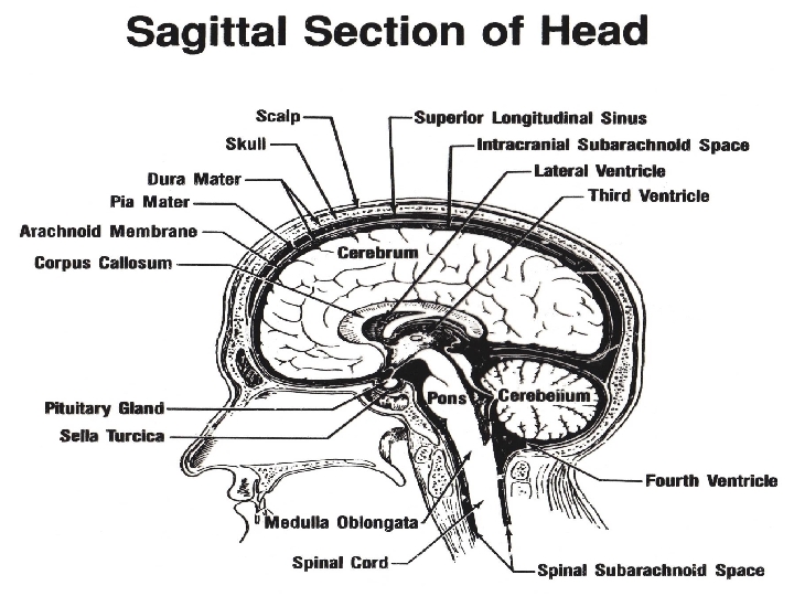
 membranous layers between the brain and the skull") Meninges Three (3) membranous layers between the brain and the skull "The meninges P. A. D. the brain. “ n Pia; Arachnoid; Dura.
Meninges Three (3) membranous layers between the brain and the skull "The meninges P. A. D. the brain. “ n Pia; Arachnoid; Dura.
 Meninges Dura Mater n Tough shiny fibrous outermost membrane Arachnoid n Middle layer, fine membrane Pia Mater n Innermost membrane, like gossamer
 Dura Mater Tentorium Cerebelli n n A transverse fold of dura forming the roof of the posterior fossa Supports the temporal and occipital lobes of the cerebral hemispheres. Supratentorial n Structures above the tentorium Infratentorial n Structures below the tentorium
 Dura Mater Falx Cerebri Falx cerebri n A fold of the dura mater that lies in the longitudinal fissure and separates the two cerebral hemispheres
 Dura Mater Falx cerebelli n A fold of the dura mater that forms a vertical partition between the hemispheres of the cerebellum Venous sinuses n n Lie at the margins of the dural folds Drain blood from the intracranial structures into the jugular veins
 Dura Mater Venous sinuses n n Venous channels found between the layers of the dura mater They receive blood from internal and external veins of the brain and ultimately empty into the internal jugular vein.
 Arachnoid Outer surface of the arachnoid membrane adheres closely to the dura, with no space normally between the 2 membranes The inner surface of the arachnoid is separated from the pia mater beneath it by the subarachnoid space, which is filled with CSF
 Pia Mater Attaches to the gray matter and dips into the sulci and gyri n n n Sulci- furrow, groove, or slight depression Gyri- convolutions Fissures- deeper grooves Has a rich vascular network that helps form the choroid plexus of the ventricles
 n Between the cerebral") Brain Cerebrum n Largest part of the brain Midbrain (mesencephalon) n Between the cerebral hemispheres and the pons Brainstem (hindbrain) n Immediately below the midbrain
Brain Cerebrum n Largest part of the brain Midbrain (mesencephalon) n Between the cerebral hemispheres and the pons Brainstem (hindbrain) n Immediately below the midbrain
 Brain Encephalon n The brain Diencephalon n 2 nd portion of the brain, or interbrain (deep structures)
 Cerebrum Divided into right and left cerebral hemispheres by a longitudinal fissure
 Cerebrum, cont. Each hemisphere controls sensation and motor activity to and receives sensory stimuli from the opposite half of the body
 Cerebrum, cont. Each hemisphere is divided into frontal, parietal, occipital and temporal lobes, insula, rhinencephalon, basal ganglia, thalamus and hypothalamus
 Frontal lobe Higher mental functions of intellect Abstract reasoning
 Parietal lobe Make sense of speech and formulate words with emotional content Hearing and the ability to understand speech
 Parietal lobe, cont. Interpret sensory information Read printed words Visual memory Music
 Occipital lobe Posterior to the parieto-occipital fissure Receives and integrates visual impulses and registers them as meaningful images
 Temporal lobe Comprehension and verbalization of words
 Insula a. k. a. Island of Reil n n n Central lobe of the cerebral hemisphere Triangular area of the cerebral cortex lying in the floor of the lateral fissure Concerned with autonomic functions
 Insula, cont. Processes convergent information to produce an emotionally relevant context for sensory experience Has an important role in pain experience and the experience of a number of basic emotions, including anger, fear, disgust, happiness and sadness Imaging studies have also implicated the insula in conscious desires, such as food craving and drug craving
 Rhinencephalon Near the sphenoid bone n Receives and integrates olfactory impulses
 Basal Ganglia Four masses of gray matter located deep in the cerebral hemispheres
 Basal Ganglia, cont. Contribute to some of the subconscious aspects of voluntary movement Initiate stimuli for movement and provide essential links in complex motor circuits
 Thalamus Ovoid, gray nuclear mass in the lateral wall of the 3 rd ventricle Is part of the diencephalon Intimately connected to the pituitary gland
 Thalamus, cont. All sensory stimuli, with the exception of olfactory, are received, associated, integrated, and relayed to the specific cortical areas
 Hypothalamus Activates, controls and integrates the peripheral autonomic nervous system, endocrine processes, and many somatic functions such as body temperature, sleep and appetite
 Sulci and Gyri Surface of the hemispheres form convolutions called gyri and intervening furrows called sulci.
 Lateral sulcus a. k. a. fissure of Sylvius Marks off the temporal lobe.
 Central sulcus A. K. A. Fissure of Rolando Separates the motor from the sensory cortex
 Central sulcus, cont. Anterior to central sulcus is the motor area, controlling voluntary motor function
 Central sulcus, cont. Posterior to the central sulcus is the sensory area, which receives sensory impulses
 Midbrain A. K. A. Mesencephalon Between the cerebral hemispheres and the pons Connects the pons and the cerebellum with the hemispheres of the cerebrum
 Midbrain, cont. Contains reflex centers for eye and head movements in response to visual and auditory stimuli Controls the majority of eye movements
 Brainstem A. K. A. hindbrain Immediately below the midbrain Consists of the Pons and the Medulla Oblongata Surgery here is very dangerous
 Pons Origin of cranial nerves V, VII, and VIII Relays sensory information between the cerebellum and cerebrum helps regulate respiration
 Medulla Oblongata Origin of cranial nerves IX, X, XI, and XII Controls cardiovascular and respiratory regulatory centers
 Cerebellum Occupies most of the posterior fossa Has 2 lateral lobes and a medial portion called the vermis
 Cerebellum, cont. Principally concerned with balance and coordination of movement
 Cerebrospinal fluid A. K. A. CSF Clear and colorless fluid Much of it originates in the choroid plexuses of the ventricles Bathes the brain and spinal cord
 Cerebrospinal fluid functions Helps support the weight of the brain Acts as a cushion for the brain and spinal cord by absorbing some of the force of external trauma Keeps intracranial pressure (ICP) constant by variations of volume
 Ventricles Four communicating cavities filled with CSF
 One lying in each cerebral hemisphere Each has a body and") Lateral ventricles (2) One lying in each cerebral hemisphere Each has a body and three hornsfrontal, occipital, and temporal Drains into the foramen of Monro (aka interventricular foramen)
Lateral ventricles (2) One lying in each cerebral hemisphere Each has a body and three hornsfrontal, occipital, and temporal Drains into the foramen of Monro (aka interventricular foramen)
 Third ventricle Centrally located below the bodies of the lateral ventricles Communicates anteriorly with the lateral ventricles through the foramen of Monro
 Third ventricle, cont. Communicates posteriorly with the fourth ventricle through the aqueduct of Sylvius, a long narrow channel passing through the midbrain
 Fourth ventricle In the posterior fossa between the cerebellum and the brainstem CSF escapes into the subarachnoid space via the foramen of Magendie and the two foramina of Luschka
 Brain blood supply Brain requires 20% more O 2 than any other organ, and utilizes glucose as its chief source of energy The brain normally receives 20% of cardiac output
 n Anterior Vertebral arteries (2) n") Brain blood supply, cont. Internal carotid arteries (2) n Anterior Vertebral arteries (2) n Posterior
Brain blood supply, cont. Internal carotid arteries (2) n Anterior Vertebral arteries (2) n Posterior
 Brain blood supply, cont. Communicate at the base of the brain through the circle of Willis Ensures continuity of circulation if any one of the four main channels is interrupted
 Brain blood supply, cont. Cerebral veins do not parallel the arteries Located in the meninges and the deep cerebral veins
 Cranial Nerves- 12 pairs On Old Olympus' Towering Top A Finn And German Viewed Some Hops Oh Oh Oh To Touch And Feel Very Good Velvet, Ah Heaven Oh, Oh, To Touch And Feel Very Good Velvet. Such Heaven
- Olfactory Sense of smell You only have one nose…") Cranial Nerve I (#1)- Olfactory Sense of smell You only have one nose…
Cranial Nerve I (#1)- Olfactory Sense of smell You only have one nose…
- Optic Sense of sight …but you have two eyes") Cranial Nerve II (#2)- Optic Sense of sight …but you have two eyes
Cranial Nerve II (#2)- Optic Sense of sight …but you have two eyes
- Oculomotor Controls four extrinsic eye muscles (except the lateral rectus") Cranial Nerve III (#3)- Oculomotor Controls four extrinsic eye muscles (except the lateral rectus and superior oblique) Controls the intrinsic muscles (cilliary and iris)
Cranial Nerve III (#3)- Oculomotor Controls four extrinsic eye muscles (except the lateral rectus and superior oblique) Controls the intrinsic muscles (cilliary and iris)
- Trochlear Controls the superior oblique eye muscle") Cranial Nerve IV (#4)- Trochlear Controls the superior oblique eye muscle
Cranial Nerve IV (#4)- Trochlear Controls the superior oblique eye muscle
- Trigeminal Sensory supply to the head, face, nose, and mouth,") Cranial Nerve V (#5)- Trigeminal Sensory supply to the head, face, nose, and mouth, and the motor intervention for the muscles of mastication (chewing)
Cranial Nerve V (#5)- Trigeminal Sensory supply to the head, face, nose, and mouth, and the motor intervention for the muscles of mastication (chewing)
- Abducens Controls the lateral rectus muscle of the eye “Abducts”") Cranial Nerve VI (#6)- Abducens Controls the lateral rectus muscle of the eye “Abducts” the eye
Cranial Nerve VI (#6)- Abducens Controls the lateral rectus muscle of the eye “Abducts” the eye
- Facial Controls superficial muscles of the face and scalp, and") Cranial Nerve VII (#7)- Facial Controls superficial muscles of the face and scalp, and the anterior two thirds of the tongue for taste
Cranial Nerve VII (#7)- Facial Controls superficial muscles of the face and scalp, and the anterior two thirds of the tongue for taste
- Acoustic A. K. A. Vestibulocochlear Cochlear branch for hearing and") Cranial Nerve VIII (#8)- Acoustic A. K. A. Vestibulocochlear Cochlear branch for hearing and a vestibular branch for balance
Cranial Nerve VIII (#8)- Acoustic A. K. A. Vestibulocochlear Cochlear branch for hearing and a vestibular branch for balance
Glossopharyngeal Taste and sensations of the tongue, swallowing, secretions of saliva,") Cranial Nerve IX (#9)Glossopharyngeal Taste and sensations of the tongue, swallowing, secretions of saliva, and pharyngeal muscles (partially) n n Glosso- Tongue Pharyngeal. Pharynx
Cranial Nerve IX (#9)Glossopharyngeal Taste and sensations of the tongue, swallowing, secretions of saliva, and pharyngeal muscles (partially) n n Glosso- Tongue Pharyngeal. Pharynx
- Vagus Innervation of pharyngeal and laryngeal musculature, control of heart") Cranial Nerve X (#10)- Vagus Innervation of pharyngeal and laryngeal musculature, control of heart rate, regulation of acid secretions in the stomach, and peristalsis
Cranial Nerve X (#10)- Vagus Innervation of pharyngeal and laryngeal musculature, control of heart rate, regulation of acid secretions in the stomach, and peristalsis
- Spinal Accessory Motor nerve to the sternocleidomastoid and trapezius muscles") Cranial Nerve XI (#11)- Spinal Accessory Motor nerve to the sternocleidomastoid and trapezius muscles Called either Spinal Accessory or just Accessory
Cranial Nerve XI (#11)- Spinal Accessory Motor nerve to the sternocleidomastoid and trapezius muscles Called either Spinal Accessory or just Accessory
Hypoglossal Motor nerve for the tongue n n Hypo- below Glossal-") Cranial Nerve XII (#12)Hypoglossal Motor nerve for the tongue n n Hypo- below Glossal- tongue
Cranial Nerve XII (#12)Hypoglossal Motor nerve for the tongue n n Hypo- below Glossal- tongue
 Cranial Nerves- Recap
 Spinal Column Consists of 33 vertebrae n 7 cervical n 12 thoracic n 5 lumbar n n 7 5 sacral 4 coccygeal 12 5 5 4
 1 st - the atlas n Supports the") Cervical (C 1 to C 7) 1 st - the atlas n Supports the skull 2 nd – the axis n Ligaments hold the 1 st and 2 nd together but allow for considerable rotational movement
Cervical (C 1 to C 7) 1 st - the atlas n Supports the skull 2 nd – the axis n Ligaments hold the 1 st and 2 nd together but allow for considerable rotational movement
 Distinguished by the presence of costal facets for") Thoracic (T 1 to T 12) Distinguished by the presence of costal facets for the articulation of the heads of ribs Body is intermediate in size between the cervical and lumbar vertebrae
Thoracic (T 1 to T 12) Distinguished by the presence of costal facets for the articulation of the heads of ribs Body is intermediate in size between the cervical and lumbar vertebrae
 Body Spinous process Transverse process Lamina Articular facets") Lumbar (L 1 to L 5) Body Spinous process Transverse process Lamina Articular facets
Lumbar (L 1 to L 5) Body Spinous process Transverse process Lamina Articular facets
 Intervertebral disc Fibrocartilaginous cushion separating one vertebral body from another Can herniate
 Fused as one large triangular shaped bone Articulates") Sacrum (S 1 to S 5) Fused as one large triangular shaped bone Articulates with L 5, Co 1 and the two pelvic bones
Sacrum (S 1 to S 5) Fused as one large triangular shaped bone Articulates with L 5, Co 1 and the two pelvic bones
 Fused as one A. K. A. tailbone") Coccyx (Co 1 to Co 4) Fused as one A. K. A. tailbone
Coccyx (Co 1 to Co 4) Fused as one A. K. A. tailbone
 Spinal cord Develops from the embryonic neural tube Covered by the meninges Passes through a central canal in the spinal column to the level of the 1 st or 2 nd lumbar vertebrae
 Spinal cord, cont. Blood supply is from the vertebral arteries Cauda equina n Collection of spinal roots from the inferior (terminal) spinal cord which resembles a horses tail
 Spinal Nerves 31 pairs 2 pairs of spinal nerves exit at each vertebral level n n Anterior motor root Posterior sensory root
 Pathology
 of the skull. The skull cannot") Craniosynostosis Premature ossification of the sutures (and fontanelles) of the skull. The skull cannot expand as the brain grows, so it may require surgical intervention. Pre-op Post-op
Craniosynostosis Premature ossification of the sutures (and fontanelles) of the skull. The skull cannot expand as the brain grows, so it may require surgical intervention. Pre-op Post-op
 Hydrocephalus Condition characterized by abnormal accumulation of CSF within the cranial vault with subsequent dilation of the ventricles
 Hydrocephalus, cont. Results from an increase in CSF production or a decrease in CSF absorption
 Hydrocephalus, cont. TX- surgical intervention to correct the obstruction, reduce production, or shunt the excess fluid to the right atrium or to the peritoneal cavity Intervention has about an 80% success rate
 Meningitis Infection or inflammation of the meninges May elevate CSF pressure
 Encephalitis Inflammatory condition of the brain May elevate CSF pressure
 Meningocele Sac like protrusion of the meninges through a congenital defect in the skull or spinal column Forms a herniated cyst filled with CSF, but does not contain neural tissue
 Spina Bifida A congenital defect in the walls of the spinal caused by a lack of union between the lamina of the vertebrae
 Spina Bifida, cont. Commonly lumbar Membranes of the cord are pushed through the opening if it is wide enough
 Spina Bifida, cont. Contents of the spinal canal may protrude Relatively common n 10 -20 per 1000 live births
 AV Malformation Thin walled vascular channels that connect arteries and veins without the usual intervening capillaries
 AV Malformation, cont. May be microscopic or massive Fistulas may be congenital, or may result from trauma or disease
 AV Malformation, cont. Difficult to treat successfully n n n Feeding vessels can be clipped Total removal gives the best results Laser and Gamma Knife have been successful
 cranial nerve Causes episodes of intense, stabbing,") Trigeminal Neuralgia Disorder of the trigeminal (fifth) cranial nerve Causes episodes of intense, stabbing, electric shock-like pain along one or more of it’s branches, but usually along the maxillary nerve
Trigeminal Neuralgia Disorder of the trigeminal (fifth) cranial nerve Causes episodes of intense, stabbing, electric shock-like pain along one or more of it’s branches, but usually along the maxillary nerve
 Trigeminal Neuralgia, cont. Also called “tic douloureux” because it’s often accompanied by a brief facial spasm or tic Most frequently occurring of all the nerve pain disorders
 Trigeminal Neuralgia, cont. Episodes last from seconds to 2 minutes in duration Cause is thought to be pressure from blood vessels on the trigeminal nerve root
 Trigeminal Neuralgia, cont. Treatment options include: n n Medications Microvascular Decompression Surgery Placement of Teflon pad between vessel and nerve root
 Considerations Hemostatic agents n n n Bone wax Scalp clips Cottonoids Gelfoam and topical thrombin Bipolar and monopolar cautery
 Considerations Hair may be removed with electric clippers, then shaved n Hair is considered the patients property and is placed in a specimen container, labeled and documented on the operative record
 Considerations Headrest or skull clamp may be used for positioning
 Considerations Positioning varies depending upon surgical approach, and may include prone, lateral, supine, or sitting
 Considerations Patient may be positioned on a hypothermia mattress to reduce cerebral blood flow and venous pressure, and to decrease blood volume and ICP Air embolism is a potential problem when the sitting position is used. n n Brain is higher than the heart. Venous pressure may be lower than atmospheric and can allow for air entry into open venous channels
 Considerations Incision site may be marked prior to the prep, or after the prep and prior to draping Mayfield or similar table may be used for cranial procedures
 Considerations Prevent sudden movement around the surgeon and bumping of the OR table or microscope, as slips can be fatal
 to prevent shock to the brain or") Considerations Irrigation must be warm (body temperature) to prevent shock to the brain or nerves n Warming basins are available Microscope may have a camera attached to allow for anticipation Anesthesia may be local or general for craniotomy
Considerations Irrigation must be warm (body temperature) to prevent shock to the brain or nerves n Warming basins are available Microscope may have a camera attached to allow for anticipation Anesthesia may be local or general for craniotomy
 Surgical interventions
 Craniectomy Craniotomy Intracranial Aneurysm AV Malformation Transsphenoidal Hypophysectomy Cranioplasty") Surgical interventions Burr Holes (Trephination) Craniectomy Craniotomy Intracranial Aneurysm AV Malformation Transsphenoidal Hypophysectomy Cranioplasty Stereotaxis Ventriculoatrial Shunt Ventriculoperitoneal Shunt Laminectomy Anterior Cervical Fusion Carotid Artery Ligation Sympathectomy Nerve Repairs Carpal Tunnel Syndrome Ulnar Nerve Transposition
Surgical interventions Burr Holes (Trephination) Craniectomy Craniotomy Intracranial Aneurysm AV Malformation Transsphenoidal Hypophysectomy Cranioplasty Stereotaxis Ventriculoatrial Shunt Ventriculoperitoneal Shunt Laminectomy Anterior Cervical Fusion Carotid Artery Ligation Sympathectomy Nerve Repairs Carpal Tunnel Syndrome Ulnar Nerve Transposition
 Perforator - cranial burr Placed to remove a localized collection of") Burr holes (trephine) Perforator - cranial burr Placed to remove a localized collection of fluid beneath the dura May be used to tap a ventricle, or to drain or treat an abscess
Burr holes (trephine) Perforator - cranial burr Placed to remove a localized collection of fluid beneath the dura May be used to tap a ventricle, or to drain or treat an abscess
 Craniectomy Incision into the skull and removal of bone by enlarging one or more burr holes
 Craniotomy Incision into the skull Use a craniotome to make burr holes, then “connect the dots”
 Bone flap should") Craniotomy, cont. Bone flap may be left attached or removed (free) Bone flap should be wrapped in a moist sponge
Craniotomy, cont. Bone flap may be left attached or removed (free) Bone flap should be wrapped in a moist sponge
 Craniotomy, cont. Moist cottonoid strips and bone wax control bleeding Dura hook, #11 blade, and dura scissors 4 -0 Neuralon on fine needles for retraction and closure of the dura
 Craniotomy, cont. Moist cottonoids and strips help prevent air emboli Frequent irrigation to prevent drying of the brain tissue Procedure of choice done, depending upon pathology
 Craniotomy, cont. Bone flap may not be replaced if swelling is anticipated, but may be preserved for later use Always hold the bone flap with two hands Bone flap may be placed using wires or plates and screws
 Intracranial aneurysm Vascular dilation usually caused by a local defect in the arterial wall Hemorrhage into the subarachnoid space, causing sudden, severe headache, is usually the first sign
 Intracranial aneurysm, cont. Fatal hemorrhage is the greatest hazard of this condition, and of the operation Aneurysm clips are frequently used, and should only be opened using their applying forceps, and only once
 Transsphenoidal Hypophysectomy Removal of pituitary gland tumors through a transsphenoidal approach Tumors are usually benign, and are often responsible for overproduction of specific pituitary hormones
 Transsphenoidal Hypophysectomy Incision is made in the upper gum margin (or may be through the nasal cavity), nasal mucosa is elevated, floor of the sphenoid sinus is removed, and floor of the sella turcica is entered
 Transsphenoidal Hypophysectomy Performed under general anesthesia, semi-sitting position An ENT surgeon may enter and close the cavity
 Cranioplasty Repair of skull defects resulting from trauma, malformations, or a surgical procedure May use bone, cartilage, celluloid, metals, synthetic resins such as methyl methacrylate and silicone rubber
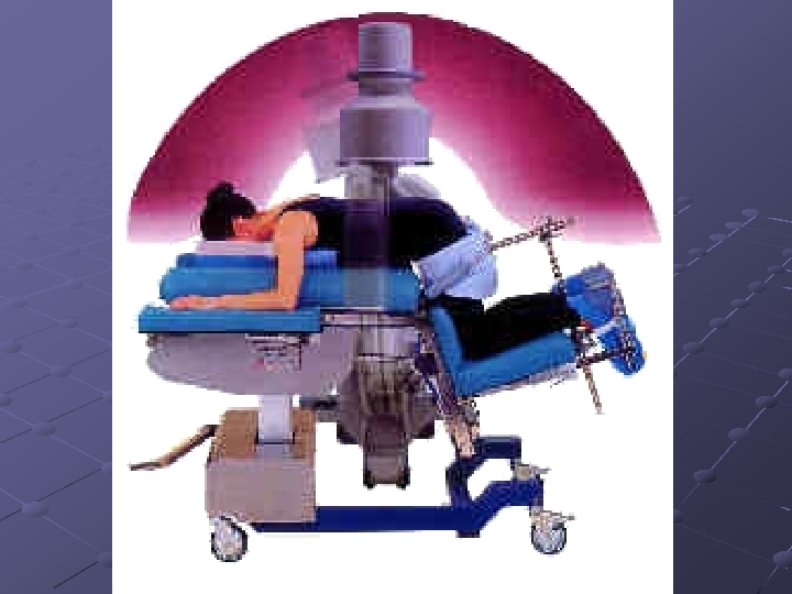
 Stereotaxis Accurate location of a definite circumscribed area within the brain from external points or landmarks on the skull Defines 3 dimensional coordinates (planes) by which to approach deep structures without damaging overlying structures
 Stereotaxis, cont. Performed under local anesthesia The patients head is placed in a stereotaxic frame Uses CT, MRI or PET to locate target site May use lasers, cryotherapy, radiation, etc.
 Ventriculoatrial or Ventriculoperitoneal Shunt Excess CSF drained to the right atrium of the heart or Peritoneal cavity Performed for treatment of hydrocephalus
 Ventriculoatrial or Ventriculoperitoneal Shunt Multi-holed ventricular catheter placed via a burr hole Reservoir and valve system directs CSF away from the ventricles Distal draining tube
 Laminectomy Removal of one or more vertebral laminae to expose the spinal canal Performed for herniated disk, compression fracture, cord tumors, etc.
, lateral or knee-chest (thoracic), sitting") Laminectomy, cont. Can be performed in the prone (lumbar), lateral or knee-chest (thoracic), sitting (cervical), or supine Spinal needle and x-ray used to check position
Laminectomy, cont. Can be performed in the prone (lumbar), lateral or knee-chest (thoracic), sitting (cervical), or supine Spinal needle and x-ray used to check position
 Laminectomy, cont. Use a moist sponge to remove tissue from the rongeur n n Bone- is discarded at most hospitals Disc- is saved and sent to pathology as a specimen
 AKA Cloward procedure Removal of the disk and") Anterior cervical disk with fusion (ACF) AKA Cloward procedure Removal of the disk and fusion of the cervical bodies Supine position, head turned slightly to the left, and right hip elevated Bone graft is taken from the iliac crest
Anterior cervical disk with fusion (ACF) AKA Cloward procedure Removal of the disk and fusion of the cervical bodies Supine position, head turned slightly to the left, and right hip elevated Bone graft is taken from the iliac crest
 Carotid artery ligation Performed to occlude the internal carotid artery Performed to control anticipated hemorrhage during intracranial surgery for vascular anomalies Permanent occlusion may be performed to control intracranial hemorrhage or for small, repeated strokes from an intracranial lesion
 Sympathectomy Excision of a portion of the sympathetic division of the autonomic nervous system Performed to treat intractable pain from certain debilitating nerve injuries, or chronic abdominal conditions (vagotomy) Position, approach, and instrumentation depends upon area to be resected
 Nerve repairs For peripheral nerve injuries Recovery will occur only if regeneration of nerve axons takes place from healthy proximal segments Nerve stimulator and microscope may be used
 Carpal tunnel syndrome Compression of the median nerve Release of the carpal ligament decompresses the nerve Usually performed as an open procedure, but may be done endoscopically
 Ulnar nerve transposition At the elbow Performed for traumatic or anatomic problems that irritate it, causing chronic discomfort Dissect the nerve free and relocate the nerve to a more anterior position

 You have now reached the END of the Neuro presentation! Have a Great Day!