

Нервная ткань1.ppt
- Количество слайдов: 59
 Нервная ткань
 Structure and function of neurons • Act through propagation of action potentials • Vary considerably in size • Dendrites, cell body and axons • May be myelinated • Synapse with other neuron or muscle cell; release neurotransmitters
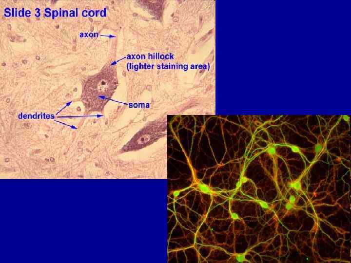
 • NEURON CELL BODY = SOMA Contains the nucleus and a nucleolus Is the major biosynthetic center Is the focal point for the outgrowth of neuronal processes Has no centrioles (hence its amitotic nature) Has well-developed Nissl bodies (rough ER) Contains an axon hillock – cone-shaped are from which axons arise
 DENDRITES – Short, unmyelinated and diffusely branched processes – They are the receptive, or input, regions of the neuron. - Conducts impulses towards the cell body – Electrical signals are conveyed as graded potentials (not action potentials) • Graded potentials (or receptor potentials when they occur in receptor cells) are short lived depolarizations or hyperpolarizations of an area of membrane. These changes cause local flows of current (current reflects the movement of ions) that decrease with distance. The magnitude of a graded potential is a direct reflection of the intensity or strength of the stimulus.
 AXONS – Arise at axon hillock : usually there is only one unbranched axon per neuron – Rare branches, if present, are called axon collaterals – Axonal terminal – branched terminus of an axon
 • • • Multipolar- many dendrites, one axon – Most neurons in CNS Bipolar- one dendrite, one axon – Sensory organs Unipolar- sensory – Axon termini extend into CNS
 • Amacrine cells are interneurons in the retina.
 Cerebellar cortex • In addition, note that the Purkinje cells are surrounded by a "basket" of axons (axons and dendrites stain black in this slide). These axons contact the Purkinje cells forming axosomatic synapses. molecular cell layer granular cell layer Purkinje cell layer

 Ventral motor neurons neuroglial cells ventral motor neuron
 Chemical synapse
 into a chemical signal back") – Is the conversion of an electrical signal (presynaptic) into a chemical signal back into an electrical signal (postsynaptic) • • • 1. nerve impulse arrives at presynaptic end bulbs 2. fusion of synaptic vesicles to PM - role for calcium 3. release of NTs 4. opening of channels in PM of postsynaptic neuron (e. g. sodium) 5. postsynaptic potential develops – depolarization & triggering of AP in postsynaptic neuron
– Is the conversion of an electrical signal (presynaptic) into a chemical signal back into an electrical signal (postsynaptic) • • • 1. nerve impulse arrives at presynaptic end bulbs 2. fusion of synaptic vesicles to PM - role for calcium 3. release of NTs 4. opening of channels in PM of postsynaptic neuron (e. g. sodium) 5. postsynaptic potential develops – depolarization & triggering of AP in postsynaptic neuron
 • Electrical Synapses – Direct physical contact between cells required – Conducted through gap junctions – Two advantages over chemical synapses • 1. faster communication – almost instantaneous • 2. synchronization between neurons or muscle fibers • Can be bidirectional • Generally associated with defensive reflexes
 1. Electrical Synapses: Communication via gap junctions between smooth muscle, cardiac muscle, and some neurons of the CNS. Provide fast, synchronized, and two-way transmission of information. 2. Chemical Synapses: Communication via chemical neurotransmitters that diffuse across a synaptic cleft. Provides slow one-way information flow
 Synapse
, segmented sheath around most long axons It functions to:") MYELIN SHEATH Whitish, fatty (protein-lipoid), segmented sheath around most long axons It functions to: – Protect the axon – Electrically insulate fibers from one another – Increase the speed of nerve impulse transmission Gaps in the myelin sheath between adjacent Schwann cells are called Nodes of Ranvier They are the sites where axon collaterals can emerge
MYELIN SHEATH Whitish, fatty (protein-lipoid), segmented sheath around most long axons It functions to: – Protect the axon – Electrically insulate fibers from one another – Increase the speed of nerve impulse transmission Gaps in the myelin sheath between adjacent Schwann cells are called Nodes of Ranvier They are the sites where axon collaterals can emerge
 Аксон моторного нейрона node of Ranvier
 Axon • Node of Ranvier. Vertical section through a node of Ranvier. The myelin sheath is interrupted at regular intervals of 2 -3 mm by the nodes of Ranvier (nr). These are the only places where the axolemma (am) (the plasma membrane surrounding the axoplasm) is exposed to the extracellular fluid. Elsewhere the axoplasm is shielded by the myelin sheath (my). Only at the nodes can ion exchange take place, and as a result myelinated nerve conducts impulses much more rapidly than unmyelinated nerve.
 • Myelin sheath. The myelin sheath is composed of hundreds of layers of adjacent membranes formed by the Schwann cell that wraps round and round the axon of the neuron. Where the two layers of Schwann cell membrane meet they form the mesaxon; as the Schwann cell grows, the mesaxon wraps round the axon of the neuron (a). Such a repeating pattern is very suitable for Xray diffraction analysis, which confirms that myelin consists of a biomolecular leaflet of phospholipids and cholesterol coated on each outer surface by a layer of protein. (b) is a transmission electron micrograph of an early stage in the formation of the myelin sheath. The mesaxon (ma) has formed a few turns round the axon (a). The outer membrane of the Schwann cells (sm) separates the extracellular space (e) from the cytoplasm of the Schwann cell (c) is a transmission electron micrograph showing a vertical section of a myelinated neuron at a later stage than (b). There are many layers of myelin (my).
 • Миелинизация аксонов в онтогенезе
 Cross Section of a Nerve Note: epineurium, perineurium, nerve fasicles
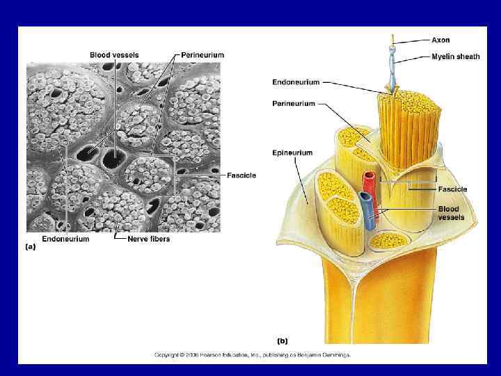
 are") • • Fibre types in peripheral nerves: • Type A fibres (myelinated) are 4 - 20 µm in diameter and conduct impulses at high velocities (15 - 120 m per second). Examples: motor fibers, which innervate skeletal muscles, and sensory fibres. • Type B fibres (myelinated) are 1 - 4 µm in diameter and conduct impulses with a velocity of 3 - 14 m per second. Example: preganglionic autonomic fibres. • Type C fibres (unmyelinated) are 0. 2 - 1 µm thick and conduct impulses at velocities ranging from 0. 2 to 2 m per second. Examples: autonomic and sensory fibres.
• • Fibre types in peripheral nerves: • Type A fibres (myelinated) are 4 - 20 µm in diameter and conduct impulses at high velocities (15 - 120 m per second). Examples: motor fibers, which innervate skeletal muscles, and sensory fibres. • Type B fibres (myelinated) are 1 - 4 µm in diameter and conduct impulses with a velocity of 3 - 14 m per second. Example: preganglionic autonomic fibres. • Type C fibres (unmyelinated) are 0. 2 - 1 µm thick and conduct impulses at velocities ranging from 0. 2 to 2 m per second. Examples: autonomic and sensory fibres.
 Аксонный транспорт
 Slow Axonal Transport: ~1 -4 mm/day Purpose: Delivery of cytosolic and cytoskeletal proteins to the nerve terminal: Fast Axonal Transport: 100 -400 mm/day Purpose: Transport organelles such as mitochondria and vesicles carrying SV and plasma membrane proteins to the nerve terminal. Also retrograde movement of vesicles containing neurotrophic factors back to the cell body.
 Cell body (“soma”) “-” “+” “Anterograde” transport Nucleus * * *") Nerve terminal (“synapse”) Cell body (“soma”) “-” “+” “Anterograde” transport Nucleus * * * “Retrograde” transport * * Microtubules Два типа аксонного транспорта
Nerve terminal (“synapse”) Cell body (“soma”) “-” “+” “Anterograde” transport Nucleus * * * “Retrograde” transport * * Microtubules Два типа аксонного транспорта
 The kinesin family: Motors for vesicle transport Transport vesicle Kinesin 2 x Light chains 2 x Heavy chains Minusend N-terminal motor domains Plusend Kinesin uses ATP hydrolysis to “walk” towards the “plus-end” of MTs similar to myosin II, may have common evolutionary origin but movement of two heads of kinesin are coordinated, unlike myosin II
 ECB 17 -18
 Аксоны в ЦНС Little regrowth after injury is possible due to the lack of a distinct tube or neurilemma
 MYELIN PNS MYELIN CNS MYELIN
 • Нейросекреторные клетки Базальная мембрана
 Neuroglial cells CNS • Much smaller than neurons and more numerous • Do not propagate action potentials • Can replace themselves (rapid mitosis in tumor formation (gliomas)
 Four types of neuroglia in CNS • Oligodendrocytes – Myelinating cells • Astrocytes – Blood-brain barrier (BBB) • Microglia – Phagocytes (from bone marrow) • Ependymal cells – Line ventricles of brain; produce cerebrospinal fluid (CSF)
 Astrocytes • • • Star-shaped cells Form blood-brain barrier by covering blood capillaries Metabolize neurotransmitters Regulate K+ balance Provide structural support
 Oligodendrocytes • Most common glial cell type • Each forms myelin sheath around more than one axons in CNS • Analogous to Schwann cells of PNS
 Microglia • Small cells found near blood vessels • Phagocytic role -- clear away dead cells • Derived from cells that also gave rise to macrophages & monocytes
 and activated (right) microglia. Note the highly ramified branches of the unactivated") Unactivated (left) and activated (right) microglia. Note the highly ramified branches of the unactivated мicroglia. The activated microglia have an enlarged cell body with shorter, stouter branches.
Unactivated (left) and activated (right) microglia. Note the highly ramified branches of the unactivated мicroglia. The activated microglia have an enlarged cell body with shorter, stouter branches.
 • This is an example of neuronophagia in which a dying neuron is surrounded by microglial cells.
 Ependymal cells • Form epithelial membrane lining cerebral cavities & central canal • Produce cerebrospinal fluid (CSF)
 Neuroglia of the PNS • Schwann cells – Myelinating cells – Help direct axon regeneration • Satellite cells – Support, protection, regulation of molecular exchange
 Schwann Cell • Cells encircling PNS axons • Each cell produces part of the myelin sheath surrounding an axon in the PNS
 • The satellite cells are concentrically located aroung the individual pseudounipolar cells of the ganglia. They stain darker than the adjacent ganglion cells. Support neurons in the PNS ganglia
 Липофусцин • Lipofuscin is the undigested residue of subcellular lytic reactions. As organelles become aged and useless, they are broken down for their components, and what is left that can't be salvaged is the pigment you see here. It tends to be greater in older animals. Since neurons do not divide, they tend to accumulate the pigment over the years.
 • Alcohol reduces new neuron dendritic growth.
 Alzheimer's disease Alois Alzheimer in 1906 performed • This photo is obtained from a brain with Alzheimer's disease. The senile plaques (white arrow) and neurofibrillary tangles (black arrow) are well demonstrated.
 • Beta-amyloid results when two enzymes, BACE and PS 1, cut a larger protein in the cell membrane of brain cells called APP. Newly formed molecules of beta-amyloid then aggregate into plaques. Caffeine reduces the level of both BACE and PS 1 enzymes, resulting in less beta-amyloid in the brain.
 Neurofibrillary Tangles Neurons have an internal support structure partly made up of microtubules. A protein called tau helps stabilize microtubules. In AD, tau changes, causing microtubules to collapse, and tau proteins clump together to form neurofibrillary tangles.
 Alzheimer’s Disease • • • Degenerative brain disorder 4 million Americans 10% of all people over 65 50% of all people over 85 19 million people are family members of an Alzheimer’s patient • 22 million people worldwide will be diagnosed by 2025
 Neuronal Storage Disease • Result from inborn errors of metabolism (deficient enzyme or abnormal lysosomal function) • Accumulation of metabolic products in the neuron – Tay Sachs disease – Neuronal ceroid lipofuscinosis – Glycogen storage disease
 • Accumulation of metabolic products in the neuron
 • β-Mannosidase, a lysosomal enzyme which acts exclusively at the last step of oligosaccharide catabolism in glycoprotein degradation, functions to cleave the unique β-linked mannose sugar found in all N-linked oligosaccharides of glycoproteins. Deficiency of this enzyme results in βmannosidosis, a lysosomal storage disease characterized by the cellular accumulation of small oligosaccharides.
 • At the left, an H and E stain demonstrates a rounded pink cytoplasmic Lewy body in a neuron of the cerebral cortex from a patient with diffuse Lewy body disease, which can be a cause for dementia. Lewy bodies can also be seen in substantia nigra with Parkinson's disease. An immunoperoxidase stain for ubiquitin, seen at the right, helps demonstrate the Lewy bodies more readily.
 as the") • Born on September 9 th, 1923 in Yonkers (New York) as the son of immigrant parents from Slovakia and Hungary. Shortly afterwards, on a trip to New Guinea, he investigated a mysterious degeneration of the brain among members of the Fore tribe. He discovered that this so-called Kuru illness was caused not by degeneration, but by a slow virus which may have been transmitted years before, and is triggered by ritual cannibalism.
• Born on September 9 th, 1923 in Yonkers (New York) as the son of immigrant parents from Slovakia and Hungary. Shortly afterwards, on a trip to New Guinea, he investigated a mysterious degeneration of the brain among members of the Fore tribe. He discovered that this so-called Kuru illness was caused not by degeneration, but by a slow virus which may have been transmitted years before, and is triggered by ritual cannibalism.
![Stanley Ben Prusiner (born May 28, 1942[1]) is an American neurologist and biochemist. •](https://present5.com/presentation/3/98148737_244148771.pdf-img/98148737_244148771.pdf-57.jpg "Stanley Ben Prusiner (born May 28, 1942[1]) is an American neurologist and biochemist. •") Stanley Ben Prusiner (born May 28, 1942[1]) is an American neurologist and biochemist. • Proteinaceous Infectious Particle PRION
Stanley Ben Prusiner (born May 28, 1942[1]) is an American neurologist and biochemist. • Proteinaceous Infectious Particle PRION
 Прионные заболевания человека • Kuru • Creutzfeldt-Jakob disease • Fatal familial insomnia
 • A haematoxylin and eosin stained section of cerebral cortex from a case of variant Creutzfeldt-Jakob disease showing the characteristic florid plaque structure, comprising a glassy centre with radiating fibrils surrounded by a halo of spongifirm change.