
Neoplasms of the Nose and Paranasal Sinuses Kevin


- Размер: 746.5 Кб
- Количество слайдов: 36
Описание презентации Neoplasms of the Nose and Paranasal Sinuses Kevin по слайдам
Neoplasms of the Nose and Paranasal Sinuses Kevin Katzenmeyer, MD Anna Pou, MD June 7,
Sinonasal Neoplasms 3% of aerodigestive malignancies 1% of all malignancies 2 to 1 males Sixth to seventh decades Symptomatology difficult
Sinonasal Neoplasms Nasal cavity (benign = malignant) Benign — inverting papilloma Malignant — SCCA Sinuses (malignant) SCCA Maxillary most common
Epidemiology Occupational exposure in >40% nickel workers — SCCA hardwood dust & leather tanning — adenoca Viral — HPV Cigarettes & alcohol
Presentation Similar sx to common problems 6 to 8 month delay in diagnosis Cranial neuropathies & proptosis RAR
Presentation Oral — 30% tooth pain, trismus, palatal fullness, erosion Nasal — 50% obstruction, epistaxis, discharge, erosion Ocular — 25% diplopia, proptosis, tearing, pain, fullness Facial V 2 numbness, asymmetry, pain Auditory — CHL
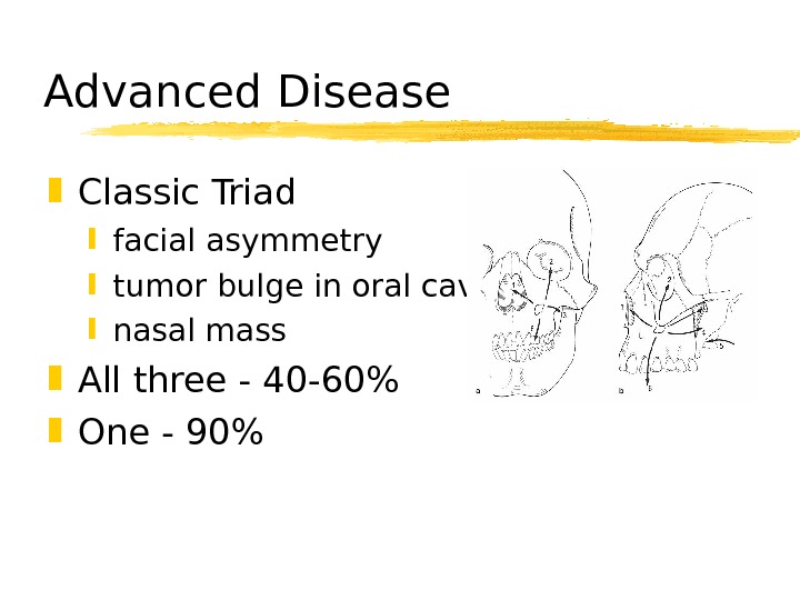
Advanced Disease Classic Triad facial asymmetry tumor bulge in oral cavity nasal mass All three — 40 -60% One — 90%
Diagnosis Physical exam Nasal endoscopy Biopsy Radiography
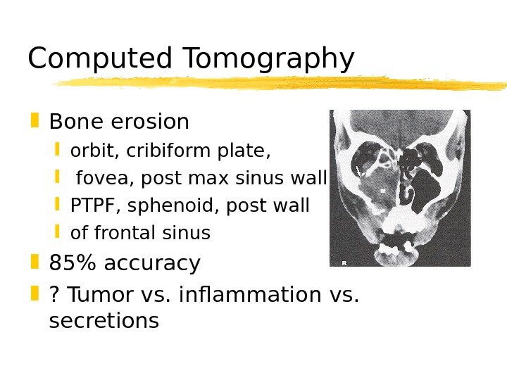
Computed Tomography Bone erosion orbit, cribiform plate, fovea, post max sinus wall, PTPF, sphenoid, post wall of frontal sinus 85% accuracy ? Tumor vs. inflammation vs. secretions
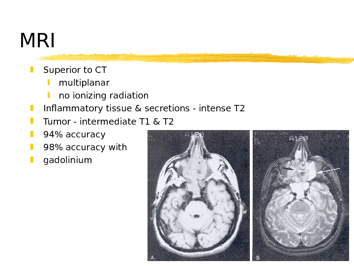
MRI Superior to CT multiplanar no ionizing radiation Inflammatory tissue & secretions — intense T 2 Tumor — intermediate T 1 & T 2 94% accuracy 98% accuracy with gadolinium
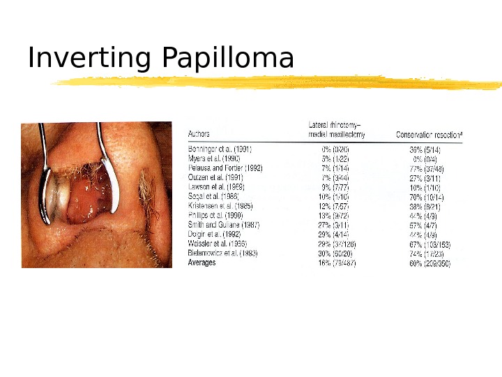
Schneiderian Papillomas Fungiform (50%) — septum Cylindrical (3%) — lateral nasal wall Inverting (47%) — lateral nasal wall recurs, locally destructive, malignant potential men, 6 th-7 th decades, unilateral SCCA — 2 -13% Recurrence — 0 -80%
Inverting Papilloma
Osteomas Benign, slow-growing 15 to 40 years frontal > ethmoid > maxillary local excision
Fibrous Dysplasia Normal bone replaced by collagen, fibroblasts, and osteoid material < 20 years ground-glass appearance treatment? No irradiation
Neurogenic tumors Schwannomas surface of nerve fibers no malignant degeneration along trigeminal & ANS Neurofibromas within nerve fibers von Recklinghausen’s disease malignant degeneration in 15% Complete excision
SCCA Most common — 80% Max > nasal cavity > ethmoids Males Sixth decade 90% have eroded walls of sinuses
Adenoid Cystic Carcinoma Palate > major salivary glands > sinuses Resistant to tx Multiple recurrences, distant mets Perineural spread Long-term followup necessary
Mucoepidermoid Carcinoma rare, widespread local invasion Adenocarcinoma 2 nd most common, 5 -20% ethmoids occupational exposures
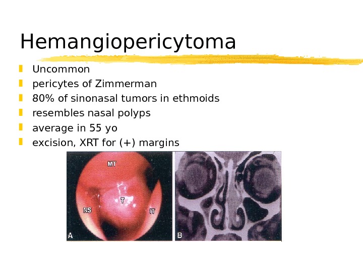
Hemangiopericytoma Uncommon pericytes of Zimmerman 80% of sinonasal tumors in ethmoids resembles nasal polyps average in 55 yo excision, XRT for (+) margins
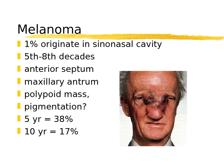
Melanoma 1% originate in sinonasal cavity 5 th-8 th decades anterior septum maxillary antrum polypoid mass, pigmentation? 5 yr = 38% 10 yr = 17%
Olfactory Neuroblastoma Neural crest origin no urinary VMA or HVA bimodal distribution at 20 and 50 locally aggressive rosettes are hallmark Kadish staging local recurrence 50 -75% metastasis 20 -30%
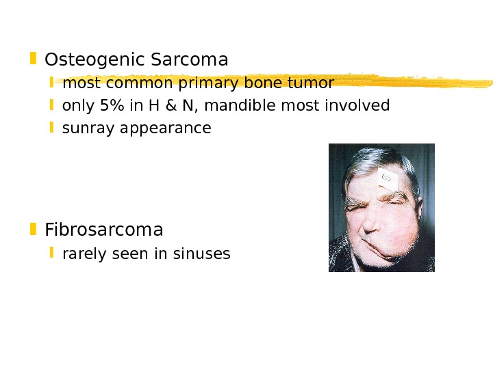
Osteogenic Sarcoma most common primary bone tumor only 5% in H & N, mandible most involved sunray appearance Fibrosarcoma rarely seen in sinuses
Chondrosarcoma 3 rd-5 th decades histologic dx difficult slow erosion of skull base, (+) margins Rhabdomyosarcoma most common in children 35 -45% in H&N, 8% in sinuses embryonal, alveolar, pleomorphic triple tx
Lymphoma bimodal presentation NHL irradiation +/- chemo Extramedullary plasmacytoma 40% in paranasal sinuses/nose “ benign” must r/o myeloma excision or irradiation
Metastatic tumors Renal cell carcinoma lungs breasts urogenital tract gastrointestinal tract Palliation necessary
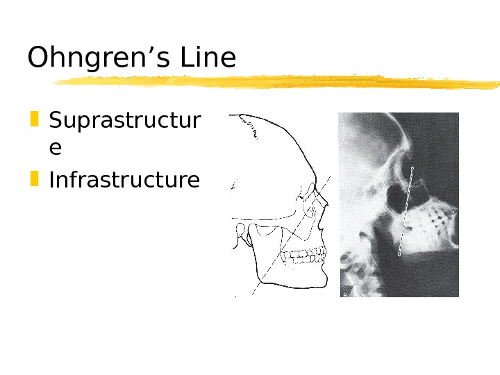
Ohngren’s Line Suprastructur e Infrastructure
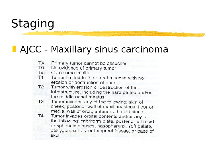
Staging AJCC — Maxillary sinus carcinoma
Treatment T 3 and T 4 60% local recurrence Surgery Irradiation Chemotherapy
Surgical resection Unresectability (Sisson) extension to frontal lobes invasion of prevertebral fascia bilateral optic nerve involvement cavernous sinus extension
Surgical resection Endoscopic excision WLE medial maxillectomy total maxillectomy radical maxillectomy +/- exenteration craniofacial resection
Orbital Preservation Harrison — proptosis, limitation of EOM, bony erosion of orbit = exenteration Conley — save eye whenever possible Sisson — preoperative XRT, decreased exenterations without change in survival Stern — nonfunctional eye without inf/med support = exenteration
Orbital preservation UVA — Mc. Cary & Levine 50 Gy preop XRT to orbit periorbital bx resect (+) periorbita functional eye
Pterygopalatine Fossa 10 -20% involvement Som — PTPF invasion = unresectable lesion Craniofacial resection (MCF) Postop XRT
Neck Dissection Retropharyngeal and jugulodigastric nodes 10% (+) necks neck dissection palpable nodes radiographic evidence of disease 40% cervical mets at 4 yrs
Radiation therapy Primary tx only for palliation 10 -15% improved 5 year survival XRT = 23% vs. Surgery + XRT = 44% preoperative vs. postoperative protection of CNS and globe XRT 12 -20% unilateral visual loss, 0 -8% bilateral visual loss Surgery 10 -20% useless globes, 2 X with XRT
Chemotherapy Palliation, unresectable disease (+) margins, perineural spread, surgical refusal, ECS Intraarterial chemotherapy Robbins — 86% response of T 4 lesions Lee — 91% satisfactory response