Нейрогенез_спин мозг_птицы-15.ppt
- Количество слайдов: 28


Нейроны: униполярный биполярный псевдоуниполярный мультиполярный веретеновидный, грушевидный треугольный многоугольный Глиальные клетки эпендимная глия макроглия танициты эпителиоидная эпендимная глия астроциты олигодендроциты


Основые этапы нейрогенеза: • • • Индукция и разметка презумптивной нервной пластинки. Рождение и миграция нейронов и глии. Определение судьбы нейронов. Рост аксонов к специфическим мишеням. Образование синаптических связей. Влияние трофических факторов на выживание и дифференцировку. • Перестройка функциональных синапсов. • Пластичность синаптических связей в течение всей жизни организма.

Строение нервной трубки: псевдомногослойный герминативный нейроэпителий

Интеркинетическая миграция ядер в нейроэпителии
. Nuclei are moved")
G 1 Fig. 4. Proposed model of interkinetic nuclear migration (INM). Nuclei are moved by actin contraction and along microtubules. Short nuclear movements may be accomplished by actin alone. Longer nuclear movements may require microtubules. Nuclei move basally during G 1 phase, reaching a peak distance during S-phase. Nuclei begin moving apically along microtubules using the dynein motor protein during G 2 phase. Tpx 2 initiates apical nuclear migration at G 2 phase. After cilia are lost, centrosomes can move to the nucleus during late G 2 to initiate nuclear envelope breakdown and apical rounding, dependent on actin. Cytoplasm of the cells are shown in green, and nuclei are shown in blue. Centrosomes are red dots and cilia are magenta. Spear, Erickson, 2012

Симметричные и несимметричные митозы в нейроэпителии

Функции интеркинетической миграции ядер: • • • морфогенез эпителиального пласта поддержание структуры и полярности пласта регуляция нейрогенеза и пролиферации


спинной мозг мозжечок головной мозг

 Diagram of a cortical")
Figure 12. 18. Neuronal migration on glial cell processes. (A) Diagram of a cortical neuron migrating on a glial cell process. (B) Electron micrograph of the region where the neuronal cell body adheres to the glial process. (C) Sequential photographs of a neuron migrating on a cerebellar glial process. The leading process has several filopodial extensions. The neuron reaches speeds around 40 mm/hr as it progresses on the glial process. (A after Rakic 1975; B from Gregory et al. 1988; C from Hatten 1990; photographs courtesy of M. Hatten. )


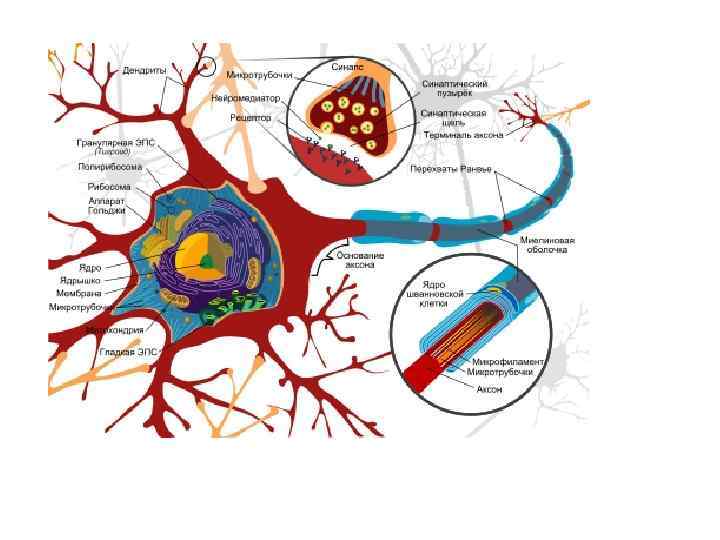
Строение мотонейрона
 The")
Развитие спинного мозга Figure 12. 17. Development of the human spinal cord. (A-D) The neural tube is functionally divided into dorsal (alar) and ventral (basal) regions, separated by the sulcus limitans. As cells from the adjacent somites form the spinal vertebrae, the neural tube differentiates into the ependymal, mantle, marginal zones, and the roof and floor plates. (E) A segment of the spinal cord with its sensory (alar) and motor (basal) roots. (After Larsen 1993. )

")
Дорсовентральная спецификация нервной трубки Figure 12. 13. Dorsal-ventral specification of the neural tube. (A) The newly formed neural tube is influenced by two signaling centers. The roof of the neural tube is exposed to BMP 4 and BMP 7 from the epidermis, and the floor of the neural tube is exposed to Sonic hedgehog protein from the notochord. (B) Secondary signaling centers are established within the neural tube. BMP 4 is expressed and secreted from the roof plate cells; Sonic hedgehog is expressed and secreted from the floor plate cells. (C) BMP 4 establishes a nested cascade of TGF-b-related factors, spreading ventrally into the neural tube from the roof plate. Sonic hedgehog diffuses dorsally as a gradient from the floor plate cells. (D) The neurons of the spinal cord are given their identities by their exposure to these gradients of paracrine factors. The amount and type of paracrine factors present cause different transcription factors to be activated in the nuclei of these cells, depending on their position in the neural tube. (E) Chick neural tube, showing areas of Sonic hedgehog (green) and Dorsalin expression (blue). Motor neurons induced by a particular concentration of Sonic hedgehog are stained orange/yellow. (Photograph courtesy of T. M. Jessell. )

Индукция хордой норма нервная трубка донная пластинка хорда дополнительная хорда= эктопическая донная пластинка 2 хорды=широкая донная пластинка

Индукция донной пластинкой хорда между нервными валиками=доп. донная пластинка вместо кроющей пластинки доп. донная пластинка= эктопическая донная пластинка доп. дорсальная донная пластинка=эктопическая донная пластинка вместо кроющей пластинки


 А – 3 дня; В – 4")
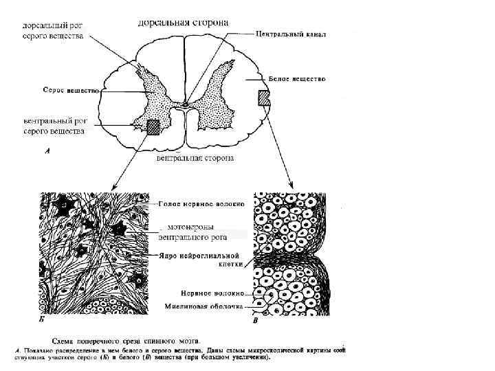
Цитоархитектоника спинного мозга куриных зародышей (метод Гольджи) А – 3 дня; В – 4 дня; С – 5 дней, двигательные (эфферентные) волокна Д 1 - Д 3 – 7 дней, разные волокна Е 1 – Е 2 – 9 дней, коллатерали в центре серого вещества, нейроглия и эпендимные клетки
Нейрогенез_спин мозг_птицы-15.ppt